Abstract
Background
Vibration-induced finger flexion reflex (VFR) in the upper extremity is inhibited by needle insertion acupuncture to the large intestine 4 (LI4) at the hand. This claim has a limitation because the inhibitory effect is deduced only from reduction in the maximum finger flexion (FF) force during the tonic flexion reflex by vibratory stimulation after acupuncture.
Methods
The study was a crossover design with two conditions—acupuncture and control—to which 16 healthy volunteers were subjected. VFR in the upper extremity was induced by applying vibratory stimulation on the volar side of the middle fingertip of the right hand, before and after acupuncture at the right LI4 in 16 healthy volunteers. We measured the area under the curve (AUC) of finger flexion force and surface electromyogram (EMG) in the flexor muscles, in addition to the maximum FF force during vibratory stimulation. We compared AUC, surface EMG and maximum FF force in the acupuncture condition with those in the control condition. We also estimated the correlation between AUC, surface EMG and maximum FF force.
Results
AUC, surface EMG and maximum FF force were significantly reduced (p <0.01) after acupuncture compared with those of the control group. A strong correlation was observed in maximum FF force versus AUC (r=0.98, p <0.01) and surface EMG (r=0.77, p <0.01).
Conclusions
Acupuncture at ipsilateral LI4 inhibited tonic activities in the finger flexor muscles during VFR, which suggests that afferent input with needle penetration has inhibitory effect on the motor neuronal activities in the reflex circuits of VFR.
Introduction
Clinically, it is well known that needle insertion during acupuncture reduces muscle tenderness. We assume this phenomenon is in part due to the suppression in motor neuronal activities of the skeletal muscles by needle insertion. Despite its well-known effectiveness, there are a few studies on effectiveness of acupuncture on the activities of motor neurons of the skeletal muscles in humans.
To evaluate the effectiveness of acupuncture on somato-motor neuronal activities, we used vibration-induced finger flexion reflex (VFR)1 in which vibration to the fingertip activates efferent neurons of the finger flexor muscles. We have previously reported that the inhibitory effect of needle insertion of acupuncture on VFR is induced by vibration on the volar side of the fingertip in the upper extremity.2–9 However, inhibitory effect on VFR was concluded only from the reduction in the maximum finger flexion (FF) force during vibratory stimulation.2 6–9 The maximum value of flexion force at a peak point during the tonic flexion reflex does not reflect whole activities of VFR. Thus, only a decrease in the maximum FF force would not be sufficient and therefore, poses a limitation to the claim that acupuncture has inhibitory effect on VFR. Further, VFR responses to acupuncture treatment in individual subjects were not evaluated in the previous study.6
In this study, we measured the area under the curve (AUC) of FF force and surface electromyogram (EMG) in the flexor muscles, in addition to the maximum FF force during vibratory stimulation, to determine the inhibitory effect of acupuncture on VFR. The aim of this study was to obtain more convincing evidence of the inhibitory effect of acupuncture on motor neuron activities of the flexor muscle in the upper extremity, thus allowing us to determine whether noxious somatosensory stimulation by acupuncture has inhibitory effect on VFR in the upper extremity.
Methods
Sixteen healthy volunteers (mean±SD, 29.9±5.4 years; eight males, eight females) who were familiar with acupuncture treatment participated in the study. This experiment had a crossover design with needle insertion of acupuncture and the control condition. All subjects gave written informed consent after the purpose and format of the study were explained to them. Each experiment was performed at approximately the same time on different days. The Ethics Committee of Showa University approved this study.
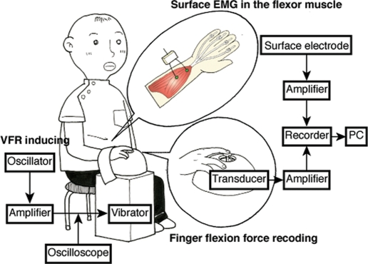
Details of VFR induction are shown in figure 1. The volar side of the middle fingertip was placed on a pressure transducer attached to a vibrator. Subjects then pressed the pressure transducer lightly as a background contraction. When the background contraction became stable, 60 Hz vibration with 1 mm displacement amplitude was applied to the volar side of the middle fingertip for 20 s.6–12 Vibration was delivered using an electromagnetic vibrator (FF 225N; Foster Electric Co, Ltd) driven by a sine-pulse generator (VP-7421A Function Generator; Matsushita Electrical Industrial Co, Ltd) coupled to a power amplifier (1706; Bose Corp). The output from the amplifier to the vibrator was monitored by a digital oscilloscope (Tectronix TDS 360P; Sony Corp) in order to measure the driving force of the vibrator, which was found to be 3.5–4.0 V. The subjects were blindfolded throughout the trial. Tonic FF force induced by the vibration was measured isometrically, using a force transducer (9E01-L2-5K; NEC San-ei Instruments, Ltd) placed between an attachment of the vibrator and the fingertip. Force measurements were then recorded using a BioLog (DL-1000; S&ME, Inc) through an amplifier (DL-170; S&ME, Inc), with EMG analyser software (m-Scope; S&ME, Inc) installed in the PC (PC-VY12MEX84EHM NEC). We measured maximum FF force of VFR during vibratory stimulation according to previous studies.2 6–9 We also measured the AUC of FF force during vibratory stimulation and simultaneously recorded the surface EMG in the flexor muscles. The raw electrical signals were recorded using a BioLog (DL-1000; S&ME, Inc) through an amplifier (DL-140; S&ME, Inc), with EMG analyser software (m-Scope; S&ME, Inc) installed in the PC (PC-VY12MEX84EHM NEC). The signals were stored at a sampling frequency of 1000 Hz and the data were analysed. The EMG analyser was fully rectified and smoothed by a low-pass filter with a time constant of 50 ms. The analysed value was used with integrated surface EMG.
Figure 1.
Illustration of experimental settings. Vibration at 60 Hz was applied to the volar side of the middle fingertip.
Respective flexion forces and surface EMG of VFRs were measured before and after acupuncture, that is, at 10 min (before 1) and 5 min before (before 2) acupuncture and every 5 min immediately (after 1) to 40 min after needle removal (after 9). Control VFR was measured using the same time course without acupuncture for the same 16 subjects. An acupuncturist applied a needle at acupoint LI4 point in the right dorsal hand of the subjects, using the alternating twirling technique (rotating the needle clockwise and counterclockwise alternately). The needle was inserted to a depth of 5 mm and remained at the place for 5 min (in-situ technique). No manipulation was performed during the in-situ technique. Sterilised stainless needles of 40 mm in length and 0.16 mm (Seirin) were used. After each needle application, we asked the subject to rate de qi on a visual analogue scale (VAS) ranging from zero (no de qi) to 100 (the most intense de qi).
Statistical analysis
The maximum FF force, AUC and integrated surface EMG at 5 min before acupuncture and at each time point after removal of acupuncture were expressed in percentages of values obtained 10 min before acupuncture, according to previous studies.7 8 12 Statistical comparisons were performed on the acupuncture and control conditions in relation to the maximum FF force, AUC and integrated surface EMG during VFR, using a two-way repeated-measures analyses of variance with ‘time’ and ‘group’ as within-subject factors. We then compared the groups at each measured time point, using paired t test. Pearson's correlation coefficient was used to indicate relationships between the maximum FF force, AUC and integrated surface EMG. Pearson's correlation coefficient was also used to indicate the relationship between de qi and the maximum FF force, AUC and integrated surface EMG, respectively. For comparison of VAS scores in de qi, Mann–Whitney's U test was used. All statistical analyses were performed using SPSS V.15.0J.
Results
Acupuncture stimulation applied to LI4 produced a prominent decrease in maximum FF force, AUC and integrated surface EMG in the flexor muscles during vibratory stimulation.
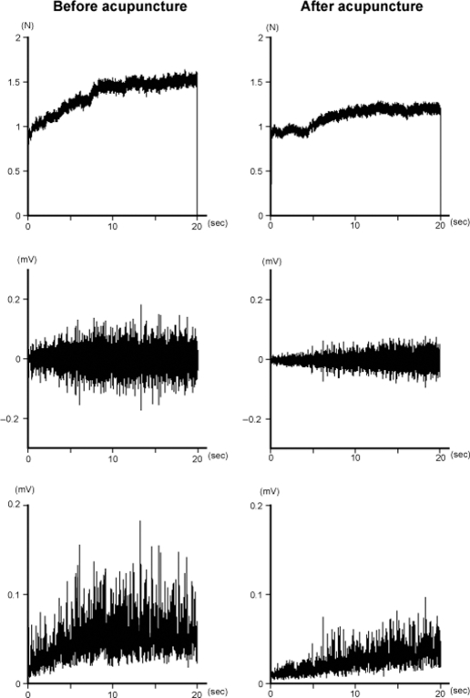
Figure 2 shows clear decrease in maximum FF force, AUC and integrated surface EMG during VFR after acupuncture as compared with the values before acupuncture in a typical subject. The maximum FF force of 1.52 N, AUC of 23.65 N/s and integrated surface EMG of 0.99 mV/s before acupuncture (before 2) decreased to 1.27 N, 18.58 N/s and 0.55 mV/s, respectively, after removal of the needle; however, those in the control condition did not decrease.
Figure 2.
Records of finger flexion force (upper), surface electromyogram (EMG) (middle) and integrated surface EMG (lower) during vibration-induced finger flexion reflex (VFR) for a subject before and after acupuncture. Maximum finger flexion force, area under the curve of finger flexion force and surface EMG during VFR decreased markedly after acupuncture compared to those before acupuncture in a typical subject.
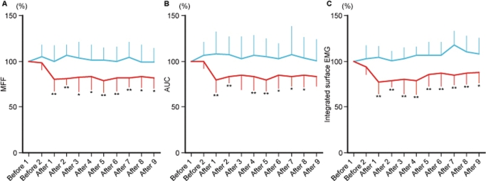
With regard to the overall mean±SD (%) changes (figure 3) in the 16 subjects, there were significant differences between the groups (Greenhouse-Geisser correction; (1) F=19.20, p=0.001; (2) F=19.20, p=0.001; (3) F=19.20, p=0.001) and time (Greenhouse-Geisser correction; (1) F=2.42, p=0.037; (2) F=19.20, p=0.001; (3) F=19.20, p=0.001), with no interaction between the two factors (Greenhouse-Geisser correction; (1) F=1.62, p=0.167; (2) F=19.20, p=0.001; (3) F=19.20, p=0.001) in (1) maximum FF force, (2) AUC and (3) integrated surface EMG, respectively. For these 16 subjects, mean±SD (%) changes in maximum FF force, AUC and integrated surface EMG for the acupuncture group at all points from after 1 to after 9 were significantly less (paired t test; p <0.05) than those for the control group except after 3 and 9 in AUC. However, mean±SD (%) change in these factors for the acupuncture group at before 2 was not significantly less (paired t test, p=0.13) than that for the control group.
Figure 3.
Changes in mean (SD) (A) maximum finger flexion force (MFF), (B) area under the curve (AUC) of finger flexion force (C) and integrated surface EMG for 16 subjects in the acupuncture condition (red line) and control condition (blue line). Vertical axis is the percentage of before 1 value of MFF, AUC and integrated surface EMG and the horizontal axis is time. *p <0.05, **p < 0.01.
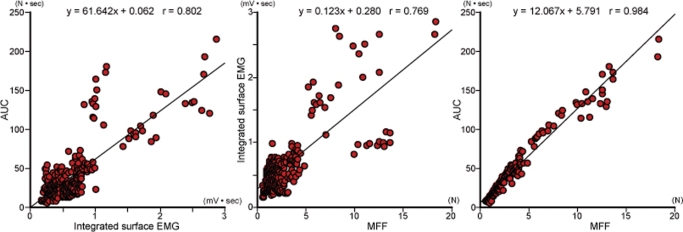
Furthermore, we tested whether there were positive correlations between maximum FF force, AUC and integrated surface EMG (figure 4) and significant positive correlation was revealed between AUC and integrated surface EMG (r=0.80, p<0.01), maximum FF force and integrated surface EMG (r=0.77, p<0.01) and maximum FF force and AUC (r=0.98, p<0.01). These results suggest that maximum FF force, used as an indication of VFR in previous studies, is an appropriate representative for VFR. However, although correlation was observed between individual maximum FF force and AUC in each subject, in seven of the 16 subjects, individual integrated surface EMGs did not correlate with maximum FF force and/or AUC (table 1). Further, both AUC and maximum FF force after acupuncture treatment did not decrease in one subject (No 3). AUC did not decrease, despite a decrease in the maximum FF force in two subjects (No 2, 9) and the maximum FF force did not decrease, despite a decrease in AUC in one subject (No 14) (table 1). We presumed these four subjects were non-responders to acupuncture treatment in VFR inhibition and the others were responders.
Figure 4.
Correlation between area under the curve (AUC) of finger flexion force and integrated surface electromyogram (EMG) (left), integrated surface EMG and maximum finger flexion force (MFF) (middle) and AUC and MFF (right).
Table 1.
Average values of MFF, AUC and IEMG from ‘after 1’ to ‘after 9’ (after acupuncture) in control condition and acupuncture condition and correlation between the MFF, AUC and IEMG for each of the 16 subjects
| MFF (%) | AUC (%) | IEMG (%) | Correlation | ||||||
|---|---|---|---|---|---|---|---|---|---|
| Subject | Control condition | Acupuncture condition | Control condition | Acupuncture condition | Control condition | Acupuncture condition | AUC and IEMG | IEMG and MFF | AUC and MFF |
| No 1 | 107.6 | 83.8 | 110.2 | 83.1 | 106.7 | 59.8 | r=0.756† | r=0.741† | r=0.974† |
| No 2 | 94.8 | 80.8 | 90.1 | 94.1 | 104.4 | 93.7 | r=0.382 | r=0.413 | r=0.920† |
| No 3 | 103.3 | 103.4 | 98.1 | 98.8 | 118.0 | 92.5 | r=0.462* | r=0.075 | r=0.710† |
| No 4 | 133.0 | 87.1 | 134.1 | 77.9 | 110.6 | 84.8 | r=0.773† | r=0.809† | r=0.979† |
| No 5 | 102.0 | 88.4 | 108.8 | 80.9 | 115.5 | 76.2 | r=0.667† | r=0.478* | r=0.953† |
| No 6 | 121.5 | 54.6 | 129.3 | 69.1 | 103.8 | 78.3 | r=0.776† | r=0.826† | r=0.981† |
| No 7 | 112.7 | 77.9 | 150.7 | 79.9 | 116.1 | 81.2 | r=0.733† | r=0.710† | r=0.962† |
| No 8 | 92.2 | 74.7 | 89.1 | 67.4 | 116.0 | 97.9 | r=0.509* | r=0.447* | r=0.983† |
| No 9 | 92.5 | 91.1 | 94.1 | 117.0 | 107.0 | 89.8 | r=0.003 | r=0.321 | r=0.759† |
| No 10 | 84.4 | 81.3 | 88.2 | 86.7 | 102.7 | 84.8 | r=0.359 | r=0.463* | r=0.932† |
| No 11 | 94.8 | 72.9 | 92.5 | 70.7 | 97.7 | 57.8 | r=0.816† | r=0.790† | r=0.974† |
| No 12 | 104.0 | 86.8 | 109.9 | 91.3 | 120.6 | 94.7 | r=0.764† | r=0.686† | r=0.945† |
| No 13 | 104.4 | 83.6 | 112.3 | 89.3 | 102.4 | 88.8 | r=0.279 | r=0.350 | r=0.965† |
| No 14 | 88.4 | 88.7 | 88.9 | 81.5 | 97.1 | 96.2 | r=0.156 | r=0.187 | r=0.904† |
| No 15 | 90.7 | 79.3 | 95.5 | 71.3 | 86.6 | 98.2 | r=0.161 | r=0.205 | r=0.955† |
| No 16 | 97.8 | 73.7 | 94.3 | 75.7 | 107.7 | 59.9 | r=0.692† | r=0.710† | r=0.974† |
p <0.05,
p <0.01.
AUC, area under the curve of finger flexion force; IEMG, integrated surface electromyogram; MFF, maximum finger flexion force.
Of the 16 subjects, nine (including subjects No 2 and 14) felt de qi but other seven subjects did not (including subjects No 3 and 9). In de qi with the needle insertion for the four non-responders, the median (mean±SD) was 1.5 (3.0±4.2) on the VAS. For the 12 responders, the median (mean±SD) was 1.5 (1.8±2.0) in de qi, which was not significantly different from that in the non-responders. There was not significant correlation between intensity of de qi and maximum FF force (r=−0.26, p=0.34), AUC (r=−0.24, p=0.38) and integrated surface EMG (r=−0.07, p=0.81), respectively.
Discussion
The maximum FF force, AUC of the FF force and surface EMG in the flexor muscles during VFR were suppressed by acupuncture at LI4 on the ipsilateral hand. The results showed that acupuncture inhibited tonic muscle activities during VFR, and maximum FF force of VFR could be a good representative of the activities in the finger flexor muscle during VFR. However, in 44% subjects, integrated surface EMG did not correlate with maximum FF force and/or AUC, despite decreases in their individual surface EMGs after acupuncture treatment. This might be because the EMG signal may still contain some noise that cannot be eliminated, even after electrical noise, mechanical artefacts and cross talk are eliminated.13 To investigate an individual's VFR response to acupuncture treatment, AUC might be more reliable and better reflect the maximum FF force than surface EMG, as maximum FF force and AUC were well correlated in all individuals.
In a healthy human, application of vibratory stimulation on the volar side of the fingertip induces a flexion reflex. Typically, FF force occurs with the onset of vibration and increases progressively during vibration.1–11 14 15 The receptor of this reflex is assumed to be the skin mechanoreceptor, because VFR is markedly reduced when the fingertip is anaesthetised or cooled.1 3 11 14 This reflex is assumed to have two reflex arcs from the cross-correlogram between vibratory stimuli and motor unit spikes in the flexor muscle, that is, the spinal short loop and supraspinal long loop.5 11 15 This flexion reflex is considered a good experimental model to investigate the mechanism of acupuncture treatment because the reflex circuit is well understood. Interestingly, the activities in both these loops are suppressed by acupuncture at the Waiguan point in the upper extremity, and suppression on the supraspinal long loop is relatively long lasting compared with that on the short loop.5 Thus, we presume that the activities in the long loop reflex circuit is mainly suppressed by sensory input from LI4 elicited by needle insertion, and therefore, a continuous decrease of VFR was observed after removal of a needle.
Current assumptions for afferent fibre participation in inducing efficacies by needle insertion are A delta or C fibres.16 Pain-eliciting transcutaneous electrical stimulation inhibits VFR, but non-pain transcutaneous electrical stimulation does not inhibit VFR.12 Needle penetration is a noxious stimulus.16 Furthermore, inhibitory effects induced by needle insertion with the manipulation giving stronger stimulation are remarkably larger than that by simple needle insertion.7 Thus, we concluded that activities in pain-conducting afferent fibres by acupuncture may participate importantly in VFR inhibition, and pain-conducting afferent fibres have an inhibitory connection to the motor neurons in the spinal cord, because high-intensity noxious transcutaneous electrical nerve stimulation inhibits motor neuronal excitability in the upper extremity.17–20
In this study we first showed the relationship between de qi, which has been considered essential for success in acupuncture treatment,16 and inhibitory effect on VFR. Considering involvement of pain-conducting afferent fibres in inducing de qi,16 it was assumed that de qi could ensure or enhance inhibitory effect on VFR. However, we have found that no significant difference in de qi between the responders and the non-responders, and no significant correlation between intensity of de qi and inhibitory effect on VFR. Moreover, five of the 12 responders did not feel de qi and two of the four non-responders felt de qi. These results indicate that de qi was not essential to induce VFR inhibition or did not enhance it as well as acupuncture analgaesia.21 We did not conduct additional needle manipulation after needle insertion to provide uniform direction and depth of needle insertion to ensure homogeneity of stimulation in all subjects in this study. Further studies are required to determine the relationship between intensity of VFR inhibition and de qi elicited during the period of post-needle insertion.
Whether acupuncture inhibited VFR by suppression onto the afferent activities or by suppression onto the motor neuron in VFR neural circuits is an interesting question. There have been several evidences that painful stimulation of the cutaneous afferents inhibits the spinal motor neurons and that there is convergence between nociceptive and non-nociceptive afferents of different origins onto the common interneurons in segment reflex pathways to α-motor neurons.22 23 Considering the evidence that sensory perception in the finger does not change by electrical stimulation to the forearm,18 we conclude that noxious somatosensory input using acupuncture suppressed the motor neurons innervated to the flexor muscles through common interneurons, which facilitates the motor neurons by vibration in the spinal cord9; however, this input did not suppress the other activities, which induce VFR in afferent pathway. This is also why VFR was inhibited, while muscle EMG activity during voluntary isometric contraction remains unchanged by acupuncture.24 Thus, if it could be true that acupuncture inhibits motor neuronal activities in the central nervous system, acupuncture treatment could have the potential to be a useful intervention for reducing muscle spasticity.25 Acupuncture treatment could be more favourable to a patient than noxious high-intensity stimulation that reduces the overall excitability of motor neurons in spastic patients,26 especially for the upper extremities.
Our study has several limitations. In case of individual responses to acupuncture treatments among subjects, there was no decrease in AUC that reflects whole activities of VFR in three subjects. We performed this experiment to confirm previous inhibitory effects of acupuncture on VFR, and therefore, made the protocol in accordance with previous studies. However, it could not totally discard possible biases, including the results produced by the unblinded subjects and the practitioner. Therefore, conclusive evidence of the inhibitory effect of acupuncture on VFR should be obtained under double-blind conditions, even if VFR is an involuntary reflex and thus, taking into account this limitation, we should be very careful when drawing conclusions from the current results.
Conclusions
Acupuncture at ipsilateral LI4 inhibited tonic activities in the finger flexor muscles during VFR, which suggests that afferent input with needle penetration has inhibitory effect on the motor neuronal activities in the reflex circuits of VFR.
Acknowledgments
We thank the all participants of Hanada College.
Footnotes
Competing interests None.
Ethics approval This study was conducted with the approval of the Ethics Committee of the Showa University, Tokyo, Japan.
Provenance and peer review Not commissioned; externally peer reviewed.
Patient consent Obtained.
References
- 1.Eklund G, Hagbarth KE, Torebjörk E. Exteroceptive vibration-induced finger flexion reflex in man. J Neurol Neurosurg Psychiatr 1978;41:438–43 [DOI] [PMC free article] [PubMed] [Google Scholar]
- 2.Homma I, Endo Y, Sakai T. Inhibitory effect of acpuncture on the vibration-induced finger flexion reflex in man. Neurosci Lett 1980;19:209–12 [DOI] [PubMed] [Google Scholar]
- 3.Motomiya Y. Physiological and clinical researches with humans into tonic vibratory and finger flexion reflexes caused by oscillation. Jikeikai Med J 1984;99:725–33 [Google Scholar]
- 4.Homma S, Nakajima Y, Toma S. Inhibitory effect of acupuncture on the vibration-induced finger flexion reflex in man. Electroencephalogr Clin Neurophysiol 1985;61:150–6 [DOI] [PubMed] [Google Scholar]
- 5.Kodachi K, Sibuya M, Endo Y, et al. Inhibitory effect of acupuncture on the finger flexion reflex induced by finger tip vibration. Neurosci Res 1987;4:419–23 [DOI] [PubMed] [Google Scholar]
- 6.Takakura N, Kanamaru A, Sibuya M, et al. Effect of acupuncture at right Hoku point on the bilateral vibration-induced finger flexion reflex in man. Am J Chin Med 1992;20:115–26 [DOI] [PubMed] [Google Scholar]
- 7.Takakura N, Ogawa H, Iijima S, et al. Effect of acupuncture at the right Hoku point on bilateral vibration-induced finger flexion reflex in man: comparison between in-situ and Sparrow Pecking technique. Am J Chin Med 1993;21:213–19 [DOI] [PubMed] [Google Scholar]
- 8.Takakura N, Ogawa H, Iijima S, et al. Effect of acupuncture at the Hoku point on vibration-induced finger flexion reflex in man: comparison between press needle technique, electroacupuncture, and in-situ technique. Am J Chin Med 1995;23:313–18 [DOI] [PubMed] [Google Scholar]
- 9.Takakura N, Iijima S, Kanamaru A, et al. Vibration-induced finger flexion reflex and inhibitory effect of acupuncture on this reflex in cervical spinal cord injury patients. Neurosci Res 1996;26:391–4 [DOI] [PubMed] [Google Scholar]
- 10.Uchida M. Vibration-induced reflexes for assessing functional aspects of nervous system. Jikeikai Med J 1985;100:111–23 [Google Scholar]
- 11.Kodachi K. Characteristics of finger flexion reflex induced by finger tip vibration. Jikeikai Med J 1987;102:1279–90 [Google Scholar]
- 12.Takakura N, Yajima H, Homma I. Inhibitory effect of pain-eliciting transcutaneous electrical stimulation on vibration-induced finger flexion reflex in the human upper limb. Jpn J Physiol 2004;54:243–8 [DOI] [PubMed] [Google Scholar]
- 13.Türker KS. Electromyography: some methodological problems and issues. Phys Ther 1993;73:698–710 [DOI] [PubMed] [Google Scholar]
- 14.Endo Y, Homma I, Koizumi H, et al. Characteristics of vibration-induced finger flexion reflex and its clinical applications. Jikeikai Med J 1981;28:187–92 [Google Scholar]
- 15.Kodachi K, Sibuya M, Homma I. Difference in electromyographic response of finger flexion muscles between tonic vibration reflex and finger flexion reflex induced by finger tip vibration. Neurosci Lett 1987;75:303–7 [DOI] [PubMed] [Google Scholar]
- 16.Stux G, Berman B, Pomeranz B. Basics of acupuncture. 5th Ed Berlin, Heidelberg, New York: Springer-Verlag, 2003:7–59 [Google Scholar]
- 17.Uncini A, Kujirai T, Gluck B, et al. Silent period induced by cutaneous stimulation. Electroencephalogr Clin Neurophysiol 1991;81:344–52 [DOI] [PubMed] [Google Scholar]
- 18.Walk D, Fisher MA. Effects of cutaneous stimulation on ipsilateral and contralateral motoneuron excitability: an analysis using H reflexes and F waves. Electromyogr Clin Neurophysiol 1993;33:259–64 [PubMed] [Google Scholar]
- 19.Rossi A, Zalaffi A, Decchi B. Interaction of nociceptive and non-nociceptive cutaneous afferents from foot sole in common reflex pathways to tibialis anterior motoneurones in humans. Brain Res 1996;714:76–86 [DOI] [PubMed] [Google Scholar]
- 20.Inghilleri M, Cruccu G, Argenta M, et al. Silent period in upper limb muscles after noxious cutaneous stimulation in man. Electroencephalogr Clin Neurophysiol 1997;105:109–15 [DOI] [PubMed] [Google Scholar]
- 21.Takakura N, Yajima H. Analgesic effect of acupuncture needle penetration: a double-blind crossover study. Open Med 2009;3:e54–61 [PMC free article] [PubMed] [Google Scholar]
- 22.Schomburg ED, Steffens H. Synaptic responses of lumbar alpha-motoneurones to selective stimulation of cutaneous nociceptors and low threshold mechanoreceptors in the spinal cat. Exp Brain Res 1986;62:335–42 [DOI] [PubMed] [Google Scholar]
- 23.Steffens H, Schomburg ED. Convergence in segmental reflex pathways from nociceptive and non-nociceptive afferents to alpha-motoneurones in the cat. J Physiol (Lond) 1993;466:191–211 [PMC free article] [PubMed] [Google Scholar]
- 24.Tough Liz. Lack of effect of acupuncture on electromyographic (EMG) activity – a randomized controlled trial in healthy volunteers. Acupunct Med 2006;24:55–60 [DOI] [PubMed] [Google Scholar]
- 25.Chan AK, Vujnovich A, Bradnam-Roberts L. The effect of acupuncture on alpha-motoneuron excitability. Acupunct Electrother Res 2004;29:53–72 [DOI] [PubMed] [Google Scholar]
- 26.Baker LL, Yeh C, Wilson D, et al. Electrical stimulation of wrist and fingers for hemiplegic patients. Phys Ther 1979;59:1495–9 [DOI] [PubMed] [Google Scholar]


