Abstract
Fournier's gangrene is a life-threatening disorder caused by aerobic and anaerobic bacterial infection. We report a case of genital infection as the initial warning sign of acute myeloid leukemia. We were able to prevent progression to Fournier's gangrene in our patient by immediate intensive therapy with incision, blood transfusions and intravenous administration of antibiotics. This case suggests that hematologists and dermatologists should keep in mind that genital infection can be a first sign of hematologic malignancy.
Key Words: Bacterial infection, Fournier's gangrene, Necrotizing fasciitis, Hematologic malignancy, Acute myeloid leukemia
Introduction
Fournier's gangrene (FG) is a life-threatening disorder caused by synergistic aerobic and anaerobic organisms [1]. The infection of the perineum, scrotum, and/or penis spreads along fascial planes, leading to soft-tissue necrosis [1]. The mortality rate for FG remains high despite antibiotics and aggressive debridement [1]. The initial signs of FG are fever, pain, swelling, and blistering in the genital area [2]. Here, we describe a case of genital infection as the first sign of acute myeloid leukemia (AML).
Case Report
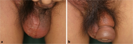
A 51-year-old male presented with fever and a painful, edematous erythema on the scrotum and penis (fig. 1a, b). The eruption had developed after the patient had ridden a bicycle 7 days earlier. Laboratory blood examination results were as follows: white blood cells 7,800/μl with neutrophil 12.0%; lymphocytes 4.7%; monocytes 0.3%; myeloblasts 83.0%; red blood cells 1.97 × 10⁶/μl; platelets 26.4 × 10⁴/μl, and C-reactive protein 4.621 mg/dl. The smear from the bone marrow indicated the presence of a massive myeloblast. The cells expressed CD7, CD11, CD13, CD14, and CD34. An incision was made in the scrotum. Corynebacterium spp. was isolated from the genital region. The patient was immediately hospitalized and intensively treated with blood transfusions and the antibiotics cefpirome sulfate 4 g and clindamycin 2,400 mg per day for 5 days and cilastatin sodium 2 g and clindamycin 2,400 mg per day for 9 days. The sign of infection diminished two weeks later. Treatment for AML was then initiated using cytarabine, aclarubicin, and granulocyte-colony stimulating factor.
Fig. 1.
a A painful and edematous erythema was present on the scrotum and associated with fever. b An edema was observed on the penis.
Discussion
FG was mainly caused by trauma and urinary tract infection [3]. In our case, we suspect that the trauma was due to the cycling activity and that the infection was worsened by leukocytopenia due to AML. Corynebacterium spp. was cultured from the scrotum. We do not think that Corynebacterium spp. was the actual pathologic bacteria because Corynebacterium spp. is part of the normal cutaneous flora in the genital area. We believe that our patient showed scrotum infection by unidentified bacteria and that intensive medication prevented progression to ulceration and the more advanced stage of necrosis as a sign of FG.
Few cases of FG have been described as a first sign of hematologic malignancies [4, 5], although cases have been reported as a complication during treatment (table 1) [2, 6,7,8,9,10,11]. Martinelli et al. [2] reported a fetal case having progressive involvement of the abdominal wall resulting in death from leukemia. They indicated that early diagnosis of the disorder and appropriate initiation of an accurate therapy can prevent progression of the acute necrotizing infection [2]. In the reported cases of FG associated with a hematologic malignancy, the edema and swelling were the initial signs [2, 5, 7, 9, 10]. The immediate start of the treatment might have prevented the present patient from being affected by FG.
Table 1.
Fournier's gangrene or genital infection associated with hematologic malignancies: a summary of the reported cases
| Case | Age/sex | Onset of Fournier's gangrenea | Hematologic malignancy | Clinical feature of the onset | Clinical feature of the severest situation | Phlogogenous bacteria |
|---|---|---|---|---|---|---|
| Naithani et al., 2008 [6] | 17/M | after diagnosis | acute promyelocytic leukemia | painful vesicular lesions in scrotum | ulcers | Staphylococcus aureus, Escherichia coli |
| Mantadakis et al., 2007 [7] | 21/M | after diagnosis | acute lymphoblastic leukemia | scrotal edema | a small necrotic scrotal eschar | Pseudomonas aeruginosa, Staphylococcus epidermidis |
| Fukuno et al., 2003 [8] | 43/M | after diagnosis | acute promyelocytic leukemia | an ulcer of 0.5 cm in diameter on the left side of the scrotum | swelling that was improved by a surgical incision | no description |
| Bakshi et al., 2003 [9] 1st case | 6/M | after diagnosis | acute myeloid leukemia | edema over the prepuce | a necrotic ulcer | Pseudomonas aeruginosa |
| Bakshi et al., 2003 [9] 2nd case | 10/M | after diagnosis | acute lymphoblastic leukemia | pain and swelling in the prepuce | abscess | Pseudomonas aeruginosa |
| Bakshi et al., 2003 [9] 3rd case | 9/M | after diagnosis | non-Hodgkin lymphoma | severe pain during micturition, erythema, and tenderness in the penile region | gangrenous changes on the prepuce and glans | no description |
| Yoshda et al., 2002 [10] | 16/M | after diagnosis | acute myelogenous leukemia | penile swelling with miction pain | gangrene in the regions of the scrotum, penis, thighs, and lower abdomen | Pseudomonas aeruginosa |
| Islamoglu et al., 2001 [5] | 33/M | before diagnosis | acute myelomonocytic leukemia | scrotum edema | complete scrotal necrosis, complete penile shaft necrosis, and a left anal ulcer that extended to the left gluteal area | Bacteroides fragilis |
| Martinelli et al., 1998 [2] 1st case | 41/M | after diagnosis | acute non-lymphocytic leukemia | genital erythema, pain, swelling and crepitation | blistering and ulceration | Pseudomonas aeruginosa |
| Martinelli et al., 1998 [2] 2nd case | 26/F | after diagnosis | acute non-lymphocytic leukemia | redness and swelling of the right labium majorum | ulceration | Pseudomonas aeruginosa |
| Martinelli et al., 1998 [2] 3rd case | 26/F | after diagnosis | acute non-lymphocytic leukemia | pain, edema, erythema and swelling of the perineal area | a necrotic ulcer | Pseudomonas aeruginosa |
| Faber et al., 1998 [4] | 50/M | before diagnosis | acute myelogenous leukemia | progressive perianal pain | a diffusely infiltrated anal region and bluish scrotum | Escherichia coli |
| Levy et al., 1998 [11] | 44/M | after diagnosis | acute promyelocytic leukemia | small indurated lesion of the right scrotum | a painful necrotic area 4×5 cm | Streptococcus faecalis, Staphylococcus coagulase negative |
The onset of Fournier's gangrene or genital infection before or after the diagnosis of a hematologic malignancy.
Hematologists and dermatologists should keep in mind that genital infection and its advanced stage of FG can be an initial sign of hematologic malignancy.
References
- 1.Ghnnam WM. Fournier's gangrene in Mansoura Egypt: a review of 74 cases. J Postgrad Med. 2008;54:106–109. doi: 10.4103/0022-3859.40776. [DOI] [PubMed] [Google Scholar]
- 2.Martinelli G, Alessandrino EP, Bernasconi P, Caldera D, Colombo A, Malcovati L, Gaviglio MR, Vignoli GP, Borroni G, Bernasconi C. Fournier's gangrene: a clinical presentation of necrotizing fasciitis after bone marrow transplantation. Bone Marrow Transplant. 1998;22:1023–1026. doi: 10.1038/sj.bmt.1701438. [DOI] [PubMed] [Google Scholar]
- 3.Bhatnagar AM, Mohite PN, Suthar M. Fournier's gangrene: a review of 110 cases for aetiology, predisposing conditions, microorganisms, and modalities for coverage of necrosed scrotum with bare testes. N Z Med J. 2008;121:46–56. [PubMed] [Google Scholar]
- 4.Faber HJ, Girbes AR, Daenen S. Fournier's gangrene as first presentation of promyelocytic leukemia. Leuk Res. 1998;22:473–476. doi: 10.1016/s0145-2126(98)00025-3. [DOI] [PubMed] [Google Scholar]
- 5.Islamoglu K, Serdaroglu I, Ozgentas E. Co-occurrence of Fournier's gangrene and pancytopenia may be the first sign of acute myelomonocytic leukemia. Ann Plast Surg. 2001;47:352–353. doi: 10.1097/00000637-200109000-00031. [DOI] [PubMed] [Google Scholar]
- 6.Naithani R, Kumar R, Mahapatra M. Fournier's gangrene and scrotal ulcerations during all-trans-retinoic acid therapy for acute promyelocytic leukemia. Pediatr Blood Cancer. 2008;51:303–304. doi: 10.1002/pbc.21549. [DOI] [PubMed] [Google Scholar]
- 7.Mantadakis E, Pontikoglou C, Papadaki HA, Aggelidakis G, Samonis G. Fatal Fournier's gangrene in a young adult with acute lymphoblastic leukemia. Pediatr Blood Cancer. 2007;49:862–864. doi: 10.1002/pbc.20695. [DOI] [PubMed] [Google Scholar]
- 8.Fukuno K, Tsurumi H, Goto H, Oyama M, Tanabashi S, Moriwaki H. Genital ulcers during treatment with ALL-trans retinoic acid for acute promyelocytic leukemia. Leuk Lymphoma. 2003;44:2009–2013. doi: 10.1080/1042819031000110982. [DOI] [PubMed] [Google Scholar]
- 9.Bakshi C, Banavali S, Lokeshwar N, Prasad R, Advani S. Clustering of Fournier (male genital) gangrene cases in a pediatric cancer ward. Med Pediatr Oncol. 2003;41:472–474. doi: 10.1002/mpo.10110. [DOI] [PubMed] [Google Scholar]
- 10.Yoshida C, Kojima K, Shinagawa K, Hashimoto D, Asakura S, Takata S, Tanimoto M. Fournier's gangrene after unrelated cord blood stem cell transplantation. Ann Hematol. 2002;81:538–539. doi: 10.1007/s00277-002-0525-9. [DOI] [PubMed] [Google Scholar]
- 11.Levy V, Jaffarbey J, Aouad K, Zittoun R. Fournier's gangrene during induction treatment of acute promyelocytic leukemia, a case report. Ann Hematol. 1998;76:91–92. doi: 10.1007/s002770050370. [DOI] [PubMed] [Google Scholar]


