Abstract
Atherosclerotic plaques tend to involve arterial localizations in which blood flow is not laminar due to arterial bends and bifurcations. A 49-year-old man was admitted to hospital with breathlessness and was subsequently diagnosed with left ventricular failure. Coronary angiography revealed three-vessel coronary artery disease and an anomalous extra left anterior descending artery taking off from the right sinus of Valsalva and spared from atherosclerosis. The absence of side branches and the relative lack of bends in arterial geometry were considered to be the cause of resistance to atherosclerosis. The present case identifies local flow conditions as an important factor determining the genesis of atherosclerosis in arterial segments.
Keywords: Atherosclerosis, Blood flow, Coronary artery anomaly, Coronary artery disease
Although atherosclerosis is a systemic disorder that may influence virtually all arteries in the human circulatory system, it is well known that some locations have a tendency for atherosclerotic plaque initiation and growth. In arteries in which the flow is laminar, with steady, pulsating shear stress, atherosclerosis is not usually observed. On the other hand, arteries with more nonlaminar or turbulent shear stress (arteries such as the inner curvature of the arcus aorta and the left coronary artery) have a predilection for atherosclerosis. Because bifurcations tend to create flow perturbations, arteries that possess many bifurcations and side-branch ostia, such as the coronary arteries, are believed to be particularly vulnerable to atherosclerosis (1).
Coronary arterial anomalies are seldom-encountered congenital heart disorders that are usually benign and discovered coincidentally in coronary arteriographic procedures. A double left anterior descending (LAD) coronary artery originating from both the right and left sinus of Valsalva is one of the rarest coronary artery anomalies, with only a few cases reported previously (2).
The present report describes a case of an additional LAD coronary artery originating from the right sinus of Valsalva that was totally free of atherosclerosis. All of the patient’s coronary arteries with normal origins had widespread atherosclerosis.
CASE PRESENTATION
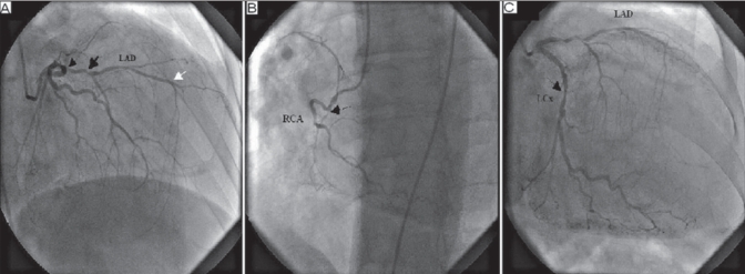
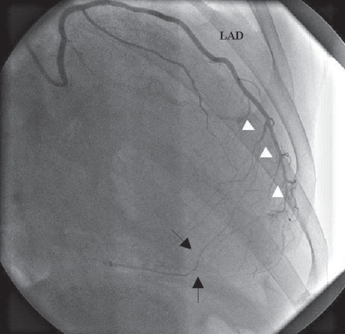
A 49-year-old man with a history of type 2 diabetes mellitus and essential hypertension was admitted to the emergency department of the Siyami Ersek Thoracic and Cardiovascular Surgery Training and Research Hospital, Istanbul, Turkey with dyspnea. He had no history of coronary artery disease. On physical examination, hypotension with a blood pressure of 85/45 mmHg and crepitant wet rales in the basilar section of the lungs were noted. An electrocardiogram was immediately obtained and revealed QS waves in leads DII, DIII and aVF, and a poor R wave progression in the precordial leads, with R/S transition shifted to lead V5. Transthoracic echocardiography showed akinetic walls in the anterior, anterolateral, inferior and inferolateral left ventricular segments in all cross-sections, with a calculated ejection fraction of 27%. The patient was immediately taken to the coronary care unit, and preload-reducing medications along with diuretics and inotropics were started. After relief of the pulmonary edema, he was transferred to the catheterization laboratory. Selective injection of the contrast media into the left main coronary artery revealed a short LAD coronary artery that terminated abruptly after giving rise to the septal and diagonal branches (Figure 1A). While performing selective right coronary angiography, another coronary ostia was engaged with a right Judkins catheter. Contrast injection showed another LAD coronary artery arising from the right aortic sinus. This vessel travelled to the left and entered into the anterior interventricular sulcus at the terminal point of the short LAD coronary artery (Figure 2). It was also noted that the anomalous vessel (long LAD coronary artery) was relatively straight, with a few side branches along its path. There were two consecutive high-grade stenoses in the proximal portion of the short LAD coronary artery and one in the left circumflex artery (Figures 1A and 1C). The right coronary artery was totally occluded from the proximal portion and was supplied in a retrograde fashion by the long LAD coronary artery (Figure 1B). Based on these angiographic views, this anomaly was considered to be a double LAD coronary artery together with three-vessel coronary artery disease. However, the anomalous coronary artery (long LAD coronary artery) was almost completely free of disease. The lesions in the normally positioned coronary arteries were not technically suitable for percutaneous angioplasty. Myocardial viability was evaluated with myocardial perfusion scintigraphy to decide whether the patient could benefit from surgical revascularization. Basal anterior, anteroseptal and all cross-sections of the inferior, inferolateral and anterolateral segments were found to be scarred. Because the only viable nonischemic segments were already supplied with an atherosclerosis-free vessel and no viability was found in the other segments supplied by atherosclerotic arteries, revascularization was not performed. Standard medical therapy for ischemic cardiomyopathy was initiated, and the patient was discharged from the hospital on the eighth day of his admission.
Figure 1).
A Coronary angiogram obtained in the right anterior oblique cranial position demonstrating a short left anterior descending (LAD) coronary artery that terminated after giving rise to the septal and diagonal branches (white arrow), and also showing two consecutive severe stenoses at the proximal portion of the LAD coronary artery (black arrows). B Right coronary angiogram obtained in the left anterior oblique caudal position showing total occlusion of the proximal right coronary artery (RCA) (arrowhead). C Left coronary angiogram obtained in the right anterior oblique caudal position demonstrating stenoses of the left circumflex (LCx) coronary artery (arrowhead)
Figure 2).
Coronary angiogram obtained in the right anterior oblique caudal position showing the anomalous left anterior descending (LAD) coronary artery from the right aortic sinus. The white arrowheads show septal branches of the LAD coronary artery and the black arrows show the collateral circulation that supplied the distal right coronary artery. Note that no atherosclerotic plaques are visible
DISCUSSION
Coronary artery anomalies are rare entities that are encountered, mostly coincidentally, in 0.6% to 1.5% of all coronary arteriograms (3). Anomalies involving the LAD coronary artery most often consist of a bifurcated dual LAD coronary artery, with bifurcation occurring somewhere in the left coronary system. Very rarely, however, a complete bifurcation with two separate ostia may be seen, with one ‘short’, ‘proper’ LAD coronary artery stemming from the left main coronary artery and the second ‘long’, ‘anomalous’ LAD coronary artery taking off from the right sinus of Valsalva. According to the anatomical classification scheme for dual LAD coronary artery proposed by Spindola-Franco et al (4), this type of LAD coronary artery anomaly is classified as type IV. Based on our patient’s coronary angiograms, we believe that the coronary anomaly seen in our patient is compatible with type IV dual LAD coronary artery.
Anomalous coronary arteries share the same embryological origins with normal coronary arteries (5). Similar to other coronary arteries, atherosclerotic plaques may also develop and obstruct blood flow in anomalous vessels. However, due to causes still largely unknown today, these arteries are less prone to atherosclerosis. In a study performed by Eid et al (6), 4616 coronary angiograms were studied, in which the incidence of coronary artery disease was found to be 50%. Of these, 17 patients had an anomalous vessel, and the incidence of atherosclerotic disease in anomalous vessels was found to be 17.65%. Interestingly, when the remaining coronary arteries of these 17 patients were studied, the incidence of coronary artery disease still remained at 50%, suggesting that anomalous vessels are somehow resistant to atherosclerosis. In our patient, we also observed such an association, in which all vessels excluding the anomalous LAD coronary artery, which had few side branches, had extensive atherosclerotic plaques. We believe that the geometric properties of the anomalous vessel may be responsible for its ability to resist atherosclerosis. It is well known that atherosclerotic lesions tend to develop in arterial locations in which flow is disturbed, such as bifurcations and ostia of side branches (1). All other coronary arteries in our case had extensive branching, which is typical for coronary arteries, but the anomalous artery was virtually free of major branches and had only a few side branches. Because the systemic risk factors can be expected to increase plaque initiation and growth in all arteries throughout the body, including the normal and anomalous coronary arteries, our hypothesis seems adequate to explain why only this vessel is spared from atherosclerosis. Moreover, this hypothesis may help to understand the absence of atherosclerosis in anomalous arteries found in other studies (6). These studies found that atherosclerosis is seen less frequently in anomalous coronary arteries and is almost exclusively seen when an anomalous coronary artery has a retroaortic course (6). Such a course may change the arterial geometry in a manner that may create altered shear stress regions that are prone to atherosclerosis. In other instances in which the anomalous artery had a relatively straightforward course toward the target myocardial area, atherosclerosis is not seen because the flow is relatively laminar, with minimal perturbations.
The embryological basis of the anomalous artery could also pose an alternative explanation for this situation because the endothelium of the vessel could have an innate resistance to atherosclerosis. However, there is no evidence that anomalous arteries originate from a different source apart from the normal arteries because both share a common developmental pathway stemming from the subepicardial primary plexus (5). Therefore, it is unlikely that developmental effects play an important role in this situation. Flow conditions and the effects of local shear stress probably have a more important role in explaining the relative freedom from atherosclerosis of the anomalous vessel.
CONCLUSION
The present article reports a rare anomaly with important aspects that may help us explain the natural resistance of anomalous coronary arteries to atherosclerosis. Moreover, the case may provide insight for the development of local atherosclerosis in the presence of ‘systemic’ risk factors and emphasizes the importance of flow conditions in the genesis of atherosclerosis.
Footnotes
CONFLICTS OF INTEREST: The authors declare that they have no commercial associations or sources of support that might pose a conflict of interest.
REFERENCES
- 1.Hahn C, Orr AW, Sanders JM, Jhaveri KA, Schwartz MA. The subendothelial extracellular matrix modulates JNK activation by flow. Circ Res. 2009;104:995–1003. doi: 10.1161/CIRCRESAHA.108.186486. [DOI] [PMC free article] [PubMed] [Google Scholar]
- 2.Voudris V, Salachas A, Saounotsou M, et al. Double left anterior descending artery originating from the left and right coronary artery: A rare coronary artery anomaly. Cathet Cardiovasc Diagn. 1993;30:45–7. doi: 10.1002/ccd.1810300112. [DOI] [PubMed] [Google Scholar]
- 3.Makaryus AN, Orlando J, Katz S. Anomalous origin of the left coronary artery from the right coronary artery: A rare case of a single coronary artery originating from the right sinus of Valsalva in a man with suspected coronary artery disease. J Invasive Cardiol. 2005;17:56–8. [PubMed] [Google Scholar]
- 4.Spindola-Franco H, Grose R, Solomon N. Dual left anterior descending coronary artery: Angiographic description of important variants and surgical implications. Am Heart J. 1983;105:445–55. doi: 10.1016/0002-8703(83)90363-0. [DOI] [PubMed] [Google Scholar]
- 5.Von Kodolitsch Y, Ito WD, Franzen O, Lund GK, Koschyk DH, Meinertz T. Coronary artery anomalies. Part I: Recent insights from molecular embryology. Z Kardiol. 2004;93:929–37. doi: 10.1007/s00392-004-0152-7. [DOI] [PubMed] [Google Scholar]
- 6.Eid AH, Itani Z, Al-Tannir M, Sayegh S, Samaha A. Primary congenital anomalies of the coronary arteries and relation to atherosclerosis: An angiographic study in Lebanon. J Cardiothorac Surg. 2009;4:58. doi: 10.1186/1749-8090-4-58. [DOI] [PMC free article] [PubMed] [Google Scholar]


