SUMMARY
Plasma cell disorders form a spectrum ranging from the asymptomatic presence of small monoclonal populations of plasma cells to conditions like plasma cell leukemia and multiple myeloma, in which the bone marrow can be replaced by the accumulation of neoplastic plasma cells. Immunophenotyping has become an invaluable tool in the management of hematological malignancies and is increasingly finding a role in the diagnosis and monitoring of plasma cell disorders. Multiparameter flow cytometry has evolved considerably during the past decade with an increasing ability to screen large numbers of events and to detect multiple antigens at the same time. This, along with a better understanding of the phenotypic heterogeneity of the clonal plasma cells in different disorders, has made immunophenotyping an indispensible tool in the diagnosis, prognostic classification and management of plasma cell disorders. This book chapter addresses the approaches taken to evaluate monoclonal plasma cell disorders, and the different markers and techniques that are important for the study of these diseases.
INTRODUCTION
Clonal plasma cell disorders make up a wide spectrum of disorders, ranging from incidental findings in asymptomatic individuals to life threatening conditions, and can present with a myriad of clinical manifestations. 1–4 Currently, we still depend primarily on morphological features and limited immunophenotypic studies for identification of the clonal plasma cells as well as the clinical manifestations for diagnosis and classification of plasma cell disorders. However, in the past decade there has been increasing appreciation of the genetic and phenotypic heterogeneities that underlie the plasma cell dyscrasias and the changes that parallel disease evolution. Increased use of multi-parameter flow cytometry and a broadening array of available reagents for surface and intracellular staining of specific antigens have enhanced our understanding of various aspects of these diseases. These improvements have led to a better insight into the disease biology and have enhanced our diagnostic and prognostic abilities. Moreover, the ability of flow cytometry to rapidly analyze a large number of cells provides the level of sensitivity of detection required for assessing disease response to treatment and for demonstrating minimal residual disease. This is a major advantage when compared to conventional methods of morphological assessment and immunohistochemistry. While flow cytometric methods have become part of the standard diagnostic approach in other hematological malignancies, such as the acute and chronic leukemias, consensus in regards to their routine use in plasma cell disorders is lacking. Much of this stems from the contradictory results seen in early studies and the lack of universally acceptable plasma cell specific markers. Some of the controversies in earlier studies most likely ensued from the wide spectrum of disease stages included in the studies, variability in the reagents used, technical differences both in terms of acquisition and gating strategies and inconsistencies in the appreciation of the high inherent autoflourescence of plasma cells. 5,6 Despite these challenges, high-sensitivity flow cytometry is evolving into an integral part of the laboratory evaluation and management of plasma cell disorders and can play an important role in the (i) diagnosis and classification, (ii) prognostic stratification, (iii) monitoring of response to therapy and minimal residual disease, (iv) understanding of the biology of disease progression, (v) studying of the role of tumor microenvironment in plasma cell disorders and (vi) identification of potential therapeutic targets on the surface of malignant plasma cells. 7,8
Classification of clonal plasma cell disorders
Monoclonal gammopathies form a large spectrum of disorders ranging from the asymptomatic presence of small clonal plasma cell populations to the near replacement of bone marrow with malignant plasma cells. 1–4 The common denominator for these disorders is the presence of a monoclonal protein, which can be in the form of intact immunoglobulin, immunoglobulin fragments, or free immunoglobulin light chains, in either the serum or urine. This is typically accompanied by the accumulation of clonal plasma cells primarily in the bone marrow or as localized soft tissue deposits or plasmacytomas, with few circulating plasma cells, except in more advance stages of the disease. A classification of clonal plasma cell and related disorders are given in Table 1.
Table 1.
Differential diagnosis of monoclonal gammopathies
| Diagnosis | Description |
|---|---|
| Monoclonal gammopathy of undetermined significance (MGUS) |
|
| Smoldering multiple myeloma (SMM) |
|
| Symptomatic multiple myeloma |
|
| Solitary Plasmacytoma |
|
| Primary Systemic Amyloidosis |
|
| POEMS syndrome (polyneuropathy, organomegaly, endocrinopathy, monoclonal protein, skin changes) |
|
| Waldenström's Macroglobulinemia |
|
Plasma cells are terminally differentiated, non-dividing, effector cells of the B-cell lineage.9– 12 They are the primary mediators of humoral immunity, secreting antigen specific immunoglobulins. As a consequence, abnormalities of these cells are responsible for a variety of autoimmune diseases in addition to plasma cell neoplasms. The development and function of plasma cells is tightly regulated. The precise etiological events that alter the normal development of B-cells into mature plasma cells, to date, are not well understood and are most likely multifactorial. The progenitor B cell undergoes an initial development process in the marrow that is antigen independent, gaining the ability to produce an intact IgM immunoglobulin; followed by migration to the spleen. A subsequent antigen dependent development phase is typically triggered by exposure to an antigen, which results in the formation of memory B cells and long lived plasma cells. 9–12 Plasma cells typically secrete an intact immunoglobulin that is made up of two identical light chains and two heavy chains. There are five major classes of heavy chains, designated mu, delta, gamma, alpha, and epsilon corresponding to the major classes of immunoglobulins; IgM, IgD, IgG, IgA, and IgE respectively. In each of the immunoglobulin molecules the heavy chains are bound to one of the two light chains (either Kappa or Lambda), but not to both.
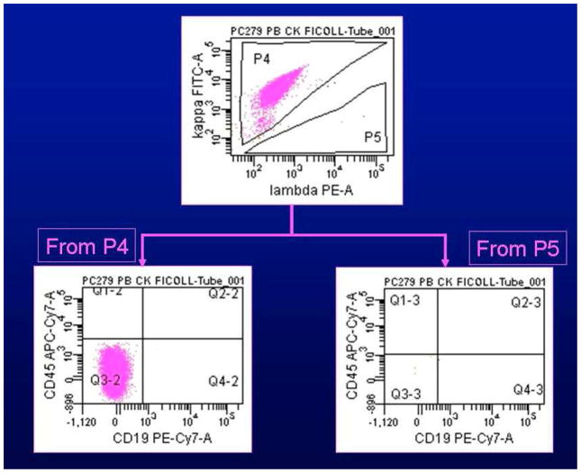
Monoclonal gammopathy of undetermined significance (MGUS) represents the majority of patients with a diagnosis of monoclonal gammopathy and is found in approximately 1 to 2 percent of adults in studies from Sweden, the United States, France, and Japan.3,13–15 (Figure 1) In fact, its prevalence increases with age and nearly 6% of individuals over 70 years have MGUS in large epidemiological studies. It is characterized by a relatively small burden of clonal plasma cells (<10%) in the bone marrow, secretion of small amounts of monoclonal protein (< 3.0 gm/dl in the serum) with small amounts detectable in the urine, and lack of any symptoms related to the clonal process. A small fraction of these patients (typically 1% every year) will undergo disease progression into a symptomatic stage, which usually presents as multiple myeloma characterized commonly by anemia, bone destruction, renal insufficiency or hypercalcemia. In contrast, patients with amyloidosis, which is much less common that myeloma, present with widespread organ deposition of light chain-derived amyloid fibrils. These patients typically have a lower burden of clonal plasma cells compared to myeloma, as is also the case in other related conditions such as light chain deposition disease or POEMS syndrome. Waldenstrom’s macroglobulinemia is another related disorder, where the clonal population is made up of lymphoplasmacytoid cells with morphological characteristics of plasma cells and B-lymphocytes. Immunophenotypic evaluation of the malignant plasma cells is an invaluable tool in the diagnosis and classification of these disorders, their prognostic assessment, the serial monitoring of disease burden and its reduction as a reflection of response to ongoing therapy.
Figure 1.
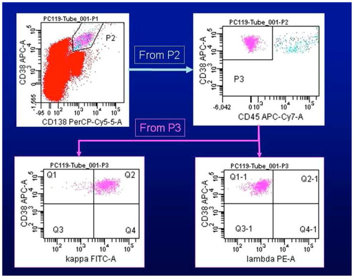
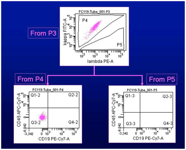
A) Gating on CD38 and CD138 positive plasma cells (upper left dotplot, gate P2) reveals them to be CD45 negative (upper right dotplot). Selective gating on this CD45 negative plasma cell population (gate P3) reveals them to be monoclonal for cytoplasmic kappa immunoglobulin light chains (lower left dotplot) and to lack expression of lambda cytoplasmic immunoglobulin light chains (lower right dotplot). B) Selective analysis of the kappa light chain restricted plasma cells (gate P4) reveals them to lack expression of CD19 as well as CD45 (lower left dotplot). No lambda light chain positive plasma cells are present in gate P5 as the plasma cells are kappa light chain restricted (lower right dotplot).
Sample preparation and analysis approaches
Sample preparation
Flow cytometric evaluation of plasma cells in whole blood/or bone marrow aspirates begins with either isotonic lysis of the erythrocytes or density gradient separation methods to isolate the mononuclear cells. Both methods involve the use of commercially available reagents, the former (isotonic lysis) is advantageous in that it involves minimal labor or reagent costs, however it does little for plasma cell enrichment as all mononuclear cells, including granulocytes, are isolated and stained. Density gradient cell isolation is typically performed using Ficoll Hypaque and provides for much greater plasma cell enrichment. This methodology also has drawbacks, however, as it is more time and labor intensive. Furthermore, Ficoll gradient separation accelerates the loss of antigens such as CD38 and CD138 from the plasma cell surface and, therefore, the cells must be quickly stained and analyzed when isolated by this method.
Plasma cell identification
Identification of the plasma cells has typically been based on the demonstration of high CD38 and CD138 expression, in conjunction with the scatter properties of the cell population.16,17 While CD38 expression is also seen in other cell populations, especially activated T cells, the bright expression by plasma cells sets it apart with reasonable specificity to suggest its routine use for identification of plasma cells.5,18 It is important to bear in mind, however, than in many abnormal plasma cell populations the surface density of CD38 is slightly decreased and, therefore, the abnormal plasma cells may have a staining intensity similar to that of normal B-cell precursors (hematogones) or activated T-cells. Therefore coincident analysis of CD138 or syndecan, another antigen characteristically expressed by plasma cells, greatly enhances the sensitivity of flow cytometry. Similar to CD38, however, heterogeneity of CD138 expression can be seen between plasma cell populations.17,19 Moreover, CD138 can be shed from the cell surface as a result of heparanase activity. The expression of CD45 and CD19 allows further refinement of the plasma cell identification process. In addition to CD38 and CD138, normal plasma cells are generally positive for these antigens whereas abnormal plasma cells characteristically lack CD19 and variably express CD45. In plasma cell dyscrasias, one typically finds a mix of CD45 positive and negative plasma cells and the proportions appear to change with disease evolution. Using 6-color flow cytometry, we were able to detect a distinct CD45 positive plasma cell fraction in the majority of bone marrow and nearly half of the peripheral blood specimens from patients with plasma cell disorders.20 The clonal nature of these cells was confirmed by light chain restriction. Interestingly, the bone marrows contained less CD45 positive than CD45 negative plasma cells, while the proportions were generally equal in the peripheral bloods. Given the higher than previously reported presence of CD45 positive cells plasma cells, additional studies were done which ruled out antibody variations or the adherence of T-cells as a potential explanation for the findings. Figure 1 shows the typical gating strategy for identification of plasma cells.
Use of controls
Although plasma cells can exhibit autofluorescence, immunoglobulin isotype controls are usually not required for accurate plasma cell analysis by flow cytometry. In all studies, it is useful to run positive and negative control cases to confirm the composition of the antibody cocktail used and to verify the instrument compensation settings. Also, for accurate analysis, cellular, viability should be assessed with a vital exclusion dye, such as 7-AAD. In addition, DDM (doublet discrimination) may be important to use to control for cell aggregates in certain situations.
Sample acquisition and event numbers
The advent of advanced flow cytometry instrumentation and analysis software has allowed for the rapid collection of very high numbers of cellular events (>500,000). This is often required in the analysis of blood and marrow specimens potentially involved by a plasma cell neoplasm as the variation between assays can be considerable when the number of neoplastic plasma cells evaluated falls below 100. The European myeloma network recommends that at least 100 neoplastic plasma cell events be acquired for accurate enumeration. This will often require acquisition of a million or more total events in assays designed for minimal residual disease assessment to provide the adequate sensitivity.
Role of flow cytometry in the diagnosis and classification of plasma cell diseases
The diagnosis of plasma cell disorders requires confirmation of the clonal nature of the plasma cells, typically against the background of increased numbers of non-malignant plasma cells. While this is most commonly performed on a bone marrow aspirate specimen, there is increasing appreciation of circulating plasma cells in these disorders due to the increased sensitivity of flow cytometry techniques and their ability to screen large number of events in a rapid manner. Given that polyclonal expansion of plasma cells can be seen in several inflammatory and infectious conditions, the ability to demonstrate clonality of a plasma cell population is critical to making the diagnosis. The pattern of surface antigens on the clonal plasma cells can help classifying the disorders and is an important adjunct to other laboratory studies and clinical features.
The quantitation of plasma cells is routinely performed using a slide-based readout of the aspirate or biopsy sections, based on morphology with or without immunohistochemistry with antibodies to plasma cell associated antigens, such as CD138 or MUM-1. The proportion of plasma cells in the aspirate can also be assessed using flow cytometry, but results have consistently shown significantly lower plasma cell numbers from flow cytometry than from slide-based morphological evaluation.21–24 The reasons for this discrepancy are not entirely clear. Various mechanisms have been suggested, including the draw sequence (the aspirate used for flow studies coming after the aspirate for the slide-based studies and resultant hemodilution), adherence of plasma cells to bone spicules and hence loss during cell isolation, clustering of plasma cells, loss of surface expression of CD138 and CD38 as a result of cell processing prior to flow cytometry, or shedding of CD138 related to cellular viability among others.21–24 While the quantity of plasma cells may be underestimated by flow cytometry compared to morphological assessment, the increased sensitivity of multi-color flow cytometry allows the detection of very small numbers of plasma cells, which may be missed by morphological or immunohistochemical evaluation.20,25 Ideally, a minimum of 1 × 10⁵ nucleated events should be screened to adequately characterize plasma cells. Use of a two-step process, involving a live gate for the acquisition of plasma cell events, allows for increased sensitivity of the assay.16,26 The consensus recommendation from the European Myeloma Network implies the use of the first bone marrow aspirate for flow cytometry and the evaluation of at least 100 neoplastic plasma cells. More recently, we have developed a lyse, no-wash method, that involves minimal processing of the aspirate sample, which in combination with enumeration beads allows for the quantitation of absolute numbers of plasma cells/μL. Approaches to distinguish abnormal from normal polyclonal plasma cells are discussed later.
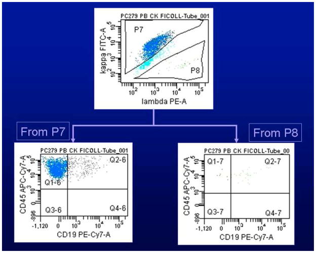
The clonality of plasma cells can be easily confirmed based on the sole presence of either kappa or lambda cytoplasmic immunoglobulin light chains in all cells. This test for light chain restriction requires the use of antibodies to kappa or lambda light chains in fixed and permeabilized cells. Plasma cell populations can be selected based on surface antigen expression, prior to determining clonality. Figure 2 explains the determination of plasma cell clonality based on kappa or lambda light chain expression. In some settings, proof of light chain restriction can be difficult; for instance, when very few abnormal plasma cells are present, when the abnormal plasma cells contain very low levels of cytoplasmic light chains, or when the disorder is bi-clonal expressing both kappa and lambda light chains. Moreover, permeabilization of plasma cells can affect their light scatter properties and, in some instances, may impair the ability to assess the expression of cell surface antigens.
Figure 2.
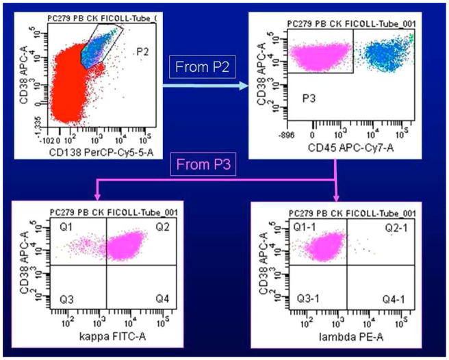
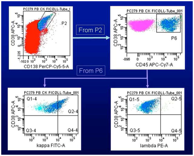
Gating on CD38 and CD138 positive plasma cells reveals both CD45 negative (Figure 2A, gate P3) and CD45 positive (Figure 2B, gate P6) subsets, both of which are kappa cytoplasmic immunoglobulin light chain restricted (lower panels). Selective analysis of the light chain restricted plasma cells reveals both the CD45 negative and CD45 positive subsets to lack expression of CD19 (Figures 2C and 2D respectively).
Differential expression of a wide variety of surface antigens helps distinguishing normal from malignant plasma cells and permits the recognition of plasma cells from different stages of disease evolution as well as from different types of plasma cell disorders.7,8 The surface antigens that have been studied in plasma cell disorders are described in Table 2. Flow cytometry allows the simultaneous assessment of multiple markers on small populations of plasma cells as well as determination of clonality.
Table 2.
Surface markers in the assessment of plasma cell disorders
| Lymphoid markers |
| CD10 |
| CD19 |
| CD20 |
| CD22 |
| CD27 |
| CD28 |
| CD52 |
| Myeloid markers |
| CD33 |
| CD117 |
| Other hematopoietic cell markers |
| CD38 |
| CD45 |
| Adhesion molecules |
| CD11 |
| CD44 |
| CD49d |
| CD49e |
| CD56 |
| CD138 |
| Other aberrantly expressed markers |
| CD32 |
| CD146 |
| CD200 |
| CD221 |
| CD307 |
Normal versus abnormal plasma cell
Non-neoplastic polyclonal plasma cells are normal components of a bone marrow aspirate and thus need to be distinguished from neoplastic, clonal plasma cells. While the majority of plasma cells in myeloma (> 95%) belong to the neoplastic population, the proportion of normal plasma cells is typically higher in MGUS and smoldering myeloma (SMM).27 Several surface antigens have been suggested to characterize individual plasma cells as malignant or normal, but no single marker can make the distinction with any degree of specificity. This distinction is based on the differential expression of a panel of antigens (Table 3). Overall, compared to normal plasma cells, abnormal plasma cells tend to be low in the expression of CD19, and CD27, have weaker expression of CD38 and CD45, increased expression of CD28, CD33 and CD56 and variable expression in a small proportion of cells of CD20 and CD117.8
Table 3.
Comparison of immunophenotypic features between normal and abnormal plasma cells
| Normal Plasma Cell | Abnormal Plasma Cell | Comments | |
|---|---|---|---|
| CD19 | Positive (>70%) | Negative (95%) | Present on all B-cells and most normal plasma cells. Clonal plasma cells have dim or no expression. |
| CD2070 | Negative | Positive (30%) | Typically seen during the maturation process of B-cells and absent from plasma cells. CD20 expression is seen on a subset of clonal plasma cells. |
| CD2732,71,72 | Positive (100%) | Negative/weak (15–45%) | This molecule is involved in the differentiation of mature B cells into plasma cells. Weakly or not expressed in myeloma. |
| CD2873,74 | Negative (<15%) | Positive (45%) | CD28 is involved in T-cell activation. Expression on plasma cells in myeloma correlates with aggressive disease. |
| CD3377,78 | Negative | Positive in a small subset of patients | This myeloid-lineage affiliated antigen may be present aberrantly in a subset of myeloma patients. |
| CD5661,96 | Negative (< 15%) | Positive (75%) | The neural-cell adhesion molecule, typically present on NK-cells. Almost invariably present on neoplastic plasma cells, but expression may be low or lost in cells in circulation and in extramedullary disease. |
| CD81 | Positive (100%) | Negative/weak | Member of CD19/CD21/Leu-13 signal transduction complex. Functions of this and other members of the tetraspan family are as yet poorly understood. |
| CD11728,97 | Negative | Positive | Typically seen on progenitors of myeloid and megakaryocytic lineage. Adapted from: Paiva et al, Clinical Cytometry, 12 Feb 2010 |
Adapted from: Paiva et al, Clinical Cytometry 12 Feb 2010
Plasma cell leukemia
Plasma cell leukemia (PCL) is characterized by circulating neoplastic plasma cells and may present de novo (primary PCL) or at the time of relapse of a previously diagnosed multiple myeloma (secondary PCL). The antigens commonly used for plasma cell identification, such as CD38 and CD138, are similarly expressed PCL and multiple myeloma.28 However, PCL cells are more likely to express CD20 and usually tend to be negative for CD56, CD117 and HLA-DR.29 Others have shown that CD28 and CD27 expression can be useful in distinguishing primary PCL from secondary PCL.30,31
Waldenström’s macroglobulinemia
Waldenstrom’s macroglobulinemia is typically associated with lymphoplasmacytoid cells secreting an IgM monoclonal protein. The lymphoplasmacytic cells are typically CD19 and CD38 positive and variably express CD138. In a study of 35 lymphoplasmacytic lymphoma cases,32 both immunohistochemistry and flow cytometry were useful in identifying the cells with lymphoid and plasmacytic features. In 19 cases, immunohistochemistry revealed a pattern in which plasma cell infiltrates were physically separate from the lymphoid infiltrates. Surface immunoglobulin light-chain restricted, mature B-cells were identified by flow cytometry in 96% of the cases and in approximately half of these the cells expressed CD5 and/or CD23. Using highly sensitive 6- color flow cytometry, clonal plasma cells were seen in over 90% of patients; and in over half of these cases, the distinctive bright CD38 and CD138 co-expression was identical to that seen in true plasma cell neoplasms, such as multiple myeloma. In contrast to typical plasma cell neoplasms, however, the plasma cells in lymphoplasmacytic lymphoma were almost always CD19 and CD45 positive.
Amyloidosis
There is limited information available on characteristic features of plasma cells in light chain (AL) amyloidosis.33–35 In a small study of 36 cases with well documented AL amyloidosis, expression of CD20, CD79a, CD56, cyclin D1 and EMA was noted in 42%, 86%, 50%, 53% and 83% of cases, respectively.34 Expression of CD56 and/or cyclin D1 was seen in 79% of cases. In 9/10 patients, small lymphoid-like plasma cells were positive for CD20 and all the 10 cases positive for cyclin D1. On the other hand, among cases without small lymphoid-like morphology, CD20 and cyclin D1 expression was seen in only a quarter of patients. More recently, expression of the low-affinity IgG Fc receptor, CD32B, was found to be elevated in plasma cells from patients with amyloidosis.35 Gene expression profiling demonstrated significantly higher expression of CD32B compared to all other Fc receptor family members. Reverse-transcriptase polymerase chain reaction (RT-PCR) using enriched CD138 positive plasma cells showed uniform expression of the stable cell surface CD32B1 isoform at diagnosis and relapse, and flow cytometry showed intense CD32B cell surface staining on 99% of CD138 positive plasma cells at diagnosis and relapse.35
Assessment of plasma cell proliferation, apoptotic rates and DNA ploidy status
The proliferative rate of malignant cells is a powerful prognostic factor in most malignancies, and this is also true for plasma cell disorders. Various methodologies have been proposed for determining proliferation, including immunohistochemistry for markers such as Ki67, labeling of the nucleus in proliferating cells with 5-bromo-2′-deoxyuridine (BrdU) or thymidine, or examination of the cells in S-phase using simple DNA stains.
Initial methods of labeling index determination in myeloma utilized the incorporation of radioactive thymidine by replicating DNA36,37. The percentage of thymidine-labeled cells was then determined by autoradiography. Plasma cells in the S-phase of the cell cycle actively incorporate BrdU. The availability of antibodies to BrdU led to the development of a slide-based immunofluorescence method for identifying dividing plasma cells that are labeled with BrdU36. This method allows for the morphological confirmation of plasma cells among all labeled cells, thereby improving specificity. By staining concurrently for cytoplasmic kappa or lambda immunoglobulin light chains, the percentage of dividing clonal plasma cells can be counted.
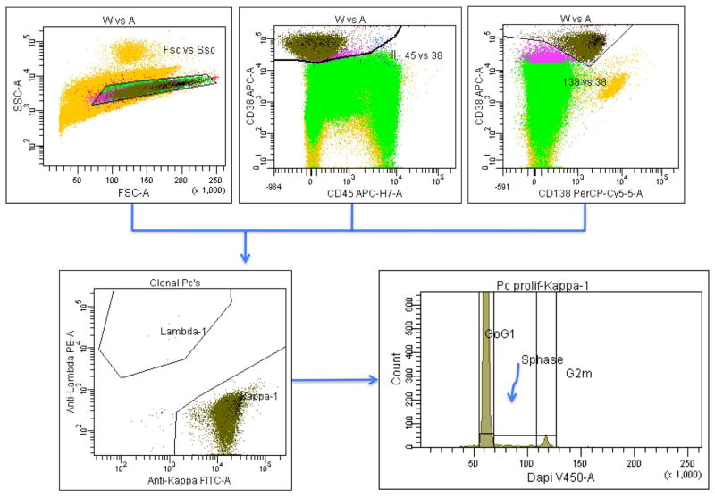
More recently, there has been increasing interest in using flow cytometry for the determination of proliferative rates. This approach involves cell fixation followed by staining with a dye, which stoichiometrically binds to DNA, such as propidium iodide. Sophisticated software is available to determine the fraction of cells currently in S-phase of the cell cycle. In addition to being less cumbersome than slide-based techniques, flow cytometric measurements also allow for the evaluation of a much greater number of cells and the simultaneous determination of the plasma cell DNA ploidy, valuable prognostic information in patients with myeloma. Figure 3 provides an illustrative example for identifying plasma cells in S-phase in the bone marrow from a patient with multiple myeloma. There are many benefits to assessing the bone marrow plasma cell labeling index in the diagnosis and management of multiple myeloma38–41. It discerns patients with stable disease not requiring treatment, including MGUS and SMM, from patients with active myeloma requiring treatment for their disease37,42. It provides valuable prognostic information in patients with SMM, where a high labeling index suggests high risk of progressing to active myeloma in the short term. In patients with active multiple myeloma, a high labeling index at diagnosis portends a shorter overall survival 43,44.
Figure 3.
The plasma cell population is identified in 3 separate gating steps using forward scatter (FSC)/side scatter (SSC), the CD45/CD38, and the CD38/CD138 gate. Subsequently, clonality of the plasma cell population is identified by using antibodies to kappa and lambda. The left lower histogram shows binding of diamino-2-phenylindole (DAPI) to the DNA in kappa monoclonal plasma cells. Based on nuclear content, a significant fraction of plasma cells is in S-phase, reflective of proliferative activity.
In addition to the proliferative rate of plasma cells, the proportion of cells undergoing apoptosis also appears to have prognostic significance in myeloma and related disorders.45 Cells in apoptosis are typically assessed using Annexin V and propidium iodide staining to determine the loss of membrane integrity. Phosphatidylserine, which in non-apoptotic cells is inside the membrane, will be exposed on the outer surface of apoptotic cells, where it can be labeled with Annexin V. The addition of propidium iodide allows for staining of DNA, which will further discriminate stages of apoptosis.
Prognostic value of immunophenotypic studies
It is clear from the discussion so far that the phenotypic features of plasma cells are quite heterogeneous, depending on the disease stage, diagnosis, a patient’s other biologic characteristics, and the type of therapies employed. Multiple studies have demonstrated the prognostic value of specific patterns of antigen expression by neoplastic plasma cells. Furthermore, the expression of specific antigens has been shown to correlate with genetic abnormalities commonly seen in plasma cell disorders. While these associations are not specific enough to propose flow cytometry in lieu of conventional genetic analyses, such as metaphase cytogenetics or fluorescence in-situ hybridization (FISH), such relations explain the prognostic value of antigen expression profiles.
Significance of normal plasma cells
It has been shown that the proportion of normal plasma cells in patients with plasma cell disorders is a powerful prognostic factor in all stages of disease as well as following therapy. 26,27 Patients with MGUS and SMM, who have >5% phenotypically normal plasma cells, have a better outcome with lower rates of disease progression to symptomatic myeloma.26,46 In a series of 594 newly diagnosed multiple myeloma patients, uniformly treated on clinical trials, those with more than 5% normal plasma cells in the marrow (14% of patients) had favorable baseline clinical characteristics and a significantly lower frequency of high-risk cytogenetic abnormalities.47 These patients had a better response to therapy and longer progression-free and overall survival compared to the rest.
Circulating Plasma Cells
Clonal plasma cells and B-lymphocytes can be detected in the peripheral blood of patients with plasma cell disorders by immunoflourescence microscopy and flow cytometry.48,49 The number of circulating plasma cells has been found to be a measure of disease activity in myeloma38 and an important, independent prognostic factor for survival in myeloma, SMM50,51 and primary amyloidosis52. Among patients with MGUS, presence of circulating plasma cells predicted for higher risk of progression to symptomatic myeloma.53
CD45
Expression of CD45 on plasma cells in myeloma has been shown to be of prognostic significance. In normal hematopoiesis, CD45 is predominantly expressed in the early stages of plasma cell development and decreasing in expression with maturation; clonal plasma cells in myeloma show a heterogeneous expression pattern. 54,55 Studies suggest the presence of two populations of myeloma cells in the marrow; early plasma cells with CD45 expression and late plasma cells with dim or no expression. 54–56 Important biological differences have been demonstrated between these two populations, including their proliferative rates, IL-6 responsiveness and dependence, chromosomal abnormalities and angiogenic capability. In plasma cell disorders, the ratio of CD45-positive to CD45-negative plasma cells appears to be altered in relation to the disease stage. Moreau et al in a study of 95 patients with myeloma undergoing high dose therapy demonstrated a shorter survival for patients lacking CD45 expression on the plasma cells57. The early stages of plasma cell proliferative diseases, namely MGUS and SMM, are characterized by a nearly equal distribution of CD45 positive and negative plasma cells, whereas in newly diagnosed and relapsed myeloma the CD45 positive population dominates. The CD45-positive fraction appears to be more responsive to IL-6 stimulation 58,59 and contains the myeloma cells with higher proliferative activity 60. The interaction between CD45, which is a protein tyrosine phosphatase, and the src kinases (lyn, hck, lyk) may mediate the effect of CD45 on IL-6 signaling 59.
CD56
Expression of CD56 (NCAM-Neural cell adhesion molecule) is commonly used to identify abnormal plasma cells, since normal plasma cells are typically CD56 negative.61,62 However, heterogeneity exists among clonal plasma cells with respect to CD56 expression, and this may have prognostic implications.63 CD56 expression has been associated with poor outcome in myeloma patients treated with conventional therapies while no such effect was seen in another group of patients undergoing autologous stem cell transplantation.64,65 In one study, CD56 negative patients had higher beta-2 microglobulin levels, a higher incidence of extramedullary disease, Bence Jones protein, renal insufficiency, and thrombocytopenia and were more likely to have a plasmablastic morphology compared to CD56 positive patients.64 CD56 expression may correlate with the ability of cells to migrate out of the bone marrow and modulation of its expression is a potential mechanism of controlling plasma cell homing to extramedullary sites.66,67 In one study, the presence of circulating plasma cells was inversely correlated with CD56 expression.68 Another study suggested that lack of CD56 expression is associated with lambda light chain expression.69
CD20
CD20 is typically not expressed by normal plasma cells. However, several studies have suggested the presence of a small proportion of neoplastic plasma cells that express CD20 on their surface. In one study in patients with multiple myeloma, CD20 expression was associated with small mature plasma cell morphology and with t(11;14).70 Although the presence of t(11;14) is associated with better outcome in patients with myeloma, the expression of CD20 does not appear to have any prognostic impact. Given the success of anti-CD20 directed monoclonal antibody therapy in lymphoma, trials have been carried out with Rituximab in myeloma, without any clear efficacy.
CD27
Expression of CD27 is uniformly seen in normal plasma cells as well as plasma cells in MGUS and its loss has been correlated with disease progression in patients with MGUS.71 Progressive loss of CD27 has also been observed with disease progression in myeloma, and lack of CD27 expression at diagnosis was associated with shorter overall survival.72 Gene expression data have demonstrated that the multiple myeloma group with the highest prevalence of poor prognostic factors had the lowest CD27 RNA levels.31,71
CD28
CD28 is a major costimulatory molecule on T-cells and aberrantly expressed on neoplastic plasma cells in a fairly consistent manner.73 Its expression level appears to increase with disease progression, with one study showing CD28 expression on plasma cells from 19% of MGUS and 41% of myeloma patients, and in 100% of human myeloma cell lines.74 Among patients with myeloma, CD28 expression seems to increase with disease evolution in that CD28 positive plasma cells were detected in 26% of myeloma patients at diagnosis, in 59% at medullary relapse, in 93% at extramedullary relapse, and in 100% of patients presenting with secondary plasma cell leukemia. In individual patients, there was increasing expression of CD28 as the disease relapsed. CD28 in multiple myeloma can up-regulate the transcription of the interleukin-8 gene, a chemokine that promotes angiogenesis, thus potentially contributing to disease progression.75 Direct activation of CD28 on myeloma cells by anti-CD28 monoclonal antibody leads to the suppression of plasma cell proliferation and protects the malignant cells from dexamethasone-induced cell death.76
CD33
CD33 is a myeloid antigen that can be aberrantly expressed by plasma cells.16,62,77 In one study, CD33 positive patients had higher beta-2 microglobulin and lactate dehydrogenase levels and a higher incidence of anemia and thrombocytopenia than did CD33 negative patients.78 The overall survival was significantly shorter in the CD33 positive group, especially due to early mortality from disease. Serial evaluation of CD33 expression showed that the amount of CD33 antigen on the surface of plasma cells significantly increased after therapy in individual patients, possibly suggesting its expression as a marker of drug resistance.
CD44
CD44 is an adhesion molecule that is encoded by a single gene but can be expressed as CD44 standard (CD44s) or variant forms (CD44v). While no significant differences have been observed in the expression of CD44s, the CD44v9 and v10 containing isoforms were differentially expressed on bone marrow plasma cells from normal individuals (predominantly CD44v9+v10+) versus myeloma patients with stable (predominantly CD44v9-v10+) or progressive (predominantly CD44v9+v10−) disease.79 Some of the CD44 isoforms play a key role in myeloma cell migration, homing and adhesion to stromal cells.80,81 Expression of CD44v6 has been associated with disease progression, with very low expression found in patients with MGUS and Stage I myeloma, and with the presence of chromosome 13 deletions, a well-known risk factor in myeloma.82
CD52
CD52, a pan-lymphoid antigen, is expressed by a proportion of plasma cells in various plasma cell disorders, including MGUS, myeloma and amyloidosis.17,83,84 In one study, 67%, 52%, and 35% of patients with MGUS, myeloma and amyloidosis, respectively, demonstrated CD52 positive plasma cells.83 The CD52 expression was predominantly confined to the clonal CD38 and CD45 positive plasma cell fraction with a median of 68%, 88%, and 82% of positive cells in MGUS, myeloma, and amyloidosis, respectively, compared with 18%, 6%, and 9% of CD52 positive but CD45 negative plasma cells, respectively. However, use of CD52 as a therapeutic target in myeloma using monoclonal antibodies has not been successful.
CD117
CD117 (c-kit) is a hematopoietic growth factor receptor with tyrosine kinase activity that is aberrantly expressed by a proportion of neoplastic plasma cells.85,86 In one study, plasma cells from normal bone marrow samples did not show any reactivity for CD117, compared with 83 – 99% of CD117 positive plasma cells found in the marrows of one-third of myeloma patients. Expression of CD117 has been associated with a good prognosis in myeloma.86,87 Recent studies have suggested that CD117 is a valuable marker to distinguish abnormal, neoplastic from normal plasma cells.16
CD200
CD200 is a membrane glycoprotein that mediates suppression of T-cell-mediated immune responses. Patients with CD200 negative myeloma cells had an improved event-free survival compared with patients with CD200 positive myeloma cells, following high-dose therapy and stem cell transplantation. In a Cox proportional-hazard model, the prognostic value of CD200 expression for event free survival was independent of International Staging System (ISS) stage and beta-2 microgloblin levels.88
Response assessment and monitoring of minimal residual disease (MRD)
Improvements in therapy are very dependent on the sensitivity of tests used for the detection of residual disease. MRD determination allows us to estimate the real impact of new drugs and treatment approaches. Chronic myelogenous leukemia (CML) offers the best example for this concept. When hematological response was considered as the endpoint for successful therapy, high response rates, albeit poorly sustained, were seen with interferon and hydroxyurea. With the advent of PCR and FISH testing for the BCR/ABL fusion, the detection of MRD in CML improved dramatically and provided an explanation for high relapse rates in these patients.
In the setting of multiple myeloma, we know that MRD is an important predictor of outcome in patients undergoing aggressive therapy26,89–93. Rawstron et al used a sensitive flow cytometry assay that quantitated normal and neoplastic plasma cells in the bone marrow of 35 patients undergoing autologous stem cell transplantation (ASCT). These investigators identified a high-risk group of patients (66%) in whom at 3 months after transplantation the flow cytometric assay confirmed the presence of neoplastic plasma cells, whereas in the low-risk group, only normal phenotype plasma cells were present at that time-point. Low-risk patients experienced significantly longer progression-free and overall survival than the high-risk group. In this study, flow cytometry demonstrated increased sensitivity for MRD when compared with the detection of paraprotein by immunofixation, which is the conventional method for the monitoring of residual disease. Among patients with detectable malignant plasma cells by flow cytometry, approximately half became immunofixation negative; however, these patients had an outcome identical to patients who remained immunofixation positive90. Bakkus et al examined the utility of detecting MRD using a quantitative allele-specific oligonucleotide PCR (ASO-qPCR) assay at 3–6 months post-transplant in 67 patients. Using specific cutoffs for the quantitative PCR results, the authors identified patients with residual disease and a short time to relapse91.
Lipinski et al retrospectively analyzed the tumor load in blood and bone marrow samples of 13 patients at the time of remission after ASCT and during progression using ASO-qPCR92. Progression was detected earlier by this method compared to monoclonal protein estimation, demonstrating the increased sensitivity of the PCR technique. Galimberti et al examined the prognostic value of MRD in 20 patients after a tandem transplant, consisting of an autologous peripheral blood stem cell transplantation (PBSCT) followed by a non-myeloablative allogeneic peripheral blood stem cell infusion (NMT)93. MRD levels were determined by PCR for immunoglobulin heavy chain rearrangements. After PBSCT only 3 patients (15%) became PCR-negative, versus 12 (60%) patients after NMT. Eradication of MRD favorably impacted on overall survival with 76% of MRD-negative patients still alive 20 months post NMT versus 34% of persistently PCR-positive cases. Although MRD evaluation by ASO-PCR is slightly more sensitive and specific than flow cytometry, it is applicable in a lower proportion of myeloma patients and is more time-consuming, while providing similar prognostic information.94 In a recent study of 295 newly diagnosed myeloma patients who received uniform treatment including stem cell transplantation, a prospective analysis by multiparameter flow cytometry showed MRD to be one of the most important predictors of outcome.95 MRD negativity on day 100 after ASCT translated into significantly improved progression-free and overall survival.
PRACTICE POINTS.
Plasma cells can be separated from other white cells based on strong CD38 and CD138 expression.
The CD38/CD138 antigen combination can also be used for the gating of abnormal plasma cells by flow cytometry; once identified, the antigen profile of gated plasma cells allows for the distinction between normal and abnormal plasma cells.
The most important findings that characterize malignant versus normal plasma cells are: absent or low expression of CD27, CD19 and/or CD45, and increased expression of CD28, CD33, CD117 and/or CD56.
In all instances, monoclonality for intracytoplasmic immunoglobulin light chains is the ultimate proof for the presence of an abnormal plasma cell population.
Some subtypes of plasma cell dyscrasias show specific immunophenotypic characteristics, such as positivity for CD19 in Waldenstrom’s macroglobulinemia, or expression of CD20 in plasma cell leukemia.
Flow cytometric MRD assessment in plasma cell disorders is easy, rapid, more sensitive and better correlated with outcome than conventional immunofixation of paraprotein.
RESEARCH AGENDA.
Large phase III trials for the various subtypes of plasma cell disorders, which employ immunophenotyping prior to treatment, are needed in order to confirm correlations of specific antigen expression profiles with outcome.
To date, only few examples exist for associations of specific antigen expression with cytogenetic aberrations in plasma cell disorders, e.g., CD44v6 with chromosome 13 deletion; given the prognostic significance of cytogenetic abnormalities in plasma cell disorders, further such surrogate markers for genetic lesions should be identified.
Considering that we are in a period of new drug developments for the treatment of plasma cell disorders, flow cytometric measurements of MRD levels will be essential for the evaluation of drug efficacy and patient response.
Footnotes
Conflict of Interest: The authors have no conflict of interest to declare.
Publisher's Disclaimer: This is a PDF file of an unedited manuscript that has been accepted for publication. As a service to our customers we are providing this early version of the manuscript. The manuscript will undergo copyediting, typesetting, and review of the resulting proof before it is published in its final citable form. Please note that during the production process errorsmaybe discovered which could affect the content, and all legal disclaimers that apply to the journal pertain.
References
- 1.Rajkumar SV, Dispenzieri A, Kyle RA. Monoclonal gammopathy of undetermined significance, Waldenstrom macroglobulinemia, AL amyloidosis, and related plasma cell disorders: diagnosis and treatment. Mayo Clin Proc. 2006;81:693–703. doi: 10.4065/81.5.693. [DOI] [PubMed] [Google Scholar]
- 2.Kyle RA. Current concepts on monoclonal gammopathies. Aust N Z J Med. 1992;22:291–302. doi: 10.1111/j.1445-5994.1992.tb02127.x. [DOI] [PubMed] [Google Scholar]
- 3.Kyle RA, Therneau TM, Rajkumar SV, et al. Prevalence of monoclonal gammopathy of undetermined significance. N Engl J Med. 2006;354:1362–1369. doi: 10.1056/NEJMoa054494. [DOI] [PubMed] [Google Scholar]
- 4.International Myeloma Working G. Criteria for the classification of monoclonal gammopathies, multiple myeloma and related disorders: a report of the International Myeloma Working Group. Br J Haematol. 2003;121:749–757. [PubMed] [Google Scholar]
- 5.Harada H, Kawano MM, Huang N, et al. Phenotypic difference of normal plasma cells from mature myeloma cells. Blood. 1993;81:2658–2663. [PubMed] [Google Scholar]
- 6.San Miguel JF, Gonzalez M, Gascon A, et al. Immunophenotypic heterogeneity of multiple myeloma: influence on the biology and clinical course of the disease. Castellano- Leones (Spain) Cooperative Group for the Study of Monoclonal Gammopathies. Br J Haematol. 1991;77:185–190. doi: 10.1111/j.1365-2141.1991.tb07975.x. [DOI] [PubMed] [Google Scholar]
- 7.Raja KR, Kovarova L, Hajek R. Review of phenotypic markers used in flow cytometric analysis of MGUS and MM, and applicability of flow cytometry in other plasma cell disorders. Br J Haematol. 2010;149:334–351. doi: 10.1111/j.1365-2141.2010.08121.x. [DOI] [PubMed] [Google Scholar]
- 8.Paiva B, Almeida J, Perez-Andres M, et al. Utility of flow cytometry immunophenotyping in multiple myeloma and other clonal plasma cell-related disorders. Cytometry B Clin Cytom. 2010;78:239–252. doi: 10.1002/cyto.b.20512. [DOI] [PubMed] [Google Scholar]
- 9.Fairfax KA, Kallies A, Nutt SL, Tarlinton DM. Plasma cell development: from B-cell subsets to long-term survival niches. Semin Immunol. 2008;20:49–58. doi: 10.1016/j.smim.2007.12.002. [DOI] [PubMed] [Google Scholar]
- 10.McHeyzer-Williams LJ, McHeyzer-Williams MG. Antigen-specific memory B cell development. Annu Rev Immunol. 2005;23:487–513. doi: 10.1146/annurev.immunol.23.021704.115732. [DOI] [PubMed] [Google Scholar]
- 11.Radbruch A, Muehlinghaus G, Luger EO, et al. Competence and competition: the challenge of becoming a long-lived plasma cell. Nat Rev Immunol. 2006;6:741–750. doi: 10.1038/nri1886. [DOI] [PubMed] [Google Scholar]
- 12.Shapiro-Shelef M, Calame K. Regulation of plasma-cell development. Nat Rev Immunol. 2005;5:230–242. doi: 10.1038/nri1572. [DOI] [PubMed] [Google Scholar]
- 13.Axelsson U, Hallen J. Review of fifty-four subjects with monoclonal gammopathy. Br J Haematol. 1968;15:417–420. doi: 10.1111/j.1365-2141.1968.tb01558.x. [DOI] [PubMed] [Google Scholar]
- 14.Kyle RA, Therneau TM, Rajkumar SV, et al. A long-term study of prognosis in monoclonal gammopathy of undetermined significance. N Engl J Med. 2002;346:564–569. doi: 10.1056/NEJMoa01133202. [DOI] [PubMed] [Google Scholar]
- 15.Saleun JP, Vicariot M, Deroff P, Morin JF. Monoclonal gammopathies in the adult population of Finistere, France. J Clin Pathol. 1982;35:63–68. doi: 10.1136/jcp.35.1.63. [DOI] [PMC free article] [PubMed] [Google Scholar]
- 16.Almeida J, Orfao A, Ocqueteau M, et al. High-sensitive immunophenotyping and DNA ploidy studies for the investigation of minimal residual disease in multiple myeloma. Br J Haematol. 1999;107:121–131. doi: 10.1046/j.1365-2141.1999.01685.x. [DOI] [PubMed] [Google Scholar]
- 17.Lin P, Owens R, Tricot G, Wilson CS. Flow cytometric immunophenotypic analysis of 306 cases of multiple myeloma. Am J Clin Pathol. 2004;121:482–488. doi: 10.1309/74R4-TB90-BUWH-27JX. [DOI] [PubMed] [Google Scholar]
- 18.Terstappen LW, Johnsen S, Segers-Nolten IM, Loken MR. Identification and characterization of plasma cells in normal human bone marrow by high-resolution flow cytometry. Blood. 1990;76:1739–1747. [PubMed] [Google Scholar]
- 19.Caraux A, Klein B, Paiva B, et al. Circulating human B and plasma cells. Age-associated changes in counts and detailed characterization of circulating normal CD138− and CD138+ plasma cells. Haematologica. 2010;95:1016–1020. doi: 10.3324/haematol.2009.018689. [DOI] [PMC free article] [PubMed] [Google Scholar]
- 20.Morice WG, Hanson CA, Kumar S, Frederick LA, Lesnick CE, Greipp PR. Novel multi-parameter flow cytometry sensitively detects phenotypically distinct plasma cell subsets in plasma cell proliferative disorders. Leukemia. 2007;21:2043–2046. doi: 10.1038/sj.leu.2404712. [DOI] [PubMed] [Google Scholar]
- 21.Smock KJ, Perkins SL, Bahler DW. Quantitation of plasma cells in bone marrow aspirates by flow cytometric analysis compared with morphologic assessment. Arch Pathol Lab Med. 2007;131:951–955. doi: 10.5858/2007-131-951-QOPCIB. [DOI] [PubMed] [Google Scholar]
- 22.Paiva B, Vidriales MB, Perez JJ, et al. Multiparameter flow cytometry quantification of bone marrow plasma cells at diagnosis provides more prognostic information than morphological assessment in myeloma patients. Haematologica. 2009;94:1599–1602. doi: 10.3324/haematol.2009.009100. [DOI] [PMC free article] [PubMed] [Google Scholar]
- 23.Paiva B, Almeida J, Perez-Andres M, et al. Utility of flow cytometry immunophenotyping in multiple myeloma and other clonal plasma cell-related disorders. Cytometry B Clin Cytom. 2010 doi: 10.1002/cyto.b.20512. [DOI] [PubMed] [Google Scholar]
- 24.Nadav L, Katz BZ, Baron S, et al. Diverse niches within multiple myeloma bone marrow aspirates affect plasma cell enumeration. Br J Haematol. 2006;133:530–532. doi: 10.1111/j.1365-2141.2006.06068.x. [DOI] [PubMed] [Google Scholar]
- 25.Rawstron AC, Orfao A, Beksac M, et al. Report of the European Myeloma Network on multiparametric flow cytometry in multiple myeloma and related disorders. Haematologica. 2008;93:431–438. doi: 10.3324/haematol.11080. [DOI] [PubMed] [Google Scholar]
- 26.Perez-Persona E, Vidriales MB, Mateo G, et al. New criteria to identify risk of progression in monoclonal gammopathy of uncertain significance and smoldering multiple myeloma based on multiparameter flow cytometry analysis of bone marrow plasma cells. Blood. 2007;110:2586–2592. doi: 10.1182/blood-2007-05-088443. [DOI] [PubMed] [Google Scholar]
- 27.Ocqueteau M, Orfao A, Almeida J, et al. Immunophenotypic characterization of plasma cells from monoclonal gammopathy of undetermined significance patients. Implications for the differential diagnosis between MGUS and multiple myeloma. Am J Pathol. 1998;152:1655–1665. [PMC free article] [PubMed] [Google Scholar]
- 28.Linden MD, Fishleder AJ, Tubbs RR, Park H. Immunophenotypic spectrum of plasma cell leukemia. Cancer. 1989;63:859–862. doi: 10.1002/1097-0142(19890301)63:5<859::aid-cncr2820630511>3.0.co;2-b. [DOI] [PubMed] [Google Scholar]
- 29.Garcia-Sanz R, Orfao A, Gonzalez M, et al. Primary plasma cell leukemia: clinical, immunophenotypic, DNA ploidy, and cytogenetic characteristics. Blood. 1999;93:1032– 1037. [PubMed] [Google Scholar]
- 30.Pellat-Deceunynck C, Barille S, Jego G, et al. The absence of CD56 (NCAM) on malignant plasma cells is a hallmark of plasma cell leukemia and of a special subset of multiple myeloma. Leukemia. 1998;12:1977–1982. doi: 10.1038/sj.leu.2401211. [DOI] [PubMed] [Google Scholar]
- 31.Morgan TK, Zhao S, Chang KL, et al. Low CD27 expression in plasma cell dyscrasias correlates with high-risk disease: an immunohistochemical analysis. Am J Clin Pathol. 2006;126:545–551. doi: 10.1309/ELGMGX81C2UTP55R. [DOI] [PubMed] [Google Scholar]
- 32.Morice WG, Chen D, Kurtin PJ, Hanson CA, McPhail ED. Novel immunophenotypic features of marrow lymphoplasmacytic lymphoma and correlation with Waldenstrom’s macroglobulinemia. Mod Pathol. 2009;22:807–816. doi: 10.1038/modpathol.2009.34. [DOI] [PubMed] [Google Scholar]
- 33.Matsuda M, Gono T, Shimojima Y, Hoshii Y, Ikeda S. Phenotypic analysis of plasma cells in bone marrow using flow cytometry in AL amyloidosis. Amyloid. 2003;10:110–116. doi: 10.3109/13506120309041732. [DOI] [PubMed] [Google Scholar]
- 34.Deshmukh M, Elderfield K, Rahemtulla A, Naresh KN. Immunophenotype of neoplastic plasma cells in AL amyloidosis. J Clin Pathol. 2009;62:724–730. doi: 10.1136/jcp.2009.065474. [DOI] [PubMed] [Google Scholar]
- 35.Zhou P, Comenzo RL, Olshen AB, et al. CD32B is highly expressed on clonal plasma cells from patients with systemic light-chain amyloidosis and provides a target for monoclonal antibody-based therapy. Blood. 2008;111:3403–3406. doi: 10.1182/blood-2007-11-125526. [DOI] [PMC free article] [PubMed] [Google Scholar]
- 36.Greipp PR, Witzig TE, Gonchoroff NJ. Immunofluorescent plasma cell labeling indices (LI) using a monoclonal antibody (BU-1) Am J Hematol. 1985;20:289–292. doi: 10.1002/ajh.2830200311. [DOI] [PubMed] [Google Scholar]
- 37.Greipp PR, Kyle RA. Clinical, morphological, and cell kinetic differences among multiple myeloma, monoclonal gammopathy of undetermined significance, and smoldering multiple myeloma. Blood. 1983;62:166–171. [PubMed] [Google Scholar]
- 38.Witzig TE, Gonchoroff NJ, Katzmann JA, Therneau TM, Kyle RA, Greipp PR. Peripheral blood B cell labeling indices are a measure of disease activity in patients with monoclonal gammopathies. J Clin Oncol. 1988;6:1041–1046. doi: 10.1200/JCO.1988.6.6.1041. [DOI] [PubMed] [Google Scholar]
- 39.Witzig TE, Dhodapkar MV, Kyle RA, Greipp PR. Quantitation of circulating peripheral blood plasma cells and their relationship to disease activity in patients with multiple myeloma. Cancer. 1993;72:108–113. doi: 10.1002/1097-0142(19930701)72:1<108::aid-cncr2820720121>3.0.co;2-t. [DOI] [PubMed] [Google Scholar]
- 40.Greipp PR, Witzig TE, Gonchoroff NJ, et al. Immunofluorescence labeling indices in myeloma and related monoclonal gammopathies. Mayo Clin Proc. 1987;62:969–977. doi: 10.1016/s0025-6196(12)65066-6. [DOI] [PubMed] [Google Scholar]
- 41.Witzig TE, Gonchoroff NJ, Ahmann GA, Katzmann JA, Greipp PR. T cell depletion using anti-CD2 coated magnetic beads simplifies the detection of peripheral blood plasma cells. J Immunol Methods. 1991;144:253–256. doi: 10.1016/0022-1759(91)90093-u. [DOI] [PubMed] [Google Scholar]
- 42.Durie BG, Salmon SE, Moon TE. Pretreatment tumor mass, cell kinetics, and prognosis in multiple myeloma. Blood. 1980;55:364–372. [PubMed] [Google Scholar]
- 43.Greipp PR, Katzmann JA, O’Fallon WM, Kyle RA. Value of beta 2-microglobulin level and plasma cell labeling indices as prognostic factors in patients with newly diagnosed myeloma. Blood. 1988;72:219–223. [PubMed] [Google Scholar]
- 44.Greipp PR, Lust JA, O’Fallon WM, Katzmann JA, Witzig TE, Kyle RA. Plasma cell labeling index and beta 2-microglobulin predict survival independent of thymidine kinase and C-reactive protein in multiple myeloma. Blood. 1993;81:3382–3387. [PubMed] [Google Scholar]
- 45.Witzig TE, Timm M, Larson D, Therneau T, Greipp PR. Measurement of apoptosis and proliferation of bone marrow plasma cells in patients with plasma cell proliferative disorders. Br J Haematol. 1999;104:131–137. doi: 10.1046/j.1365-2141.1999.01136.x. [DOI] [PubMed] [Google Scholar]
- 46.Perez-Persona E, Mateo G, Garcia-Sanz R, et al. Risk of progression in smouldering myeloma and monoclonal gammopathies of unknown significance: comparative analysis of the evolution of monoclonal component and multiparameter flow cytometry of bone marrow plasma cells. Br J Haematol. 2010;148:110–114. doi: 10.1111/j.1365-2141.2009.07929.x. [DOI] [PubMed] [Google Scholar]
- 47.Paiva B, Vidriales MB, Mateo G, et al. The persistence of immunophenotypically normal residual bone marrow plasma cells at diagnosis identifies a good prognostic subgroup of symptomatic multiple myeloma patients. Blood. 2009;114:4369–4372. doi: 10.1182/blood-2009-05-221689. [DOI] [PubMed] [Google Scholar]
- 48.Witzig TE, Kimlinger TK, Ahmann GJ, Katzmann JA, Greipp PR. Detection of myeloma cells in the peripheral blood by flow cytometry. Cytometry. 1996;26:113–120. doi: 10.1002/(SICI)1097-0320(19960615)26:2<113::AID-CYTO3>3.0.CO;2-H. [DOI] [PubMed] [Google Scholar]
- 49.Bergsagel PL, Smith AM, Szczepek A, Mant MJ, Belch AR, Pilarski LM. In multiple myeloma, clonotypic B lymphocytes are detectable among CD19+ peripheral blood cells expressing CD38, CD56, and monotypic Ig light chain. Blood. 1995;85:436–447. [PubMed] [Google Scholar]
- 50.Witzig TE, Kyle RA, O’Fallon WM, Greipp PR. Detection of peripheral blood plasma cells as a predictor of disease course in patients with smouldering multiple myeloma. Br J Haematol. 1994;87:266–272. doi: 10.1111/j.1365-2141.1994.tb04908.x. [DOI] [PubMed] [Google Scholar]
- 51.Witzig TE, Gertz MA, Lust JA, Kyle RA, O’Fallon WM, Greipp PR. Peripheral blood monoclonal plasma cells as a predictor of survival in patients with multiple myeloma. Blood. 1996;88:1780–1787. [PubMed] [Google Scholar]
- 52.Pardanani A, Witzig TE, Schroeder G, et al. Circulating peripheral blood plasma cells as a prognostic indicator in patients with primary systemic amyloidosis. Blood. 2002 doi: 10.1182/blood-2002-06-1698. [DOI] [PubMed] [Google Scholar]
- 53.Kumar S, Rajkumar SV, Kyle RA, et al. Prognostic value of circulating plasma cells in monoclonal gammopathy of undetermined significance. J Clin Oncol. 2005;23:5668–5674. doi: 10.1200/JCO.2005.03.159. [DOI] [PubMed] [Google Scholar]
- 54.Pellat-Deceunynck C, Bataille R. Normal and malignant human plasma cells: proliferation, differentiation, and expansions in relation to CD45 expression. Blood Cells Mol Dis. 2004;32:293–301. doi: 10.1016/j.bcmd.2003.12.001. [DOI] [PubMed] [Google Scholar]
- 55.Asosingh K, De Raeve H, Van Riet I, Van Camp B, Vanderkerken K. Multiple myeloma tumor progression in the 5T2MM murine model is a multistage and dynamic process of differentiation, proliferation, invasion, and apoptosis. Blood. 2003;101:3136–3141. doi: 10.1182/blood-2002-10-3000. [DOI] [PubMed] [Google Scholar]
- 56.Bataille R, Robillard N, Pellat-Deceunynck C, Amiot M. A cellular model for myeloma cell growth and maturation based on an intraclonal CD45 hierarchy. Immunol Rev. 2003;194:105–111. doi: 10.1034/j.1600-065x.2003.00039.x. [DOI] [PubMed] [Google Scholar]
- 57.Moreau P, Robillard N, Avet-Loiseau H, et al. Patients with CD45 negative multiple myeloma receiving high-dose therapy have a shorter survival than those with CD45 positive multiple myeloma. Haematologica. 2004;89:547–551. [PubMed] [Google Scholar]
- 58.Mahmoud MS, Ishikawa H, Fujii R, Kawano MM. Induction of CD45 expression and proliferation in U-266 myeloma cell line by interleukin-6. Blood. 1998;92:3887–3897. [PubMed] [Google Scholar]
- 59.Ishikawa H, Tsuyama N, Abroun S, et al. Requirements of src family kinase activity associated with CD45 for myeloma cell proliferation by interleukin-6. Blood. 2002;99:2172–2178. doi: 10.1182/blood.v99.6.2172. [DOI] [PubMed] [Google Scholar]
- 60.Pope B, Brown R, Gibson J, Joshua D. The bone marrow plasma cell labeling index by flow cytometry. Cytometry. 1999;38:286–292. doi: 10.1002/(sici)1097-0320(19991215)38:6<286::aid-cyto5>3.0.co;2-7. [DOI] [PubMed] [Google Scholar]
- 61.Drach J, Gattringer C, Huber H. Expression of the neural cell adhesion molecule (CD56) by human myeloma cells. Clin Exp Immunol. 1991;83:418–422. doi: 10.1111/j.1365-2249.1991.tb05654.x. [DOI] [PMC free article] [PubMed] [Google Scholar]
- 62.Leo R, Boeker M, Peest D, et al. Multiparameter analyses of normal and malignant human plasma cells: CD38++, CD56+, CD54+, cIg+ is the common phenotype of myeloma cells. Ann Hematol. 1992;64:132–139. doi: 10.1007/BF01697400. [DOI] [PubMed] [Google Scholar]
- 63.Kraj M, Sokolowska U, Kopec-Szlezak J, et al. Clinicopathological correlates of plasma cell CD56 (NCAM) expression in multiple myeloma. Leuk Lymphoma. 2008;49:298– 305. doi: 10.1080/10428190701760532. [DOI] [PubMed] [Google Scholar]
- 64.Sahara N, Takeshita A, Shigeno K, et al. Clinicopathological and prognostic characteristics of CD56-negative multiple myeloma. Br J Haematol. 2002;117:882–885. doi: 10.1046/j.1365-2141.2002.03513.x. [DOI] [PubMed] [Google Scholar]
- 65.Hundemer M, Klein U, Hose D, et al. Lack of CD56 expression on myeloma cells is not a marker for poor prognosis in patients treated by high-dose chemotherapy and is associated with translocation t(11;14) Bone Marrow Transplant. 2007;40:1033–1037. doi: 10.1038/sj.bmt.1705857. [DOI] [PubMed] [Google Scholar]
- 66.Pellat-Deceunynck C, Barille S, Puthier D, et al. Adhesion molecules on human myeloma cells: significant changes in expression related to malignancy, tumor spreading, and immortalization. Cancer Res. 1995;55:3647–3653. [PubMed] [Google Scholar]
- 67.Dahl IM, Rasmussen T, Kauric G, Husebekk A. Differential expression of CD56 and CD44 in the evolution of extramedullary myeloma. Br J Haematol. 2002;116:273–277. doi: 10.1046/j.1365-2141.2002.03258.x. [DOI] [PubMed] [Google Scholar]
- 68.Rawstron A, Barrans S, Blythe D, et al. Distribution of myeloma plasma cells in peripheral blood and bone marrow correlates with CD56 expression. Br J Haematol. 1999;104:138–143. doi: 10.1046/j.1365-2141.1999.01134.x. [DOI] [PubMed] [Google Scholar]
- 69.Van Camp B, Durie BG, Spier C, et al. Plasma cells in multiple myeloma express a natural killer cell-associated antigen: CD56 (NKH-1; Leu-19) Blood. 1990;76:377–382. [PubMed] [Google Scholar]
- 70.Robillard N, Avet-Loiseau H, Garand R, et al. CD20 is associated with a small mature plasma cell morphology and t(11;14) in multiple myeloma. Blood. 2003;102:1070–1071. doi: 10.1182/blood-2002-11-3333. [DOI] [PubMed] [Google Scholar]
- 71.Guikema JE, Hovenga S, Vellenga E, et al. CD27 is heterogeneously expressed in multiple myeloma: low CD27 expression in patients with high-risk disease. Br J Haematol. 2003;121:36–43. doi: 10.1046/j.1365-2141.2003.04260.x. [DOI] [PubMed] [Google Scholar]
- 72.Moreau P, Robillard N, Jego G, et al. Lack of CD27 in myeloma delineates different presentation and outcome. Br J Haematol. 2006;132:168–170. doi: 10.1111/j.1365-2141.2005.05849.x. [DOI] [PubMed] [Google Scholar]
- 73.Pellat-Deceunynck C, Bataille R, Robillard N, et al. Expression of CD28 and CD40 in human myeloma cells: a comparative study with normal plasma cells. Blood. 1994;84:2597–2603. [PubMed] [Google Scholar]
- 74.Robillard N, Jego G, Pellat-Deceunynck C, et al. CD28, a marker associated with tumoral expansion in multiple myeloma. Clin Cancer Res. 1998;4:1521–1526. [PubMed] [Google Scholar]
- 75.Shapiro VS, Mollenauer MN, Weiss A. Endogenous CD28 expressed on myeloma cells up-regulates interleukin-8 production: implications for multiple myeloma progression. Blood. 2001;98:187–193. doi: 10.1182/blood.v98.1.187. [DOI] [PubMed] [Google Scholar]
- 76.Bahlis NJ, King AM, Kolonias D, et al. CD28-mediated regulation of multiple myeloma cell proliferation and survival. Blood. 2007;109:5002–5010. doi: 10.1182/blood-2006-03-012542. [DOI] [PMC free article] [PubMed] [Google Scholar]
- 77.Robillard N, Wuilleme S, Lode L, Magrangeas F, Minvielle S, Avet-Loiseau H. CD33 is expressed on plasma cells of a significant number of myeloma patients, and may represent a therapeutic target. Leukemia. 2005;19:2021–2022. doi: 10.1038/sj.leu.2403948. [DOI] [PubMed] [Google Scholar]
- 78.Sahara N, Ohnishi K, Ono T, et al. Clinicopathological and prognostic characteristics of CD33-positive multiple myeloma. Eur J Haematol. 2006;77:14–18. doi: 10.1111/j.1600-0609.2006.00661.x. [DOI] [PubMed] [Google Scholar]
- 79.van Driel M, Gunthert U, Stauder R, Joling P, Lokhorst HM, Bloem AC. CD44 isoforms distinguish between bone marrow plasma cells from normal individuals and patients with multiple myeloma at different stages of disease. Leukemia. 1998;12:1821–1828. doi: 10.1038/sj.leu.2401179. [DOI] [PubMed] [Google Scholar]
- 80.Asosingh K, Gunthert U, Bakkus MH, et al. In vivo induction of insulin-like growth factor-I receptor and CD44v6 confers homing and adhesion to murine multiple myeloma cells. Cancer Res. 2000;60:3096–3104. [PubMed] [Google Scholar]
- 81.Van Driel M, Gunthert U, van Kessel AC, et al. CD44 variant isoforms are involved in plasma cell adhesion to bone marrow stromal cells. Leukemia. 2002;16:135–143. doi: 10.1038/sj.leu.2402336. [DOI] [PubMed] [Google Scholar]
- 82.Liebisch P, Eppinger S, Schopflin C, et al. CD44v6, a target for novel antibody treatment approaches, is frequently expressed in multiple myeloma and associated with deletion of chromosome arm 13q. Haematologica. 2005;90:489–493. [PubMed] [Google Scholar]
- 83.Kumar S, Kimlinger TK, Lust JA, Donovan K, Witzig TE. Expression of CD52 on plasma cells in plasma cell proliferative disorders. Blood. 2003;102:1075–1077. doi: 10.1182/blood-2002-12-3784. [DOI] [PubMed] [Google Scholar]
- 84.Rawstron AC, Laycock-Brown G, Hale G, et al. CD52 expression patterns in myeloma and the applicability of alemtuzumab therapy. Haematologica. 2006;91:1577–1578. [PubMed] [Google Scholar]
- 85.Kraj M, Poglod R, Kopec-Szlezak J, Sokolowska U, Wozniak J, Kruk B. C-kit receptor (CD117) expression on plasma cells in monoclonal gammopathies. Leuk Lymphoma. 2004;45:2281–2289. doi: 10.1080/10428190412331283279. [DOI] [PubMed] [Google Scholar]
- 86.Bataille R, Pellat-Deceunynck C, Robillard N, Avet-Loiseau H, Harousseau JL, Moreau P. CD117 (c-kit) is aberrantly expressed in a subset of MGUS and multiple myeloma with unexpectedly good prognosis. Leuk Res. 2008;32:379–382. doi: 10.1016/j.leukres.2007.07.016. [DOI] [PubMed] [Google Scholar]
- 87.Mateo G, Montalban MA, Vidriales MB, et al. Prognostic value of immunophenotyping in multiple myeloma: a study by the PETHEMA/GEM cooperative study groups on patients uniformly treated with high-dose therapy. J Clin Oncol. 2008;26:2737–2744. doi: 10.1200/JCO.2007.15.4120. [DOI] [PubMed] [Google Scholar]
- 88.Moreaux J, Hose D, Reme T, et al. CD200 is a new prognostic factor in multiple myeloma. Blood. 2006;108:4194–4197. doi: 10.1182/blood-2006-06-029355. [DOI] [PubMed] [Google Scholar]
- 89.Rawstron AC. Minimal residual disease detection in myeloma: no more molecular remissions? Haematologica. 2005;90:1300B. [PubMed] [Google Scholar]
- 90.Rawstron AC, Davies FE, DasGupta R, et al. Flow cytometric disease monitoring in multiple myeloma: the relationship between normal and neoplastic plasma cells predicts outcome after transplantation. Blood. 2002;100:3095–3100. doi: 10.1182/blood-2001-12-0297. [DOI] [PubMed] [Google Scholar]
- 91.Bakkus MH, Bouko Y, Samson D, et al. Post-transplantation tumour load in bone marrow, as assessed by quantitative ASO-PCR, is a prognostic parameter in multiple myeloma. Br J Haematol. 2004;126:665–674. doi: 10.1111/j.1365-2141.2004.05120.x. [DOI] [PubMed] [Google Scholar]
- 92.Lipinski E, Cremer FW, Ho AD, Goldschmidt H, Moos M. Molecular monitoring of the tumor load predicts progressive disease in patients with multiple myeloma after high-dose therapy with autologous peripheral blood stem cell transplantation. Bone Marrow Transplant. 2001;28:957–962. doi: 10.1038/sj.bmt.1703276. [DOI] [PubMed] [Google Scholar]
- 93.Galimberti S, Benedetti E, Morabito F, et al. Prognostic role of minimal residual disease in multiple myeloma patients after non-myeloablative allogeneic transplantation. Leuk Res. 2005;29:961–966. doi: 10.1016/j.leukres.2005.01.017. [DOI] [PubMed] [Google Scholar]
- 94.Sarasquete ME, Garcia-Sanz R, Gonzalez D, et al. Minimal residual disease monitoring in multiple myeloma: a comparison between allelic-specific oligonucleotide real-time quantitative polymerase chain reaction and flow cytometry. Haematologica. 2005;90:1365–1372. [PubMed] [Google Scholar]
- 95.Paiva B, Vidriales MB, Cervero J, et al. Multiparameter flow cytometric remission is the most relevant prognostic factor for multiple myeloma patients who undergo autologous stem cell transplantation. Blood. 2008;112:4017–4023. doi: 10.1182/blood-2008-05-159624. [DOI] [PMC free article] [PubMed] [Google Scholar]
- 96.Harrington AM, Hari P, Kroft SH. Utility of CD56 immunohistochemical studies in follow-up of plasma cell myeloma. Am J Clin Pathol. 2009;132:60–66. doi: 10.1309/AJCPOP7TQ3VHHKPC. [DOI] [PubMed] [Google Scholar]
- 97.Ocqueteau M, Orfao A, Garcia-Sanz R, Almeida J, Gonzalez M, San Miguel JF. Expression of the CD117 antigen (c-Kit) on normal and myelomatous plasma cells. Br J Haematol. 1996;95:489–493. doi: 10.1111/j.1365-2141.1996.tb08993.x. [DOI] [PubMed] [Google Scholar]


