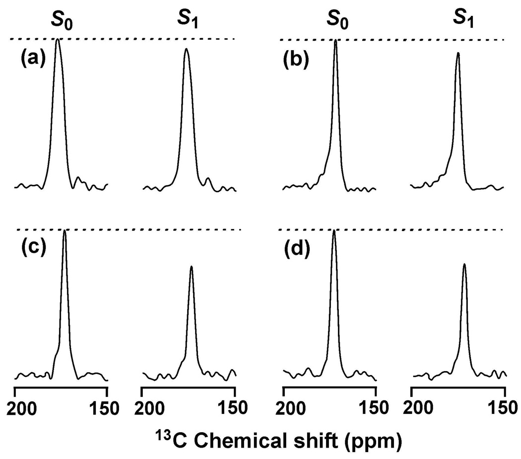
Figure 2.
REDOR S0 and S1 13C SSNMR spectra at 32.2 ms dephasing time for (a) HFP-NC, (b) HFP-P, (c) HFP-A, or (d) HFP-AP. Each spectrum was processed with 200 Hz line broadening and baseline correction and was the sum of: (a) 38624; (b) 23488; (c) 24914; or (d) 14240 scans. Relatively narrow 13CO signals were observed in the HFP-P, HFP-A, and HFP-AP samples because the HFPs were membrane-associated with predominant β sheet conformation at the labeled 13CO site. A broader 13CO signal was observed in the HFP-NC sample because there was no membrane and there were populations of lyophilized HFP with either α helical or β sheet conformation at the labeled 13CO site.

