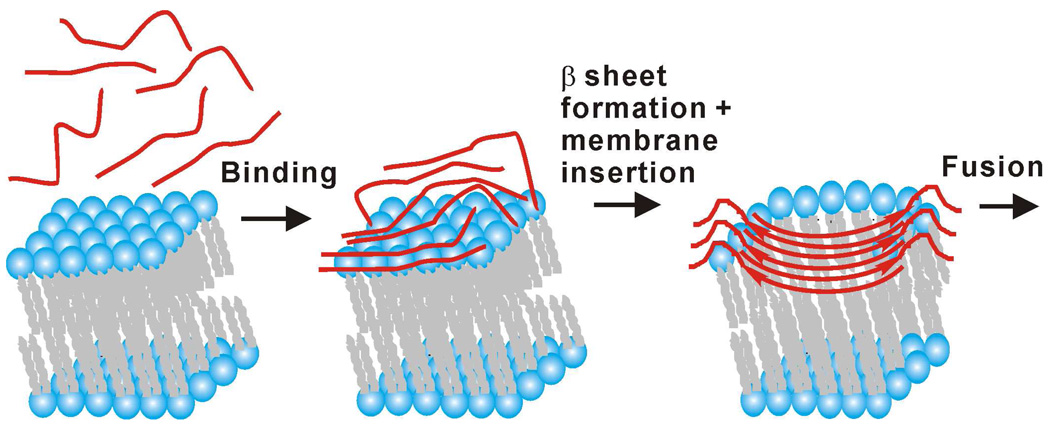
Figure 7.
Pictorial model of HFP (red lines) binding to membranes followed by antiparallel β sheet formation and membrane insertion and then fusion. Time increases from left-to-right. For reasons of clarity, some lipids are not shown in the right-most picture. Although there are no data yet on fusion peptide structure during HIV/host cell fusion, the antiparallel β sheet structure of the right-most picture is plausible because: (1) the structure is consistent with multiple trimers at the fusion site; and (2) the structure is membrane-inserted with deeper insertion positively correlated with increased membrane perturbation and vesicle fusion rate.

