Abstract
Rift Valley fever (RVF) is an epizootic viral disease of sheep that can be transmitted from sheep to humans, particularly by contact with aborted fetuses. A capripoxvirus (CPV) recombinant virus (rKS1/RVFV) was developed, which expressed the Rift Valley fever virus (RVFV) Gn and Gc glycoproteins. These expressed glycoproteins had the correct size and reacted with monoclonal antibodies (MAb) to native glycoproteins. Mice vaccinated with rKS1/RVFV were protected against RVFV challenge. Sheep vaccinated with rKS1/RVFV twice developed neutralizing antibodies and were significantly protected against RVFV and sheep poxvirus challenge. These findings further document the value of CPV recombinants as ruminant vaccine vectors and support the inclusion of RVFV genes encoding glycoproteins in multivalent recombinant vaccines to be used where RVF occurs.
Rift Valley fever (RFV) virus (RVFV) is a mosquito-borne member of the genus Phlebovirus, family Bunyaviridae. It is widely distributed in Africa, causing endemic and epidemic disease in both humans and livestock, including sheep, cattle, and goats. RVF was first described in Kenya and was shown to be caused by a filterable virus transmissible via blood (9). Acute RVF in lambs is characterized by fever and death within 24 to 48 h of being detected (43). Signs in adult sheep include fever, mucopurulent nasal discharge, hemorrhagic diarrhea, and abortion in pregnant ewes (43). RVFV can be transmitted from infected sheep to humans, particularly when humans are exposed to aborted sheep fetuses and blood.
Attenuated live RVFV vaccines are available for use in livestock. A mutagen-attenuated RVFV vaccine induces protective immune responses in lambs and appears to be safe (25); however, other studies documented teratogenic effects on lambs from vaccinated pregnant ewes similar to those caused by the attenuated RVFV strain Smithburn (18). An inactivated RVFV vaccine induces neutralizing antibody responses in humans (33), and its use in sheep would not induce teratogenic effects or abortions. However, the inactivated vaccine requires 3 doses (33) and is expensive to produce. Efforts to make RVFV vaccines without these disadvantages include an attenuated RVFV developed by reverse genetics and lacking the NSs and NSm genes (4) and other new-generation RVFV vaccines (reviewed in reference 19) that protect mice against virus challenge (7, 16, 24, 27).
The middle (M) RNA segment of the RVFV genome encodes the viral glycoproteins Gn and Gc (8, 20), and recombinant vaccinia virus expressing these glycoproteins induces neutralizing antibody and protective immunity to RVFV in mice (7). Vaccinia virus is safe for animals, but there is some risk to humans, as it was reported previously to spread from human vaccinees to contacts (28, 55) and to cause serious clinical disease in human immunodeficiency virus-infected patients (36). Although modified vaccinia virus Ankara is a safer alternative for humans (6, 57), there are animal poxviruses with naturally restricted host ranges for vaccine vectors in animals (1, 13, 30, 31, 40, 46, 47, 52, 53).
For ruminants, the genus Capripoxvirus (CPV) of the family Poxviridae has been an effective recombinant vector to induce protective immunity against several other viruses (3, 17, 29, 32, 40, 41, 51). This genus has three closely related species causing sheep pox, goat pox, and lumpy skin disease (LSD) of cattle. A recombinant LSD vaccine expressing the Gn and Gc glycoproteins of RVFV induced protection against RVFV challenge in mice (52, 53) and sheep (52). The three species of CPV have 96 to 97% nucleotide identity (49) and are restricted to ruminants, with no evidence of human infections (10, 11). Furthermore, attenuated CPV vaccines are in use in Africa and the Middle East to control ruminant poxvirus disease (11, 21). The use of a CPV vector to deliver virus vaccines to ruminants also induces immunity to the CPV vector, thus increasing the valence of the vaccine (3, 17, 39, 40). We report here the construction of a recombinant CPV that expresses the RVFV Gn and Gc glycoproteins and induces protective immunity against RVFV and sheep poxvirus (SPV) challenge in sheep.
MATERIALS AND METHODS
Animal care and biosafety.
Animal experiments were performed at the Kenya Agricultural Research Institute (KARI) research facilities at Kabete, Kenya, and were approved by the Director of KARI and by the Washington State University Animal Care and Use Committee. The animals were kept in insect-proof animal facilities, and the animal care and animal and laboratory experiments were performed by staff vaccinated for RVFV using a vaccine obtained from the U.S. Department of the Army, U.S. Army Medical Material Development Activity, Fort Detrick, Frederick, MD. The animal facilities were close to the laboratory facilities, and there was 24-h security during the animal and laboratory experiments. The research and animal containment facilities were also inspected as part of the Initial Environmental Examination by the U.S. Agency for International Development (USAID) Regional Natural Resources Advisor and the USAID Mission Agricultural Development Officer, and the containment facility, procedures for disposal of biohazards, and protection of humans were found to be compatible with guidelines of the United States. The signs of RVFV and capripoxvirus challenge in experimental animals were mild and did not require treatment or euthanasia. The use of recombinant DNA, RVFV, and capripoxvirus in laboratory and animal experiments was further approved by the KARI Biosafety Committee, the Director of KARI, and the Washington State University Institutional Biosafety Committee.
Viruses and cells.
Capripoxvirus (CPV) strain KS1 was used for vector construction. It was isolated during an outbreak of sheep pox, attenuated, and used as a live attenuated vaccine for sheep pox and goat pox in Kenya (10, 11). The RVFV used was the Smithburn strain, a live attenuated virus (45) currently used as an animal vaccine in Kenya. CPV was propagated in primary lamb testis (LT) cells at a passage of 12 or less, and RVFV was propagated in baby hamster kidney cells (BHK-21; ATCC CCL-10) using RPMI 1640 medium containing 10% fetal bovine serum (FBS), 2 mM l-glutamine, 100 units/ml penicillin, and 100 μg/ml streptomycin. Virus-containing medium was harvested when the cytopathic effect (CPE) exceeded 75%, and the viral infectivity titer was determined by limiting dilution (37).
Construction of CPV insertion plasmid pLSDRV.
Insertion plasmid pLSDRV was constructed as described below to contain an expression cassette with RVFV glycoprotein genes flanked by lumpy skin disease virus (LSDV) TK gene sequences. To make pLSDRV, the 2.5-kb SalI-XbaI fragment from plasmid p1114 containing the P7.5 promoter, a multiple-cloning site, and the P19 promoter followed by the Escherichia coli gpt (Ecogpt) gene (p1114 was obtained from M. Mackett, Patterson Institute for Cancer Research, Manchester, England) was excised and treated with Klenow DNA polymerase (22). The fragment with the Ecogpt gene for later recombinant virus selection (14) was ligated into pLSDTK3c digested with KpnI and treated with T4 DNA polymerase to make blunt ends (23). pLSDTK3c was obtained from Anna-Lise Williamson, Department of Medical Microbiology, University of Cape Town, Cape Town, South Africa, and it contained the 2.5-kb HindIII S fragment of LSDV, including the TK gene (1). The ligation mixture (blunt-ended SalI-XbaI p1114 fragment and blunt-ended KpnI-digested pLSDTK3) was used to transform competent E. coli DH5α cells (23), plasmids from ampicillin-resistant colonies were evaluated by restriction enzyme analysis, and one with an insert in the correct orientation was selected and designated pLSDKgpt. A 3.4-kb NcoI-SspI fragment was then excised from plasmid pSCRV-6 (obtained from M. Collett, Molecular Vaccines Inc., Gaithersburg, MD), which contained the entire coding sequence for the Gn and Gc glycoproteins of RVFV (7). This 3.4-kb fragment was treated with Klenow DNA polymerase and blunt-end ligated into SmaI-digested pLSDTKgpt downstream of the P7.5 promoter. Plasmids from ampicillin-resistant transformed bacteria were evaluated by restriction enzyme analysis, and one with an insert in the correct orientation was selected and designated pLSDRV.
Generation of rKS1/RVFV.
Subconfluent LT cells in six-well plates were infected with CPV KS1, and 1 h later, the cells were rinsed twice with warm serum-free RPMI 1640 medium. A mixture of 2 μg of insertion plasmid pLSDRV DNA and 20 μg of Lipofectin (Gibco BRL) was added in a volume of 0.5 ml and incubated at 37°C for 36 h. Maintenance medium was added, and incubation continued until the CPE was >75%. The remaining cells were scraped, virus was released by three freeze-thaw cycles, and the virus-containing supernatant was then collected after centrifugation at 1,000 × g for 10 min. LT cell monolayers were then pretreated overnight with MXHAT medium (25 μg/ml mycophenolic acid, 250 μg/ml xanthine, 10−4 M hypoxanthine, 4 × 10⁻⁷ M aminopterin, and 1.6 × 10⁻⁷ M thymidine) and infected with 10-fold dilutions of the supernatant (23). After adsorption for 2 h at 37°C, the monolayers were washed twice with serum-free RPMI 1640 medium, overlaid with MXHAT medium containing 1% low-melting agarose, and incubated at 37°C with 5% CO2 (23). A well-isolated plaque was picked 3 to 7 days later and resuspended in 500 μl of medium, and the virus was immediately replaqued without amplification. After three rounds of plaque purification, the recombinant virus was amplified in the presence of MXHAT medium and designated rKS1/RVFV.
IFA test.
LT cells were infected with rKS1/RVRF, rKS1/gpt, or KS1 at a multiplicity of infection (MOI) of 0.1. Two days later, the cells were acetone fixed or processed for either surface membrane or cytoplasmic indirect fluorescent antibody (IFA) testing (23). For surface membrane IFA testing, infected cells were fixed with 10% buffered formalin (pH 7.5) for 30 min at room temperature. For cytoplasmic IFA testing, formalin-fixed cells were permeabilized with 0.1% Triton X-100 and 0.25% bovine serum albumin (BSA) in phosphate-buffered saline (PBS) for 30 min. Nine monoclonal antibodies (MAbs) to discrete noncompeting epitopes located on glycoprotein Gc and seven MAbs to Gn of RVFV were obtained as mouse ascites from Jonathan Smith, USAMRIID, Fort Detrick, MD, and used as primary antibodies. The binding of MAbs was detected by using an anti-mouse IgG-fluorescein isothiocyanate (FITC) conjugate.
Western immunoblots.
Cell lysates were obtained from LT cells infected with rKS1/RVFV, rKS1/gpt, and KS1 at an MOI of 10 for 2 days by scraping and boiling in SDS sample buffer (23). RVFV was obtained from infected BHK-21 cells. Lysates were subjected to electrophoresis on discontinuous 7.5-to-15% gradient SDS-PAGE gels and electroblotted onto nitrocellulose membranes. After blocking for 30 min at room temperature with 5% nonfat milk in Tris-buffered saline (TBS), the membranes were probed with either MAb RVI-4D4 (15 μg/ml) or mouse antiserum to RVFV (1:25 dilution) for 1 h. The antiserum was made by multiple intraperitoneal inoculations of mice with the RVFV Smithburn strain (45). The membranes were then washed three times for 10 min each and incubated for 30 min with diluted peroxidase-conjugated antibodies to mouse IgG. After washing three times, bound conjugate was detected with 0.01% diaminobenzidine (Sigma) containing H2O2.
Southern blots.
DNAs prepared from virus (12), plasmid (35), and LT cell (38) samples were digested with either HindIII or PstI, and the fragments were separated on 0.8% agarose gels. Fragments were transferred onto nylon membranes (26) and probed with digoxigenin-11-dUTP-labeled DNA fragments containing CPV TK (2.5-kb HindIII fragment from pLSDTK3c), the Ecogpt gene (1.8-kb HindIII fragment from pTKgptF1s [14], obtained from B. Moss, NIH, Bethesda, MD), or the RVFV M segment (3.4-kb NcoI-SspI fragment from pSCRV-6). Bound probes were detected according to the manufacturer's instructions (Boehringer Mannheim). Freedom from wild-type virus contamination was confirmed by the absence of the 2.5-kb HindIII fragment in Southern blots when probed with the LSDV TK fragment.
Growth curve of recombinant and wild-type CPV.
LT cells in petri dishes were infected at an MOI of 0.02 with either the recombinant or parental KS1. At 0, 4, 8, and 24 h postinfection and at five 24-h intervals thereafter, infected cells were frozen and thawed three times at −70°C to release the virus. The virus harvested was clarified, dilutions of 10−1 to 10−7 were evaluated for infectivity in eight wells per dilution in LT cells, and the 50% tissue culture infective dose (TCID50) was calculated (37).
Vaccination of mice with rKS1/RVFV.
Three groups of 10 mice each were vaccinated once with 104.5 TCID50 rKS1/RVFV either subcutaneously, intramuscularly, or intraperitoneally, and three other groups of 10 mice were similarly vaccinated with 107.25 TCID50. All six groups were challenged with 100 50% lethal doses (LD50) of virulent RVFV 21 days after vaccination, and surviving mice were bled for serum collection 21 days after RVFV challenge. In a third experiment, two groups of 10 mice each were vaccinated three times either intraperitoneally or intramuscularly with 107, 106, and 106 TCID50 of rKS1/RVFV at 3-week intervals for the first, second, and third vaccinations, respectively. These two mouse groups were challenged with 100 LD50 of virulent RVFV 21 days after the third vaccination. The mice were bled for serum collection before vaccination, 14 days after the third vaccination, and 17 days after RVFV challenge in surviving mice.
Vaccination of sheep with rKS1/RVFV.
Twenty-two male and female Dorper breed sheep without serum neutralization (SN) antibodies to sheep poxvirus (SPV) or RVFV were obtained from a farm with no recent history of sheep pox or RVF vaccinations or outbreaks. The sheep were divided into four groups of five sheep each and one group (group 5) with two sheep, attempting to divide the females among the groups except for group 3. Groups 1, 2, and 5 were vaccinated with 108 TCID50 rKS1/RVFV subcutaneously near the prescapular lymph node. Group 3 was similarly vaccinated with 108 TCID50 of control rKS1/gpt, whereas group 4 was vaccinated with saline. Three weeks later, the sheep were similarly vaccinated a second time and monitored for 4 weeks before virus challenge. The groups were monitored daily for skin lesions at the vaccination site and for fever, and during this time one sheep from group 3 and one from group 4 died due to unrelated causes. Sheep from groups 1 and 3 were challenged subcutaneously with 107 TCID50 of RVFV, whereas sheep from groups 2 and 4 were challenged subcutaneously with 107 TCID50 of the Kedong strain of SPV. The challenged sheep were monitored twice daily, and plasma was obtained for virus isolation from the RVFV-challenged sheep daily for 8 days. Three weeks later, group 1 sheep initially challenged with RVFV were challenged with SPV, and group 2 sheep initially challenged with SPV were challenged with RVFV. Group 5 sheep were inoculated with 107 TCID50 SPV and 1 month later were vaccinated with rKS1/RVFV and challenged with RVFV as described above for group 1. Serum was obtained weekly from all sheep for antibody evaluation.
SN assays.
SN assays were done using constant virus and diluted serum in 96-well, flat-bottom plates. Sera were heated for 30 min at 56°C before use. For RVFV, 100 μl of diluted serum was mixed with 100 μl containing 100 TCID50 RVFV and incubated at 37°C for 1 h and at 4°C overnight. Fifty microliters containing 16,000 BHK cells was then added to each well, and the wells were incubated at 37°C with 5% CO2 for 5 days. Wells were evaluated, and the neutralization titer was the highest serum dilution causing >50% inhibition of CPE in at least one of two wells. The same procedure was used for poxvirus except that the input virus was 50 TCID50 of SPV, the cells used were LT cells at 10,000 cells/well, and the CPE was evaluated after 10 days.
RESULTS
Demonstration of RVFV genes within the TK gene of rKS1/RVFV.
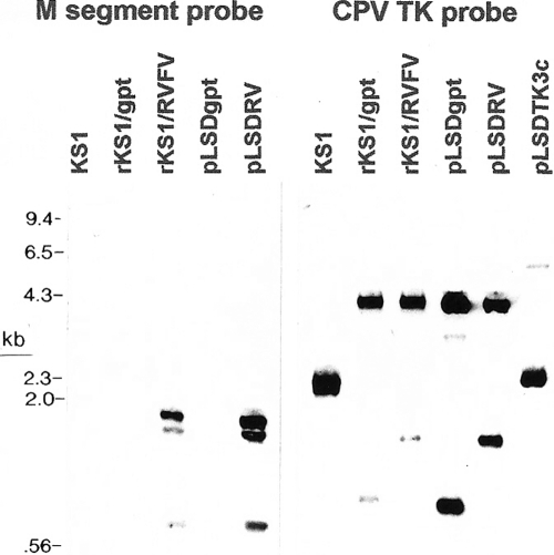
RVFV genes were demonstrated in rKS1/RVFV and parent plasmid pLSDRV DNAs by fragments migrating at 1.8, 1.7, and 0.7 kb hybridizing with the RVFV M segment (Fig. 1, left). There was no hybridization of the RVFV M segment with HindIII-digested control DNA from KS1, rKS1/gpt, and pLSDgpt. To verify that the RVFV genes were inserted into the TK gene, HindIII-digested KS1 and rKS1/RVFV DNAs were hybridized with the capripoxvirus (CPV) TK gene (Fig. 1, right). This resulted in a predicted 2.5-kb band in KS1 and plasmid pLSDTK3c DNAs. Two bands migrating at 4.2 and 1.7 kb were found in recombinant rKS1/RVFV and in pLSDRV DNAs. In contrast, two bands migrating at 4.2 and 1.0 kb were detected in rKS1/gpt and in pLSDgpt DNAs (Fig. 1, right). Finally, results of hybridization with the gpt gene did not detect any band with KS1 DNA, but a 4.2-kb fragment was detected in both the rKS1/gpt and KS1/RVFV DNAs (data not shown).
FIG. 1.
Southern blot analysis of HindIII-digested DNAs (labeled above each probed lane). The five lanes on the left were probed with the labeled RVFV M segment, and the six lanes on the right were probed with the labeled CPV TK gene. The migrations of standards in kilobases are indicated on the left.
Expression of RVFV glycoproteins in rKS1/RVFV CPV-infected cells.
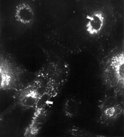
RVFV Gn and Gc glycoprotein antigens were detected by fluorescence in the cytoplasm of acetone-fixed LT cells infected with rKS1/RVFV using nine different MAbs to RVFV Gc and seven to Gn glycoproteins. A similar fluorescence occurred with IFA in infected cells fixed with formalin and permeabilized with Triton X-100 (Fig. 2); however, the infected cells did not stain when the permeabilization step was omitted. LT cells infected with either KS1 virus or rKS1/gpt did not fluoresce with the MAbs in IFA testing (data not shown).
FIG. 2.
Photograph of LT cells infected with rKS1/RVFV, fixed in formalin, permeabilized with Triton X-100, and used in an indirect fluorescent antibody test. The primary antibody was a single MAb (RVI-4D4) to the RVFV Gn glycoprotein, and the secondary antibodies used were FITC-labeled antibodies to mouse IgG. The white arrow (top right) is pointing to the nucleus of a cell with cytoplasmic fluorescence; the white arrow (bottom left) is pointing to the nucleus of another cell with cytoplasmic fluorescence surrounded by other cells with similar fluorescences. LT cells infected with either KS1 or rKS1/gpt and evaluated with the same indirect fluorescent antibody test did not have cytoplasmic fluorescence (data not shown).
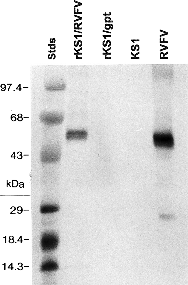
Western immunoblots also demonstrated the expression of the RVFV glycoprotein in LT cells infected with rKS1/RVFV. RVFV Gn glycoprotein MAb (RVI-4D4) bound a 55-kDa antigen in lanes containing lysate from rKS1/RVFV-infected cells and purified RVFV but not antigens in rKS1/gpt and KS1 (Fig. 3). In another Western immunoblot (data not shown), mouse antiserum to RVFV reacted with 63- and 55-kDa antigens in purified rKS1/RVFV, and these were interpreted as the expressed RVFV Gn and Gc glycoproteins, respectively.
FIG. 3.

Western immunoblots of lanes containing rKS1/RVFV, rKS1/gpt, KS1, and RSFV were reacted with a single MAb (RVI-4D4) to the RVFV Gn glycoprotein. A major band of approximately 55 kDa was detected in the lanes containing rKS1/RVFV and RVFV but not in control lanes containing rKS1/gpt and KS1. The migrations of the protein standards (kDa) are on the left.
In vitro growth pattern of recombinant CPV KS1/RVFV.
The growth pattern of rKS1/RVFV was compared with that of wild-type KS1 by inoculating LT cells with each virus at an MOI of 0.02. Titration of progeny virus harvested at regular intervals demonstrated that the viruses replicated to the same titer (Fig. 4), indicating that the insertion of RVFV genes into the TK gene of rKS1/RVFV did not detectably alter in vitro replication.
FIG. 4.
In vitro growth comparison of rKS1/RVFV with wild-type KS1. To obtain the log10 TCID50, eight wells of LT cells were inoculated with each dilution (10−1 to 10−7) of the virus harvested from each time point.
rKS1/RVFV protected mice against RVFV challenge.
In preliminary experiments, increasing a single vaccine dose from 105.4 to 107.25 TCID50 rKS1/RVFV increased survival after RVFV challenge from 20 to 70% in mice vaccinated intraperitoneally and from 0 to 60% in mice vaccinated intramuscularly (Table 1). All mouse groups vaccinated once made SN antibody to RVFV except those vaccinated subcutaneously with 104.5 TCID50. Finally, 100% and 90% of mice vaccinated three times intraperitoneally and intramuscularly with rKS1/RVFV survived RVFV challenge (Table 2). Vaccinated mice had SN antibodies to RVFV 14 days after the second immunization (Table 2).
TABLE 1.
Survival and serum antibody titers of mice vaccinated with a single dose of rKS1/RVFV and challenged with RVFVa
| Route of vaccination | Dose (TCID50) | No. of surviving mice/no. of challenged mice (%) | Prevaccination RVFV IFA titer | RVFV IFA titer (SN titer)b at 21 days postvaccination |
|---|---|---|---|---|
| Intraperitoneal | 104.5 | 2/10 (20) | <16 | 64 (4) |
| Intramuscular | 104.5 | 0/10 | <16 | 64 (4) |
| Subcutaneous | 104.5 | 0/10 | <16 | 64 (<4) |
| Intraperitoneal | 107.25 | 7/10 (70) | <16 | 64 (4) |
| Intramuscular | 107.25 | 6/10 (60) | <16 | 32 (4) |
| Subcutaneous | 107.25 | 2/10 (20) | <16 | 32 (8) |
Mice were challenged subcutaneously with 100 LD50 of RVFV at 21 days postvaccination.
SN titers to RVFV are in parentheses. The SN titer was not determined with prevaccination sera. IFA (indirect fluorescence antibody) and SN titers are the reciprocal of the last positive dilution determined with pooled sera from each group of mice.
TABLE 2.
Survival and RVFV SN antibody titers of mice vaccinated three times with rKS1/RVFV and then challenged with RVFVa
| Route of vaccination | No. of surviving mice/no. of challenged mice (%) | RVFV SN titerb |
||
|---|---|---|---|---|
| Prevaccination | 14 days after 3rd vaccination | 17 days after RVFV challenge | ||
| Intraperitoneal | 10/10 (100) | <4 | 8 | 16 |
| Intramuscular | 9/10 (90) | <4 | 4 | 32 |
Mice were challenged subcutaneously with 100 LD50 of virulent RVFV 21 days after the third vaccination.
SN titers are the reciprocal of the last positive dilution determined with pooled sera from each group of mice.
rKS1/RVFV protected sheep against RVFV challenge.
No RVFV was detected in the blood of rKS1/RVFV-vaccinated group 1 sheep challenged with RVFV 28 days after the second vaccination. There was a significant difference in viremia (P < 0.05 by chi-square test) in control group 2, as RVFV was detected on the first and second days after challenge in three of five sheep (Table 3). There was also a significant difference in fever (P < 0.01 by chi-square test), as one group 1 sheep (sheep 3248) and all group 2 sheep were febrile on day 1 after RVFV challenge (Table 3). Protection against RVFV viremia and fever in group 1 sheep was associated with SN antibodies to RVFV present by 15 days after the first vaccination and increasing after the second vaccination and after RVFV challenge (Table 4). No SN antibodies were detected in control group 2 sheep following vaccinations; however, they were detected in all five of these sheep following RVFV challenge (Table 4).
TABLE 3.
Body temperatures and RVFV titers of vaccinated sheep challenged with RVFV
| Sheep (sex) | Group (inoculum) | Body temp (°C) (RVFV titer [log10 TCID50/ml]) at day post-RVFV challengea: |
||||
|---|---|---|---|---|---|---|
| 0 | 1 | 2 | 3 | 4 | ||
| 2517 (F) | 1 (rKS1/RVFV) | 39.0 | 39.8 (Neg) | 39.1 (Neg) | 38.8 (Neg) | 39.0 (Neg) |
| 2518 (M) | 1 (rKS1/RVFV) | 39.2 | 39.7 (Neg) | 39.1 (Neg) | 39.3 (Neg) | 39.2 (Neg) |
| 2519 (F) | 1 (rKS1/RVFV) | 39.3 | 39.5 (Neg) | 39.5 (Neg) | 39.6 (Neg) | 40.1 (Neg) |
| 3244 (M) | 1 (rKS1/RVFV) | 39.3 | 40.0 (Neg) | 38.7 (Neg) | 38.8 (Neg) | 39.0 (Neg) |
| 3248 (F) | 1 (rKS1/RVFV) | 39.1 | 40.6 (Neg) | 39.4 (Neg) | 39.0 (Neg) | 39.6 (Neg) |
| 2523 (M) | 2 (rKS1/gpt) | 39.0 | 41.4 (101) | 39.8 (101) | 39.4 (Neg) | 40.0 (Neg) |
| 3264 (F) | 2 (rKS1/gpt) | 39.1 | 40.5 (101) | 39.5 (101) | 39.0 (Neg) | 39.6 (Neg) |
| 3266 (F) | 2 (rKS1/gpt) | 39.0 | 41.0 (101) | 39.7 (101) | 39.3 (Neg) | 40.3 (Neg) |
| 3267 (M) | 2 (rKS1/gpt) | 39.4 | 40.3 (Neg) | 39.5 (Neg) | 39.0 (Neg) | 39.0 (Neg) |
| 3269 (M) | 2 (rKS1/gpt) | 39.2 | 41.0 (Neg) | 40.2 (Neg) | 39.3 (Neg) | 39.5 (Neg) |
| 2520 (M) | 3 (rKS1/RVFV) | 39.1 | 40.0 (Neg) | 39.5 (Neg) | 39.2 (Neg) | 39.6 (Neg) |
| 2521 (M) | 3 (rKS1/RVFV) | 38.7 | 39.3 (Neg) | 40.0 (Neg) | 37.2 (Neg) | ND (Neg) |
| 3252 (M) | 3 (rKS1/RVFV) | 39.0 | 40.1 (Neg) | 40.2 (Neg) | 39.9 (Neg) | 39.3 (Neg) |
| 3255 (M) | 3 (rKS1/RVFV) | 39.0 | 38.8 (Neg) | 39.6 (Neg) | 40.3 (Neg) | 40.1 (Neg) |
| 2524 (F) | 4 (saline) | 38.5 | 39.0 (Neg) | 39.5 (Neg) | 39.0 (Neg) | 38.9 (Neg) |
| 3271 (M) | 4 (saline) | 39.2 | 40.2 (Neg) | 39.3 (Neg) | 39.9 (Neg) | 40.5 (Neg) |
| 3273 (M) | 4 (saline) | 39.3 | 41.0 (102) | 39.7 (102) | 40.3 (102) | 40.0 (Neg) |
| 3274 (F) | 4 (saline) | 39.4 | 40.3 (102) | 41.6 (104) | 41.7 (101) | 39.7 (Neg) |
| 3246 (M) | 5 (rKS1/RVFV) | 39.2 | 40.0 (Neg) | 39.0 (Neg) | 38.8 (Neg) | 39.0 (Neg) |
| 3263 (F) | 5 (rKS1/RVFV) | 39.6 | 40.7 (Neg) | 40.2 (Neg) | 39.3 (Neg) | 39.0 (Neg) |
RVFV titers (in parentheses) are log10 TCID50/ml, and Neg indicates a virus titer of less than 101 TCID50/ml. Temperatures in boldface type are ≥40.3°C, which was the threshold for fever determined as 1.2°C (5 standard deviations) above the mean of 39.1°C for the 20 sheep at time zero in this table. ND indicates not done.
TABLE 4.
RVFV SN antibody titers in sheep sera after rKS1/RVFV or rKS1/gpt inoculation and after RVFV challenge
| Sheep (sex) | Group (inoculum) | RVFV SN antibody titera |
||||||||
|---|---|---|---|---|---|---|---|---|---|---|
| Preinoculation | 7 dpi | 15 dpi | 21 dpi | 7 dpb | 15 dbp | 28 dpb | 10 dpc | 28 dpc | ||
| 2517 (F) | 1 (rKS1/RVFV) | Neg | 8 | 2 | 4 | 8 | 8 | 8 | 256 | 32 |
| 2518 (M) | 1 (rKS1/RVFV) | Neg | NT | 4 | 4 | 16 | 8 | 16 | 32 | 128 |
| 2519 (F) | 1 (rKS1/RVFV) | Neg | 4 | 8 | 8 | 16 | 16 | 16 | 256 | 128 |
| 3244 (M) | 1 (rKS1/RVFV) | Neg | 4 | 4 | Neg | 8 | 16 | 8 | 32 | 128 |
| 3248 (F) | 1 (rKS1/RVFV) | Neg | Neg | 4 | 8 | 8 | 16 | 256 | 128 | 128 |
| 2523 (M) | 2 (rKS1/gpt) | Neg | Neg | Neg | Neg | Neg | Neg | Neg | 16 | 32 |
| 3264 (F) | 2 (rKS1/gpt) | Neg | Neg | Neg | Neg | Neg | Neg | Neg | 32 | 64 |
| 3266 (F) | 2 (rKS1/gpt) | Neg | Neg | Neg | Neg | Neg | Neg | Neg | 16 | 32 |
| 3267 (M) | 2 (rKS1/gpt) | Neg | Neg | Neg | Neg | Neg | Neg | Neg | 2 | 64 |
| 3269 (M) | 2 (rKS1/gpt) | Neg | Neg | Neg | Neg | Neg | Neg | Neg | 4 | 32 |
| 3246 (M) | 5 (rKS1/gpt) | Neg | Neg | Neg | Neg | 16 | 16 | 8 | >256 | >256 |
| 3263 (F) | 5 (rKS1/gpt) | Neg | Neg | Neg | Neg | 8 | 16 | 16 | >256 | 256 |
dpi, days post-rKS1/RVFV or rKS1/gpt initial inoculation; dpb, days post-rKS1/RVFV or rKS1/gpt second vaccination; dpc, days post-RVFV challenge; Neg, no SN antibody detected at a 1:2 dilution of serum (all other titers are the reciprocal of the last positive serum dilution); NT, not tested.
To obtain additional data, group 3 sheep vaccinated with rKS1/RVFV and group 4 sheep inoculated with saline and challenged with CPV (see below) were subsequently challenged with RVFV 85 days after the second vaccination. None of the group 3 sheep had fever or detectable viremia, but two of four group 4 sheep became febrile and viremic (Table 3); however, these differences between groups 3 and 4 were not significant (P > 0.05 by chi-square test).
Finally, a preliminary evaluation of recent poxvirus infection on the immunity induced by the subsequent inoculation of rKS1/RVFV was performed. Group 5 sheep challenged with SPV 1 month before the first vaccination with rKS1/RVFV were subsequently challenged with RVFV. No RVFV was detected in the two sheep, even though one was febrile (40.7°C) 1 day after RVFV challenge (Table 3). Compared to group 1 sheep, the appearance of SN antibody in the two group 5 sheep was delayed until after the second vaccination (Table 4).
Taken together, none of 11 sheep (groups 1, 3, and 5) inoculated with rKS1/RVFV and challenged with RVFV became viremic. In contrast, five of nine control sheep (groups 3 and 4) became viremic following RVFV challenge. The difference in the viremias of the combined groups of rKS1/RVFV-inoculated sheep and the combined control groups was significant (P < 0.01 by chi-square test).
rKS1/RVFV protected sheep against SPV challenge.
The protection against SPV challenge in rKS1/RVFV-vaccinated sheep (group 3) compared with control group 4 (saline inoculated) was significant for fever (P < 0.01) and skin lesions (P < 0.01) (Table 5). The skin lesions were raised, 3- to 5-cm circumscribed areas that were painful to the touch and exuded serum by day 7 but disappeared by day 10. Poxvirus was isolated from biopsy specimens of skin lesions from three of four group 4 sheep (no virus from sheep 3271) on day 10 after SPV challenge. Protection against SPV in group 3 sheep was associated with SN antibodies to SPV that developed after the first vaccination (Table 6). Group 4 saline-inoculated sheep did not have detectable SN antibodies until after the SPV challenge (Table 6).
TABLE 5.
Body temperatures of vaccinated sheep after SPV challenge
| Sheep (sex) | Group (inoculum) | Body temp (°C) at dpca: |
|||||||||
|---|---|---|---|---|---|---|---|---|---|---|---|
| 0 | 3 | 4 | 5 | 6 | 7 | 8 | 9 | 10 | 11 | ||
| 2520 (M) | 3 (rKS1/RVFV) | 39.4 | 39.5 | 39.3 | 39.5 | 39.8 | 39.7 | 40.1 | 40.0 | 40.0 | 39.1 |
| 2521 (M) | 3 (KS1/RVFV) | 39.1 | 39.4 | 39.5 | 39.3 | 39.7 | 40.0 | 40.0 | 39.9 | 39.7 | 39.6 |
| 3252 (M) | 3 (KS1/RVFV) | 38.6 | 38.7 | 39.0 | 39.1 | 39.7 | 39.8 | 39.6 | 40.0 | 40.0 | 39.0 |
| 3255 (M) | 3 (KS1/RVFV) | 38.8 | 39.0 | 39.2 | 39.4 | 39.8 | 39.5 | 39.2 | 39.8 | 39.2 | 38.9 |
| 2524 (F) | 4 (saline) | 39.6 | 39.6 | 39.5 | 41.0 | 41.0 | 40.6 | 40.8 | 40.3 | 40.4 | 39.7 |
| 3271 (M) | 4 (saline) | 39.4 | 40.2 | 40.1 | 40.7 | 41.2 | 41.0 | 40.0 | 40.0 | 39.5 | 39.3 |
| 3273 (M) | 4 (saline) | 40.0 | 40.1 | 40.9 | 41.3 | 40.8 | 40.7 | 40.2 | 40.2 | 40.0 | 40.0 |
| 3274 (F) | 4 (saline) | 39.6 | 39.4 | 40.2 | 40.3 | 41.1 | 40.5 | 40.3 | 40.3 | 40.6 | 39.5 |
dpc, days post-SPV challenge. Temperatures in boldface type are ≥40.3°C, which was the threshold for fever determined as 1.2°C (5 standard deviations) above the mean of 39.1°C for the 20 sheep at time zero in Table 3.
TABLE 6.
CPV SN antibody titers in sheep sera after rKS1/RVFV or saline inoculations and after SPV challenge
| Sheep (sex) | Group (inoculum) | CPV SN antibody titera |
||||||||
|---|---|---|---|---|---|---|---|---|---|---|
| Preinoculation | 7 dpi | 15 dpi | 21 dpi | 7 dpb | 15 dbp | 28 dpb | 10 dpc | 28 dpc | ||
| 2520 (M) | 3 (rKS1/RVFV) | Neg | Neg | 4 | 4 | >64 | >64 | 32 | 16 | 32 |
| 2521 (M) | 3 (rKS1/RVFV) | Neg | 4 | 8 | 16 | 8 | 32 | 32 | >64 | >64 |
| 3252 (M) | 3 (rKS1/RVFV) | Neg | 4 | 2 | 4 | 16 | 8 | 16 | >64 | 16 |
| 3255 (M) | 3 (rKS1/RVFV) | Neg | 2 | 8 | 4 | 32 | 32 | 32 | 16 | >64 |
| 2524 (F) | 4 (saline) | Neg | Neg | Neg | Neg | Neg | Neg | Neg | 32 | >64 |
| 3271 (M) | 4 (saline) | Neg | Neg | Neg | Neg | Neg | Neg | Neg | 2 | 4 |
| 3273 (M) | 4 (saline) | Neg | Neg | Neg | Neg | Neg | Neg | Neg | 2 | 8 |
| 3274 (F) | 4 (saline) | Neg | Neg | Neg | Neg | Neg | Neg | 2 | 32 | 16 |
dpi, days post-rKS1/RVFV or saline initial inoculation; dpb, days post-rKS1/RVFV or saline second vaccination; dpc, days post-wild-type KS1 virus challenge; Neg, no SN antibody detected at a 1:2 dilution of serum (all other titers are the reciprocal of the last positive serum dilution).
Group 1 sheep inoculated with rKS1/RVFV and challenged with RVFV as described above were then challenged with SPV 88 days after the second vaccination with rKS1/RVFV. None developed fever or skin lesions (data not shown).
DISCUSSION
The recombinant vaccine rKS1/RVFV was constructed by inserting the RVFV Gn and Gc glycoprotein genes into the TK gene of wild-type KS1 capripoxvirus (CPV). KS1 was also used previously by others for the construction of recombinant vectors expressing genes from rinderpest, peste-des-petits-ruminants, and bluetongue viruses (3, 32, 40, 51). The RVFV glycoproteins expressed by sheep cells infected with rKS1/RVFV were of sizes similar to those of the native RVFV glycoproteins Gn and Gc when evaluated by Western immunoblotting. The immunological fidelity of the expressed glycoproteins was further demonstrated by using nine MAbs to nonoverlapping epitopes on the RVFV glycoprotein Gc and seven MAbs to Gn (42). All 16 MAbs reacted with the recombinant expressed glycoproteins in IFA using formalin-fixed, permeabilized cells, indicating that the epitopes recognized by these MAbs were present in the recombinant expressed glycoproteins. These MAbs did not react with formalin-fixed, nonpermeabilized cells, indicating that the expressed RVFV glycoproteins were not transported to the cell surface in enough of a quantity for detection by IFA, consistent with data from a previous report using recombinant vaccinia virus to express the RVFV M segment in Vero cells (54). In addition, rKS1/RVFV replicated to the same level as KS1 in LT cells, indicating no loss of replication potential and helping justify evaluation as a vaccine in mice.
rKS1/RVFV inoculation of mice resulted in protection against RVFV challenge, with protection varying based on the amount, route, and number of inoculations; however, three intraperitoneal inoculations of mice with 107.25 TCID50 rKS1/RVFV resulted in 100% survival from RVFV challenge. Since protective immune responses were stimulated despite host restriction, it is possible that these responses were to expressed exogenous gene products carried by rKS1/RVFV, similar to that described previously for vaccinia virus recombinants (15). However, it is likely that immunity was due to RVFV protein expression in mouse cells taking up rKS1/RVFV in the absence of productive infection, as described previously for immunity induced with avipoxvirus vectors in nonavian species (34, 46, 47, 48). Regardless of how host restriction was overcome, the demonstration of protective immune responses in rKS1/RVFV-inoculated mice further verified the integrity of the expressed RVFV glycoproteins and justified testing in sheep.
Sheep inoculated with rKS1/RVFV were significantly protected against RVFV challenge. The most important evidence of protection was the absence of viremia following RVFV challenge in rKS1/RVFV-inoculated group 1 sheep compared to the number of viremic sheep in control group 2 (P < 0.05), and this was supported by a significant difference between the number of febrile sheep in group 1 compared to that in group 2 (P < 0.01). There were no RVFV SN antibodies in the control sheep before RVFV challenge, so the reason for the lack of viremia in some of the control sheep was presumed to be because of a low RVFV challenge dose. These results demonstrating protection of mice and sheep with the rKS1/RVFV vaccine are similar to previously obtained results with mice and sheep with a recombinant LSD vaccine expressing the RVFV glycoprotein Gn and Gc genes (52, 53). However, viremia was not measured in sheep challenged with RVFV in those studies with the recombinant LSD vaccine (52). In addition, the inoculation of sheep with rKS1/RVFV induced significant protective immunity against challenge with SPV, evidenced by a significant absence of fever and skin lesions compared with the control group which could not be evaluated with the recombinant LSD vaccine (52). Recombinant CPV vectors expressing other ruminant viruses also induced protective immunity to CPV challenge (3, 17, 39, 40). The bivalent protection against both RVFV and SPV challenge in rKS1/RVFV-vaccinated sheep was associated with the presence of SN antibodies, although this was the only potentially protective immune response that was measured.
Vaccination of sheep with rKS1/RVFV would avoid the abortigenic complications of live attenuated RVFV vaccines. Furthermore, the RVFV glycoproteins expressed by rKS1/RVFV have the potential to induce protective immunity against most field isolates in Africa. This assumption is based on observations that minimal genetic variance has occurred in the RVFV glycoprotein-encoding region of 22 African isolates collected over a period of 34 years (2), that RVFV strains evaluated by neutralization are of a single serotype (44), and that RVFV vaccines based on the Smithburn strain have been used extensively in Kenya and South Africa without reports of breakthroughs (56). The addition of other virus genes that express proteins that induce protective immunity to the rKS1/RVFV vector should further increase the number of viruses for which protection could be induced. A major advantage of this and other CPV vectors, including those made with LSD, is their minimal danger to humans due to host restriction. The inactivation of the TK gene during the preparation of rKS1/RVRF (5) should further reduce this vector's virulence for natural ruminant hosts. Sheep infected with wild-type RVFV make antibody to the nucleocapsid protein (50), and tests for this antibody are candidates for differentiating RVFV-infected sheep and rKS1/RVFV-vaccinated sheep.
Acknowledgments
This research was supported in part by U.S. Agency for International Development grant DAN-1328-6-00-0046-00 in collaboration with the Kenya Agricultural Research Institute, Washington State University, and Colorado State University.
We thank Jonathan Smith for the monoclonal antibodies, Anna-Lise Williamson for the LSD vector, and D. Black for important early discussion about the project.
Footnotes
Published ahead of print on 28 September 2010.
REFERENCES
- 1.Aspden, K., A. A. van Dijk, J. Bingham, D. Cox, J. A. Passmore, and A. L. Williamson. 2002. Immunogenicity of a recombinant lumpy skin disease virus (neethling vaccine strain) expressing the rabies virus glycoprotein in cattle. Vaccine 20:2693-2701. [DOI] [PubMed] [Google Scholar]
- 2.Battles, J. K., and J. M. Dalrymple. 1988. Genetic variation among geographic isolates of Rift valley fever virus. Am. J. Trop. Med. Hyg. 39:617-631. [DOI] [PubMed] [Google Scholar]
- 3.Berhe, G., C. Minet, C. Le Goff, T. Barrett, A. Ngangnou, C. Grillet, G. Libeau, M. Fleming, D. N. Black, and A. Diallo. 2003. Development of a dual recombinant vaccine to protect small ruminants against peste-des-petits-ruminants virus and capripoxvirus infections. J. Virol. 77:1571-1577. [DOI] [PMC free article] [PubMed] [Google Scholar]
- 4.Bird, B. H., C. G. Albarino, A. L. Hartman, B. R. Erickson, T. G. Ksiazek, and S. T. Nichol. 2008. Rift Valley fever virus lacking the NSs and NSm genes is highly attenuated, confers protective immunity from virulent virus challenge, and allows for differential identification of infected and vaccinated animals. J. Virol. 82:2681-2691. [DOI] [PMC free article] [PubMed] [Google Scholar]
- 5.Buller, R. M., G. L. Smith, K. Cremer, A. L. Notkins, and B. Moss. 1985. Decreased virulence of recombinant vaccinia virus expression vectors is associated with a thymidine kinase-negative phenotype. Nature 317:813-815. [DOI] [PubMed] [Google Scholar]
- 6.Chen, Z., L. Zhang, C. Qin, L. Ba, C. E. Yi, F. Zhang, Q. Wei, T. He, W. Yu, J. Yu, H. Gao, X. Tu, A. Gettie, M. Farzan, K. Y. Yuen, and D. D. Ho. 2005. Recombinant modified vaccinia virus Ankara expressing the spike glycoprotein of severe acute respiratory syndrome coronavirus induces protective neutralizing antibodies primarily targeting the receptor binding region. J. Virol. 79:2678-2688. [DOI] [PMC free article] [PubMed] [Google Scholar]
- 7.Collett, M. S., K. Keegan, S. Hu, P. Sridhar, A. F. Purchio, W. H. Ennis, and J. M. Dalrymple. 1987. Protective subunit immunogens to Rift Valley fever virus from bacteria and recombinant vaccinia virus, p. 321-329. In B. Mahy and D. Kolakofsky (ed.), The biology of negative strand viruses. Elsevier, New York, NY.
- 8.Collett, M. S., A. F. Purchio, K. Keegan, S. Frazier, W. Hays, D. K. Anderson, M. D. Parker, C. Schmaljohn, J. Schmidt, and J. M. Dalrymple. 1985. Complete nucleotide sequence of the M RNA segment of Rift Valley fever virus. Virology 114:228-245. [DOI] [PubMed] [Google Scholar]
- 9.Daubney, R., and J. R. Hudson. 1931. Enzootic hepatitis or Rift Valley fever. An un-described virus disease of sheep, cattle and man from East Africa. J. Pathol. Bacteriol. 34:545-579. [Google Scholar]
- 10.Davies, F. G. 1976. Characteristics of a virus causing a pox disease in sheep and goats in Kenya, with observations on the epidemiology and control. J. Hyg. (Camb.) 76:163-171. [DOI] [PMC free article] [PubMed] [Google Scholar]
- 11.Davies, F. G. 1981. Sheep and goat pox, p. 733-749. In E. P. J. Gibbs (ed.), Virus diseases of food animals, vol. 2. Academic Press, New York, NY. [Google Scholar]
- 12.Esposito, J., R. Condit, and J. Obijeski. 1981. The preparation of orthopoxvirus DNA. J. Virol. Methods 2:175-179. [DOI] [PubMed] [Google Scholar]
- 13.Esposito, J. J., J. C. Knight, J. H. Shaddock, J. F. Novembre, and G. M. Baer. 1988. Successful oral rabies vaccination of raccoons with raccoon poxvirus recombinants expressing rabies virus glycoprotein. Virology 165:313-316. [DOI] [PubMed] [Google Scholar]
- 14.Falkner, F. G., and B. Moss. 1988. Escherichia coli gpt gene provides dominant selection for vaccinia virus open reading frame expression vectors. J. Virol. 62:1849-1854. [DOI] [PMC free article] [PubMed] [Google Scholar]
- 15.Franke, C. E., and D. E. Hruby. 1987. Association of non-viral proteins with recombinant vaccinia virus virions. Arch. Virol. 94:347-351. [DOI] [PubMed] [Google Scholar]
- 16.Holman, D. H., A. Penn-Nicholson, D. Wang, J. Woraratanadharm, M. K. Harr, M. Luo, E. M. Maher, M. R. Holbrook, and J. Y. Dong. 2009. A complex adenovirus-vectored vaccine against Rift Valley fever virus protects mice against lethal infection in the presence of preexisting vector immunity. Clin. Vaccine Immunol. 16:1624-1632. [DOI] [PMC free article] [PubMed] [Google Scholar]
- 17.Hosamani, M., S. K. Singh, B. Mondal, A. Sen, V. Bhanuprakash, S. K. Bandyopadhyay, M. P. Yadav, and R. K. Singh. 2006. A bivalent vaccine against goat pox and peste des petits ruminants induce protective immune response in goats. Vaccine 24:6058-6084. [DOI] [PubMed] [Google Scholar]
- 18.Hunter, P., B. J. Erasmus, and J. H. Vorster. 2002. Teratogenicity of a mutagenized Rift Valley fever virus (MVP 12) in sheep. Onderstepoort J. Vet. Res. 69:95-98. [PubMed] [Google Scholar]
- 19.Ikegami, T., and S. Makino. 2009. Rift Valley fever vaccines. Vaccine 27(Suppl. 4):D69-D72. [DOI] [PMC free article] [PubMed] [Google Scholar]
- 20.Kakach, L. T., T. L. Wasmoen, and M. S. Collett. 1988. Rift Valley fever virus M segment: use of recombinant vaccinia viruses to study Phlebovirus gene expression. J. Virol. 61:826-833. [DOI] [PMC free article] [PubMed] [Google Scholar]
- 21.Kitching, R. P., J. M. Hammond, and W. P. Taylor. 1987. A single vaccine for the control of capripox infection in sheep and goats. Res. Vet. Sci. 42:53-60. [PubMed] [Google Scholar]
- 22.Mackett, M., G. L. Smith, and B. Moss. 1982. Vaccinia virus: a selectable eucaryotic cloning and expression vector. Proc. Natl. Acad. Sci. U. S. A. 79:7415-7419. [DOI] [PMC free article] [PubMed] [Google Scholar]
- 23.Mackett, M., G. L. Smith, and B. Moss. 1985. The construction and characterization of vaccinia virus recombinant expressing foreign genes, p. 191-211. In D. M. Glover (ed.), DNA cloning: a practical approach, vol. 2. IRL Press, Oxford, United Kingdom. [Google Scholar]
- 24.Mandell, R. B., R. Koukuntia, L. J. Mogler, A. K. Carzoli, A. N. Freiberg, M. R. Holbrook, B. K. Martin, W. R. Staplin, N. N. Vahanian, C. J. Link, and R. Flick. 2010. A replication-incompetent Rift Valley fever vaccine: chimeric virus-like particles protect mice and rats against lethal challenge. Virology 397:187-198. [DOI] [PMC free article] [PubMed] [Google Scholar]
- 25.Morrill, J. C., L. Carpenter, D. Taylor, H. H. Ramsburg, J. Quance, and C. J. Peters. 1991. Further evaluation of a mutagen-attenuated Rift Valley fever vaccine in sheep. Vaccine 9:35-41. [DOI] [PubMed] [Google Scholar]
- 26.Nakano, H., H. Tucker, K. A. Oka, and W. V. Brown. 1990. A simple semi-dry capillary transfer of DNA. Biotechniques 8:173-174. [PubMed] [Google Scholar]
- 27.Naslund, J., N. Lagerqvist, M. Habjan, A. Lundkvist, M. Evander, C. Ahlm, F. Weber, and G. Bucht. 2009. Vaccination with virus-like particles protects mice from lethal infection of Rift Valley fever virus. Virology 385:409-415. [DOI] [PubMed] [Google Scholar]
- 28.Neff, J. M., J. M. Lane, V. A. Fulginiti, and D. A. Henderson. 2002. Contact vaccinia—transmission of vaccinia from smallpox vaccination. JAMA 288:1901-1905. [DOI] [PubMed] [Google Scholar]
- 29.Ngichabe, C. K., H. M. Wamwayi, E. K. Ndungu, P. K. Mirangi, C. J. Bostock, D. N. Black, and T. Barrett. 2002. Long term immunity in African cattle vaccinated with a recombinant capripox-rinderpest virus vaccine. Epidemiol. Infect. 128:343-349. [DOI] [PMC free article] [PubMed] [Google Scholar]
- 30.Ogawa, R., N. Yanagida, S. Saeki, S. Saito, S. Ohkawa, H. Gotoh, K. Kodama, K. Kamogawa, K. Sawaguchi, and Y. Iritani. 1990. Recombinant fowlpox viruses inducing protective immunity against Newcastle disease and fowlpox viruses. Vaccine 8:486-490. [DOI] [PubMed] [Google Scholar]
- 31.Panicali, D., S. W. Davis, R. L. Weinberg, and E. Paoletti. 1983. Construction of live vaccines by using genetically engineered poxviruses: biological activity of recombinant vaccinia virus expressing influenza virus hemagglutinin. Proc. Natl. Acad. Sci. U. S. A. 80:5364-5368. [DOI] [PMC free article] [PubMed] [Google Scholar]
- 32.Perrin, A., E. Albina, E. Breard, C. Sailleau, S. Prome, C. Grillet, O. Kwiatek, P. Russo, R. Thiery, S. Zientara, and C. Cetre-Sossah. 2007. Recombinant capripoxviruses expressing proteins of bluetongue virus: evaluation of immune responses and protection in small ruminants. Vaccine 25:6774-6783. [DOI] [PubMed] [Google Scholar]
- 33.Pittman, P. R., C. T. Liu, T. L. Cannon, R. S. Makuch, J. A. Mangiafico, P. H. Gibbs, and C. J. Peters. 1999. Immunogenicity of an inactivated Rift Valley fever vaccine in humans: a 12-year experience. Vaccine 18:181-189. [DOI] [PubMed] [Google Scholar]
- 34.Poulet, H., S. Brunet, C. Boularand, A. L. Guiot, V. Leroy, J. Tartaglia, J. Minke, J. C. Audonnet, and P. Desmettre. 2003. Efficacy of a canarypox virus-vectored vaccine against feline leukaemia. Vet. Rec. 153:141-145. [DOI] [PubMed] [Google Scholar]
- 35.Reddy, S. V., K. Hamsabhushanam, and P. Jagadeeswaran. 1989. A rapid method for large scale isolation of plasmid DNA by boiling in a plastic bag. Biotechniques 7:820-822. [PubMed] [Google Scholar]
- 36.Redfield, R. R., D. C. Wright, W. D. James, T. S. Jones, C. Brown, and D. S. Burke. 1987. Disseminated vaccinia in a military recruit with human immunodeficiency virus disease. N. Engl. J. Med. 316:673-676. [DOI] [PubMed] [Google Scholar]
- 37.Reed, L. J., and H. Muench. 1938. A simple method of estimating 50% end points. Am. J. Hyg. 27:493-497. [Google Scholar]
- 38.Reynolds, C. D. 1987. A rapid method for the preparation of multiple samples of eucaryotic DNA. Nucleic Acids Res. 15:8118. [DOI] [PMC free article] [PubMed] [Google Scholar]
- 39.Romero, C. H., T. Barrett, R. W. Chamberlain, R. P. Kitching, M. Fleming, and D. N. Black. 1994. Recombinant capripoxvirus expressing the hemagglutinin protein gene of rinderpest virus: protection of cattle against rinderpest and lumpy skin disease viruses. Virology 204:425-429. [DOI] [PubMed] [Google Scholar]
- 40.Romero, C. H., T. Barrett, S. A. Evans, R. P. Kitching, P. D. Gershon, C. Bostock, and D. N. Black. 1993. Single capripoxvirus recombinant vaccine for the protection of cattle against rinderpest and lumpy skin disease. Vaccine 11:737-742. [DOI] [PubMed] [Google Scholar]
- 41.Romero, C. H., T. Barrett, R. P. Kitching, C. Bostock, and D. N. Black. 1995. Protection of goats against peste des petits ruminants with recombinant capripoxviruses expressing the fusion and haemagglutinin protein genes of rinderpest virus. Vaccine 13:36-40. [DOI] [PubMed] [Google Scholar]
- 42.Saluzzo, J. F., G. W. Anderson, L. A. Hodgson, J. P. Digoutte, and J. F. Smith. 1989. Antigenic and biological properties of Rift Valley fever virus isolated during the 1987 Mauritanian epidemic. Res. Virol. 140:155-164. [DOI] [PubMed] [Google Scholar]
- 43.Shimshony, A., and R. Barzilai. 1983. Rift Valley fever. Adv. Vet. Sci. Comp. Med. 27:347-425. [PubMed] [Google Scholar]
- 44.Shope, R. E., C. J. Peters, and J. S. Walker. 1980. Serological relation between Rift Valley fever virus and viruses of the phlebotomus fever serogroup. Lancet i:886-887. [DOI] [PubMed] [Google Scholar]
- 45.Smithburn, K. C. 1949. Rift Valley fever; the neurotropic adaptation of the virus and the experimental use of this modified virus as a vaccine. Br. J. Exp. Pathol. 30:1-16. [PMC free article] [PubMed] [Google Scholar]
- 46.Taylor, J., R. Weinberg, B. Languet, P. Desmettre, and E. Paoletti. 1987. Recombinant fowlpox virus inducing protective immunity in non-avian species. Vaccine 6:497-503. [DOI] [PubMed] [Google Scholar]
- 47.Taylor, J., C. Trimarch, R. Weinberg, B. Languet, F. Guillemin, P. Desmetre, and E. Paoletti. 1990. Efficacy studies on canarypox-rabies recombinant virus. Vaccine 9:190-193. [DOI] [PubMed] [Google Scholar]
- 48.Taylor, J., R. Weinberg, B. Languet, J. Tartaglia, C. Richardson, G. Alkhatib, D. Briedis, M. Appel, E. Norton, and E. Paoletti. 1992. Non replicating viral vectors as potential vaccines: recombinant canarypoxvirus expressing measles virus fusion (F) and hemagglutinin (HA) glycoproteins. Virology 187:321-328. [DOI] [PubMed] [Google Scholar]
- 49.Tulman, E. R., C. L. Afonso, Z. Lu, L. Zsak, J. H. Sur, N. T. Sandybaev, U. Z. Kerembekova, V. L. Zaitsev, G. F. Kutish, and D. L. Rock. 2002. The genomes of sheeppox and goatpox viruses. J. Virol. 76:6054-6061. [DOI] [PMC free article] [PubMed] [Google Scholar]
- 50.van Vuren, P. J., A. C. Potgieter, J. T. Paweska, and A. A. van Dijk. 2007. Preparation and evaluation of a recombinant Rift Valley fever virus N protein for detection of IgG and IgM antibodies in humans and animals by indirect ELISA. J. Virol. Methods 140:106-114. [DOI] [PubMed] [Google Scholar]
- 51.Wade-Evans, A. M., C. H. Romero, P. Mellow, H. Takamatsu, J. Anderson, J. Thevasagayam, M. J. Fleming, P. P. C. Mertens, and D. N. Black. 1996. Expression of the major core structural protein (VP7) of bluetongue virus, by a recombinant capripoxvirus, provides partial protection of sheep against a virulent heterotypic bluetongue virus challenge. Virology 220:227-231. [DOI] [PubMed] [Google Scholar]
- 52.Wallace, D. B., C. E. Ellis, A. Espach, S. J. Smith, R. R. Greyling, and G. J. Viljoen. 2006. Protective immune responses induced by different recombinant vaccine regimes to Rift Valley fever. Vaccine 24:7181-7189. [DOI] [PubMed] [Google Scholar]
- 53.Wallace, D. B., and G. J. Viljoen. 2005. Immune responses to recombinants of the South African vaccine strain of lumpy skin disease virus generated by using thymidine kinase gene insertion. Vaccine 23:3061-3067. [DOI] [PubMed] [Google Scholar]
- 54.Wasmoen, T. L., L. T. Kakach, and M. S. Collett. 1988. Rift Valley fever virus M segment: cellular localization of M segment-encoded proteins. Virology 166:275-280. [DOI] [PubMed] [Google Scholar]
- 55.Williams, W. L., H. T. Cook, and C. T. Caraway. 1985. Contact spread of vaccinia from a recently vaccinated marine. JAMA 251:883-8845. [PubMed] [Google Scholar]
- 56.World Health Organization. 1983. The use of veterinary vaccines for prevention and control of Rift Valley fever: memorandum from a WHO/FAO meeting. Bull. World Health Organ. 61:261-268. [PMC free article] [PubMed] [Google Scholar]
- 57.Wyatt, L. S., M. W. Carroll, C. P. Czerny, M. Merchlinsky, J. R. Sisler, and B. Moss. 1998. Marker rescue of the host range restriction defects of modified vaccinia virus Ankara. Virology 251:334-342. [DOI] [PubMed] [Google Scholar]


