Figure 5.
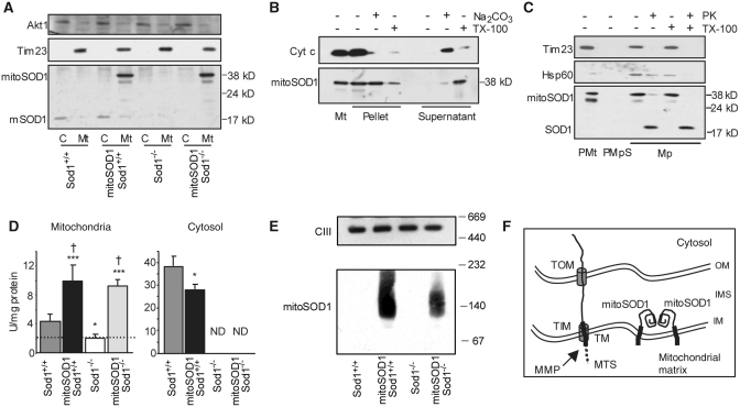
MitoSOD1 localization and enzymatic activity. (A) Western blot of spinal cord mitochondria (Mt) and cytosolic fractions (C) demonstrating mitochondrial localization of mitoSOD1. (B) Protein alkaline extraction. Western blot of the pellet and supernatant of mitoSOD1,Sod+/+ brain mitochondria ± Na2CO3 or Triton X-100 (TX-100). (C) Proteinase K (PK) digestion. Western blot of mitoplasts (Mp) ± proteinase K or TX-100. Mitoplasts were prepared from mitoSOD1,Sod1+/+ brain purified mitochondria (PMt) treated with digitonin (PMpS, post-mitoplast supernatant). (D) KCN-sensitive SOD activity in mitochondrial and cytosolic fractions from spinal cord (*P < 0.05 and ***P < 0.0005 versus Sod1+/+, †P < 0.05 versus Sod1−/−; ANOVA with Fisher post hoc test; n = 3–5 animals per group). The dashed line defines the inhibition of cyt c reduction in mitochondria that is not dependent on SOD1. (E) Complex III (CIII), detected by an antibody against core II protein, was used as a loading control representative of high molecular weight mitochondrial protein complexes. (F) Schematic of the proposed mechanism of mitoSOD1 import and maturation in mitochondria. OM = outer membrane; IM = inner membrane; TOM and TIM = translocators of the outer membrane and inner membrane, respectively; MTS = mitochondrial targeting sequence; MMP = matrix metalloproteinases; TM = transmembrane domain; Tim23 = mitochondrial inner membrane protein; Akt1 = cytosolic soluble protein; cyt c = soluble intermembrane space protein; Hsp60 = soluble mitochondrial matrix protein; ND = activity not detectable.

