Abstract
Aim: In spite of available recommendations, therapeutic procedures of locally recurrent breast cancer are very different. In a retrospective study, the possibilities and results of complete, full-thickness chest wall resection are presented.
Methods: Between 1985 and 2004, 51 women underwent complete, full-thickness chest wall resection with primary coverage. Primary surgical therapy of breast cancer had been mastectomy in 88%. Median age of patients undergoing surgery for a local recurrence was 57 (29 - 81) years. The median interval between surgery of the primary tumour and of the local recurrence was 70.3 (10.7 - 327.2) months; median follow-up was 29.4 (1.8 - 230.9) months. 40 (78.4%) patients required rib resections, 15 (29.4%) of them in combination with partial sternal resection. In 4 (7.8%) patients complete and in 7 (13.7%) patients partial sternal resection without additional rib resection were performed.
Coverage was mainly realized using latissimus dorsi myocutaneous flaps (n=44; 86.3%). Survival rates were calculated by means of the Kaplan-Meier method, the relative risk using univariate and multivariate Cox-regression analysis.
Results: In the total collective, cumulative 5-, 10- and 15-year survival (YS) rates were 39%, 31% and 23%, respectively, median survival 46.4 months. R0 resection was associated with a 5-YS of 42%. Prognostic factors were age at the time of primary surgery, disease-free interval and tumour invasion of bony structures. Mortality was 2%, morbidity 35%.
Conclusion: Full-thickness chest wall resection of locally recurrent breast cancer is possible in almost any patient when performed by a team of thoracic and plastic surgeons. Only radical resection provides good long-term results with low mortality and morbidity.
Keywords: chest wall resection, breast cancer, local recurrence, compartment resection, thoracic surgery, plastic surgery
Abstract
Vorhaben: Die Therapie des Lokalrezidives des Mammakarzinoms wird trotz vorliegender Empfehlungen sehr uneinheitlich durchgeführt. Mit der retrospektiven Untersuchung sollen die Möglichkeiten und Ergebnisse der kompletten, allschichtigen Brustwandresektion aufgezeigt werden.
Methode: Zwischen 1985 und 2004 wurde bei 51 Frauen eine komplette, allschichtige Thoraxwandresektion mit primärer Deckung durchgeführt. Die primäre, operative Therapie des Mammakarzinoms bestand bei 88% in einer Mastektomie. Das Alter betrug bei der Operation des Lokalrezidives im Median 57 (29 - 81) Jahre. Das mediane Intervall zwischen Primärtumoroperation und Lokalrezidivoperation betrug 70,3 (10,7 - 327,2) Monate, die mediane Follow-up Zeit betrug 29,4 (1,8 - 230,9) Monate. Rippen wurden bei 40 (78,4%) Patienten reseziert, bei 15 (29,4%) in Kombination mit einer Sternumresektion. Bei 4 (7,8%) Patientinnen wurde eine komplette und bei 7 (13,7%) eine partielle Sternumresektion ohne zusätzliche Rippenresektion durchgeführt. Die Deckung wurde hauptsächlich mittels muskulokutaner Latissimus dorsi Lappen durchgeführt (86,3%, n=44). Die Überlebensdaten wurden mit der Kaplan-Meier Methode, das relative Risiko mit der univariaten und multivariaten Cox-Regression berechnet.
Ergebnisse: Die kumulative 5, 10 und 15-Jahresüberlebenszeit (Jü) beträgt für die gesamte Gruppe 39%, 31% und 23%, das mediane Überleben 46,4 Monate. Bei R0 Resektion beträgt die 5-Jü 42%. Prognostische Faktoren waren das Alter der Patientinnen bei der Primäroperation, das krankheitsfreie Intervall und die Tumorinfiltration von knöchernen Strukturen. Die Letalität betrug 2%, die Morbidität 35%.
Schlussfolgerung: Die allschichtige Brustwandresektion des Lokalrezidives des Mammakarzinoms ist im Team von Thorax- und plastischen Chirurgen nahezu immer möglich. Nur durch die radikale Resektion lassen sich gute Langzeiterfolge bei geringer Letalität und Morbidität erzielen.
Introduction
The locoregional recurrence of breast cancer is defined as the recurrence of a tumour in a previously operated or irradiated area of treated breast cancer. The incidence of local recurrences after mastectomy and breast-conserving therapy varies between 5% and 40% depending on risk factors and primary therapy [1]. So far, a standard therapy of local recurrences has not been defined. It is recommended to excise the visible tumour with subsequent radiation therapy. In many cases, however, irradiation or chemotherapy is performed primarily. Surgical therapy of local recurrences or chest wall metastases of breast cancer is either not performed at all or with an insufficient safety margin. Local recurrence is often misinterpreted as the first indication of a systemic dissemination of the disease and curative approach is therefore abandoned [2]. Still today large ulcerated chest wall tumours are seen. Whether radical resection of local recurrence offers palliative or curative approach or major prolongation of survival is still unclear. Moreover, chest wall resection is often regarded as a mutilating intervention with high morbidity and mortality. The possibility of chest wall resection with plastic coverage for complete resection of chest wall recurrences is almost unknown. Given a palliative approach, full-thickness chest wall resection provides a markedly improved quality of life and given a curative approach, a good chance for long-term survival. There are, however, very few reports on results of full-thickness complete chest wall resections for locally recurrent breast cancer with sufficient safety margins.
Patients and methods
Between 1985 and 2004, 51 women underwent complete, full-thickness chest resection with primary coverage. The term "local recurrence" was defined as a relapse of tumour manifestations after breast-conserving procedure in mammary region (primary surgical region), after radical therapy in chest wall region, covering skin, scar tissue and remaining glandular tissue. Regional recurrence also includes the involvement of ipsilateral lymph nodes. We included only patients with local recurrences who met this definition.
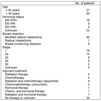
Patient data were obtained retrospectively from the medical records. The present condition of the patients was documented either by regular outpatient follow-up or by obtaining information from the family physician, the patient herself or from relatives. Descriptive data on primary tumour treatment are listed in Table 1 (Tab. 1). 45 (88.2%) patients had primary breast resection, 6 (11.8%) breast conserving surgery. Tumour stages 1 and 2 were present in 76.5% of the patients, while it was unknown in 5 (9.8%) patients. The primary hormone-receptor status was unknown in 43.1% (n=22). 17 (33.3%) patients were known to have an adjuvant therapy.
Table 1. Characteristics of the primary breast cancer and treatment.
Resection was performed according to following standards: The visible tumour was resected with safety margin of at least 2 cm, if possible 5 cm. If tumour invasion of a rib was suspected, cranial and caudal ribs were resected as well. In case of doubt of extension of tumour, frozen sections of the margins were performed.
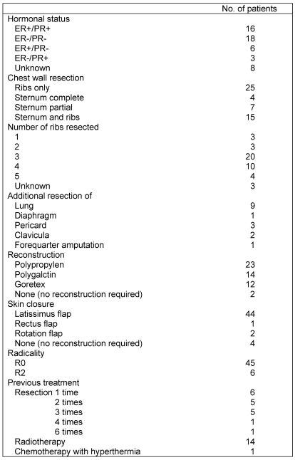
At the time of surgical intervention of the local recurrence, median age was 57 (range 29-81) years. Disease free interval (DFI) was 70.3 months (M) (range 10.7-327.2 M). Median follow-up was 29.4 months (range 1.8-230.9 M), median follow-up of survivors 36.7 M (range 1.8-230.9 M). Except of 5 patients, in whom replacement of chest wall soft tissue was not necessary, all interventions were performed by a team of one thoracic and one plastic surgeon. Surgical data are detailed in Table 2 (Tab. 2). A mean of 3.2 (range 1-5) ribs were resected in 40 (78.4%) patients, in 15 (29.4%) of them in combination with a sternal resection. In 4 (7.8%) patients complete sternal resection and in 7 (13.7%) patients partial sternal resection without additional rib resection were performed. Median size of resected material was 100 (range 36-360) cm². Chest wall stability was restored mainly by non-absorbable polypropylene mesh (n=23, 45.1%) or by absorbable polygalactin mesh (n=14, 27.5%), rarely (n=12, 23.5%) by Gore-tex. In two cases (3.9%), no patch was used. Coverage was mainly realized using a latisssimus dorsi myocutaneous flap (n=44, 86.3%), in one case (2%) by a rectus abdominis myocutaneous flap, and in two cases (3.9%) a pectoralis major myocutaneous flap. No additional flap coverage was necessary in four cases (7.8%).
Table 2. Characteristics of the locally recurrent breast cancer and treatment.
Survival was calculated using the Kaplan-Meier method, the impact on survival was determined for the different factors univariate by means of the log-rank test. Calculation was started at the time of chest wall resection and included postoperative fatalities. Relative risk and multivariate analyses were calculated by Cox Regression analysis. Proportional hazards as well as possible colinearities and interdependencies were considered.
The following factors were included: bone infiltration (yes-no); disease free interval (≤2 years - >2 years); metastases (yes - no); multilocular chest wall recurrence (yes - no); tumor diameter (≤3 cm - >3 cm); age at primary resection (≤45 years - >45 years).
Statistical analyses were computed using SAS (Vers. 8; SAS Institute, Cary, USA); Kaplan-Meier analyses and plots were performed using SPSS (Vers. 12; Chicago, USA).
Results
All patients were in good general condition (WHO grade IV). In all cases, the local recurrence was completely resected. Because of R1 resection one patient had to undergo a second sternal resection after 10 days. Palliative R2 resection because of pleural carcinosis or dissemination of pulmonary metastases was performed in 6 patients. The diameter of resected tumour was 1-11 cm, median 3 cm, in case of multilocular tumour the largest diameter was considered.
Hospital mortality was 2%, i.e. one patient died postoperatively from pulmonary complications. Complications were observed in 33% (n=17). Bleedings needing revision were found in 14% (n=7), flap necroses in 3.9% (n=2), and wound infections in 7.8% (n=4), myocardial and respiratory insufficiency, recurrent nerve palsy and empyema in one patient respectively.
At the time of follow-up, 24 patients (47.1%) were still alive, 21 (87.5%) of them without any recurrence. Cumulative 5-year survival (5-YS) for the total collective was 41.1%, 10-year survival (10-YS) 28.5%, and 15-year survival 21.3%, median survival (MS) was 50.1 months.
Comparing patients with R0 resection to those with R2 resections, according to the above mentioned definition, 5- and 10-YS of completely resected patients (n=45) amount to 41.4 and 36.8%, respectively, with a MS of 46.7 months in contrast to a 5- and 10-YS in patients with R2 resections (n=6) of 41.7 and 0% with a MS of 57.2 months (p=0.33).
The following subgroup analyses of apparently prognostically relevant parameters included only R0 resected patients.
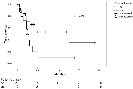
If the recurrence did not invade bony structures (n=28), 5- and 10-YS were 52.6% with a MS of 154.6 months. If there was a histologically proven bone invasion (n=17), 5-YS was 19.8% and 10-YS 9.9% with a MS of 29.4 months (p=0.03) (Figure 1 (Fig. 1)).
Figure 1. Cumulative survival of complete resected (R0) patients by presence of bone infiltration.
If additional metastases of lung, pericardium or diaphragm were resected (n=9), 5- and 10-YS were 19.4% with a median MS of 37.3 months. The group without metastases (n=36) had a 5- and 10-YS of 47.3% and 41.4%, respectively, with a MS of 56.6 months (p=0.30).
If tumour size increases above 3 cm, survival apparently tends to deteriorate. A tumour size below 3 cm (n=23) was associated with a 5-YS of 49.3%, above 3 cm (n=22) with only 34.2% with a MS of 46.7 and 37.3 months, respectively (p=0.43).
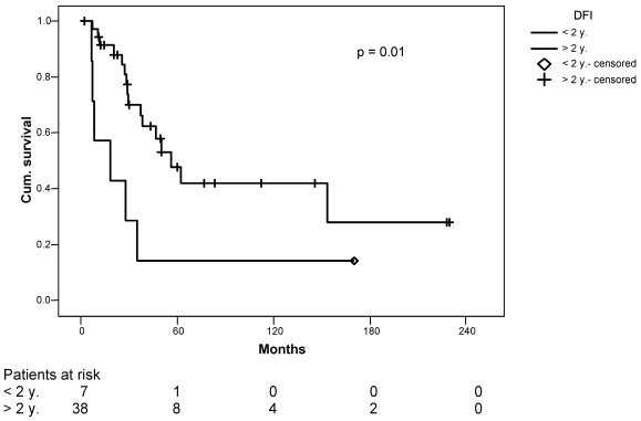
With an DFI equal or more than 24 months (n=38), 5- and 10-YS are 47.8% and 41.8% with a MS of 56.6 months compared to a 14.3% 5- and 10-YS with a MS of 27.7 months if DFI was less than 24 months (n=7) (Figure 2 (Fig. 2)) (p=0.04).
Figure 2. Cumulative survival of complete resected (R0) patients according to disease free interval.
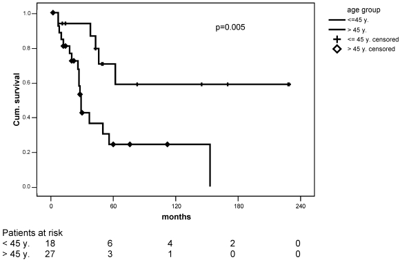
Another important factor is patient age at the time of primary surgery. Patients below 45 years of age at the time of primary therapy of breast cancer (n=18) had a better prognosis with a 5-YS of 70.5% and a 10-YS of 58.8%, with the median survival not being achieved so far. In contrast, women above 45 years of age at the time of primary therapy (n=27) had a 5- and 10-YS of 23.4%, with a MS of 29.4 months (p=0.005) (Figure 3 (Fig. 3)).
Figure 3. Cumulative survival of complete resected (R0) patients according to age groups at the time of primary therapy.
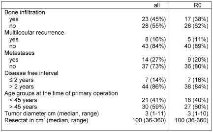
Comparing the p-values of survival probabilities calculated by Cox-Regression, there are demonstrative results for the expected prognostic factors bone invasion and disease-free interval with p-values < 0.05. Additional metastases, multilocular manifestation of the recurrence and largest tumour diameter do not have impact on prognosis. Descriptive data are given in Table 3 (Tab. 3).
Table 3. Characteristics of prognostic factors.
Risk ratio of univariate Cox analysis shows that patients with bone invasion had a 2.5 fold higher risk of premature death, patients with a disease-free interval of ≤ 2 years had a 3-fold increased risk and patients with additional metastases still a 1.6-fold increased risk. Patients above 45 years of age at the time of primary therapy had a 3.9-fold increased of risk of death. For tumour diameter and multilocular recurrence the best estimator of risk ratio is 1.5 and 2.3 respectively. Since the relevant confidence intervals are very broad, these data only indicate a trend, which however might become significant given a higher number of cases (Table 4 (Tab. 4)).
Table 4. Univariate statistics of prognostic factors (patients with complete resection).
The factors DFI, bone invasion and age at the time of primary therapy were analysed using multivariate Cox analysis, additionally. Only bone invasion and age at the time of primary therapy showed demonstrative p values below 0.05 and a risk of premature death which is 2.8 and 6.1 fold higher for patients with bone invasion and age > 45 years at the time of primary treatment respectively. The best estimator for the risk ratio of DFI was 2.6 (Table 5 (Tab. 5)).
Table 5. Multivariate statistics of prognostic factors (patients with complete resection).
Discussion
A review of the literature showed that a complete chest wall resection is only performed in very rare cases. The largest collective reported on in the last 20 years includes 69 patients [3]. In our opinion, there are major therapeutic shortcomings in this field. Unfortunately, many patients are only referred for complete chest wall resection after multiple previous therapeutic procedures, not few patients came on their own initiative. In our group, 27 patients received a mean of 2.1 therapeutic procedures before final resection. One patient each had undergone 5 and 6 surgical interventions previously. 24 patients (47%) were primarily presented at our institution.
Guidelines on therapy of locally recurrent breast cancer do not provide a clear therapeutic strategy. Surgical resection is recommended, if possible, otherwise radiotherapy. Astonishingly, there is no information on safety margins defining R0 resection [4]. In most cases, only local excision of tumour with surrounding smooth tissue is performed which then often proves of be R1 or R2 resection. As a consequence of these incomplete resections, radiotherapy is applied in most cases. Capgar found radiotherapy to be of great importance in a study of 130 very inhomogenously treated patients with local recurrence. However, only 25% of the patients had a complete resection of the tumour1 [5]. Complete chest wall resection is usually performed first after exhausting all alternative therapies [6]. In addition, many physicians consider the local recurrence as a harbinger of progressive systemic metastatic disease leading to nihilistic attitude towards further treatments. Although several reports have shown that the incidence of metastases after local recurrences is higher, the opinion that the local recurrence is a marker of metastatic formation mix the principle of cause and effect up [7]. Fortin demonstrated that the incidence of metastases increases both after manifestation of a local recurrence and after therapy of a primary tumour after the same time. In both cases, there is a peak after about 2 years with a subsequent decrease in the incidence of new metastases [8]. Thus, the local recurrence has to be regarded as a repeated episode of a disease with an increased risk of subsequent metastases and not vice versa. The curve of metastatic incidence might be flattened markedly by a radical resection with sufficient safety margins.
One of the most important factors is the complete resection of the tumour with sufficient safety margins of 2-5 cm. This is in most cases only possible by performing a complete chest wall resection, which almost always is possible if thoracic and plastic surgeons are cooperating. Fortin found that resection with insufficient safety margin always bears a high risk of local recurrence. Adjuvant radiotherapy did not reduce the risk (8). Treating local recurrence by resecting soft tissue solely shows a re-recurrence rate of 45% [9] to 62% (10). Radiotherapy exclusively has a re-recurrence rate of 83% [10].
Mortality after chest wall resection is reported to be 1.6%-4.5% [11]. Hospital mortality in our collective was 2%. A similar rate of 1.6% is reported by Henderson in a group of 61 patients [11]. Morbidity is reported to be 20%-50%. Faneyte described a complication rate of 40% in his collective with surgical revision being necessary in 22% [6]. A complication rate of 41% was reported by Kluiber in a group of 12 patients [12]. Our morbidity of 35% is within these ranges.
In our series following factors showed influence in predicting differences of long term survival: Age at primary treatment, tumour invasion of bony structures, additional metastases and disease free interval.
As reported by Fortin, risk factors affecting long-term survival were diameter of the local recurrence > 1.5 cm, disease-free interval < 2 years, skin invasion and initial tumour stage T2 or positive lymph nodes [8]. Faneyte found age at the time of primary resection and the disease-free interval to be prognostic factors. 5-YS in the whole collective was 45%, after R0 resection without additional metastases 58%, in the palliative setting still 21%. Patients who were less than 35 years old at the time of primary therapy had significantly lower survival rates after resection of chest wall recurrence. However, this group consists of 4 patients only, and the 5-YS is not provided [6]. Miyauchi reported on a 5-YS of 48% in 23 patients. Prognostic factors were disease-free interval and positive mediastinal lymph nodes [13]. Henderson reported on a 5-YS of only 24%. However, the incidence of incomplete resections in this group was 26%. The only prognostic factor was additional metastases [11]. The Sloan Kettering Cancer Centre reported on lymph node involvement to be a risk factor. In his group of 38 patients, Downey reported on a 5-YS of 18%. If the patients with additional metastases are excluded, 5-YS was 35%. However, this group has an astonishingly high incidence of R1 and R2 resections. Downey concludes that chest wall resection primarily represents a palliative approach [2]. Our data do not support these results. Patients with metastases, who underwent complete resection, had a 5-YS of 19.4%. We do not consider additional resection of metastases per se to be a palliative intervention. In earlier reports we have shown that metastatic resection indeed offers a curative approach in the treatment of breast cancer [14], [15], [16], [17]. Furthermore, it is recommended to always exclude metastases in view of the fact that patients with additional metastases do not profit from an extended chest wall resection. In order to obtain an adequate oncological concept a complete staging is required before starting any therapy to assess the status of the tumour disease. On the basis of these results, the adequate therapeutic approach is then selected. In most cases, however, metastatic disease does not represent a contraindication for chest wall resection.
Even in cases of multilocular metastases, resection of the local recurrence with a palliative purpose may contribute to a markedly improved quality of life. Palliative resections are mostly applied in primarily irradiated patients with a re-recurrence. Some of the tumours are ulcerated or the patients suffer from severe pain. Even in the palliative setting 5-YS rates of 21%-41.7% are reported [2], [6].
The most ostentatious prognostic factor in our group, however, was the age of women at the time of primary surgery. In contrast to the common prognosis, younger patients achieved a better prognosis by surgical therapy of the local recurrence than older patients. Whether this is really due to the therapeutic procedure or to the generally decreased life expectancy of older patients has to be subject of a more detailed investigation.
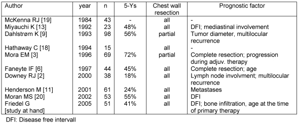
The results and the procedures of the references cited are detailed in Table 6 (Tab. 6), [18], [19], [20].
Table 6. Literature review.
Conclusion
In locally recurrent breast cancer complete chest wall resection mostly is necessary to achieve radical resection with sufficient tumour free safety margins of 2-5 cm. To enable surgical therapy and to cover large chest wall defects cooperation between thoracic and plastic surgeon is of eminent importance.
References
- 1. Bedwinek J. Natural History and Management of Isolated Local-Regional Recurrence Following Mastectomy. Semin Radiat Oncol. 1994;4(4):260–269. doi: 10.1053/SRAO00400260. [DOI] [PubMed] [Google Scholar]
- 2. Downey RJ, Rusch V, Hsu FI, Leon L, Venkatraman E, Linehan D, et al. Chest wall resection for locally recurrent breast cancer: is it worthwhile? J Thorac Cardiovasc Surg. 2000;119(3):420–428. doi: 10.1016/s0022-5223(00)70119-x. [DOI] [PubMed] [Google Scholar]
- 3. Mora EM, Singletary SE, Buzdar AU, Johnston DA. Aggressive therapy for locoregional recurrence after mastectomy in stage II and III breast cancer patients. Ann Surg Oncol. 1996;3(2):162–168. doi: 10.1007/BF02305796. [DOI] [PubMed] [Google Scholar]
- 4.Kreienberg R, Kopp I, Lorenz W, Budach W, Dunst J, Lebau A, et al. Diagnostik, Therapie und Nachsorge des Mammakarzinoms der Frau. Frankfurt: 2004. [Google Scholar]
- 5. Chagpar A, Meric-Bernstam F, Hunt KK, Ross MI, Cristofanilli M, Singletary SE, et al. Chest wall recurrence after mastectomy does not always portend a dismal outcome. Ann Surg Oncol. 2003;10(6):628–634. doi: 10.1245/aso.2003.01.004. [DOI] [PubMed] [Google Scholar]
- 6. Faneyte IF, Rutgers EJ, Zoetmulder FA. Chest wall resection in the treatment of locally recurrent breast carcinoma: indications and outcome for 44 patients. Cancer. 1997;80(5):886–891. [PubMed] [Google Scholar]
- 7. Veronesi U, Marubini E, Del Vecchio M, Manzari A, Andreola S, Greco M, et al. Local recurrences and distant metastases after conservative breast cancer treatments: partly independent events. J Natl Cancer Inst. 1995;87(1):19–27. doi: 10.1093/jnci/87.1.19. [DOI] [PubMed] [Google Scholar]
- 8. Fortin A, Larochelle M, Laverdiere J, Lavertu S, Tremblay D. Local failure is responsible for the decrease in survival for patients with breast cancer treated with conservative surgery and postoperative radiotherapy. J Clin Oncol. 1999;17(1):101–109. doi: 10.1200/JCO.1999.17.1.101. [DOI] [PubMed] [Google Scholar]
- 9. Dahlstrom KK, Andersson AP, Andersen M, Krag C. Wide local excision of recurrent breast cancer in the thoracic wall. Cancer. 1993;72(3):774–777. doi: 10.1002/1097-0142(19930801)72:3<774::aid-cncr2820720323>3.0.co;2-y. [DOI] [PubMed] [Google Scholar]
- 10. Probstfeld MR, O'Connell TX. Treatment of locally recurrent breast carcinoma. Arch Surg. 1989;124(10):1127–1130. doi: 10.1001/archsurg.1989.01410100025005. [DOI] [PubMed] [Google Scholar]
- 11. Henderson MA, Burt JD, Jenner D, Crookes P, Bennett RC. Radical surgery with omental flap for uncontrolled locally recurrent breast cancer. ANZ J Surg. 2001;71(11):675–679. doi: 10.1046/j.0004-8682.2001.02234.x. [DOI] [PubMed] [Google Scholar]
- 12. Kluiber R, Bines S, Bradley C, Faber LP, Witt TR. Major chest wall resection for recurrent breast carcinoma. Am Surg. 1991;57(8):523–530. [PubMed] [Google Scholar]
- 13. Miyauchi K, Koyama H, Noguchi S, Inaji H, Yamamoto H, Kodama K, et al. Surgical treatment for chest wall recurrence of breast cancer. Eur J Cancer. 1992;28A(6-7):1059–1062. doi: 10.1016/0959-8049(92)90456-c. [DOI] [PubMed] [Google Scholar]
- 14. Friedel G, Pastorino U, Ginsberg RJ, Goldstraw P, Johnston M, Pass H, et al. Results of lung metastasectomy from breast cancer: prognostic criteria on the basis of 467 cases of the International Registry of Lung Metastases. Eur J Cardiothorac Surg. 2002;22(3):335–344. doi: 10.1016/s1010-7940(02)00331-7. [DOI] [PubMed] [Google Scholar]
- 15. Friedel G, Linder A, Toomes H. The significance of prognostic factors for the resection of pulmonary metastases of breast cancer. Thorac Cardiovasc Surg. 1994;42(2):71–75. doi: 10.1055/s-2007-1016460. [DOI] [PubMed] [Google Scholar]
- 16. Friedel G, Pastorino U, Buyse M, Ginsberg RJ, Girard P, Goldstraw P, et al. [Resection of lung metastases: long-term results and prognostic analysis based on 5206 cases--the International Registry of Lung Metastases] Zentralbl Chir. 1999;124(2):96–103. [PubMed] [Google Scholar]
- 17. Planchard D, Soria JC, Michiels S, Grunenwald D, Validire P, Caliandro R, et al. Uncertain benefit from surgery in patients with lung metastases from breast carcinoma. Cancer. 2004;100(1):28–35. doi: 10.1002/cncr.11881. [DOI] [PubMed] [Google Scholar]
- 18. Hathaway CL, Rand RP, Moe R, Marchioro T. Salvage surgery for locally advanced and locally recurrent breast cancer. Arch Surg. 1994;129(6):582–587. doi: 10.1001/archsurg.1994.01420300020003. [DOI] [PubMed] [Google Scholar]
- 19. McKenna RJ RJ, Jr, McMurtrey MJ, Larson DL, Mountain CF. A perspective on chest wall resection in patients with breast cancer. Ann Thorac Surg. 1984;38(5):482–487. doi: 10.1016/s0003-4975(10)64189-6. [DOI] [PubMed] [Google Scholar]
- 20. Moran MS, Haffty BG. Local-regional breast cancer recurrence: prognostic groups based on patterns of failure. Breast J. 2002:81–87. doi: 10.1046/j.1524-4741.2002.08202.x. [DOI] [PubMed] [Google Scholar]


