

Abstract
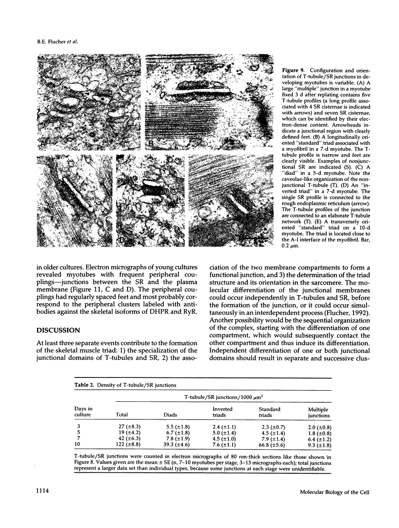
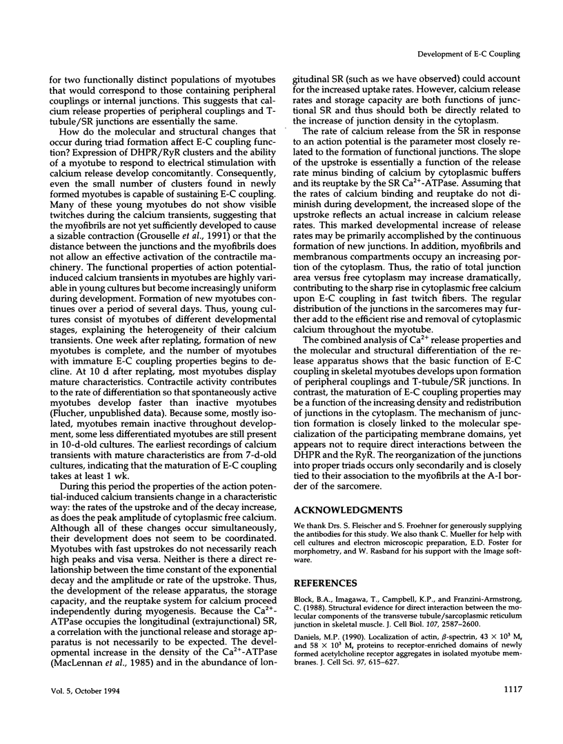
The relationship between the molecular composition and organization of the triad junction and the development of excitation-contraction (E-C) coupling was investigated in cultured skeletal muscle. Action potential-induced calcium transients develop concomitantly with the first expression of the dihydropyridine receptor (DHPR) and the ryanodine receptor (RyR), which are colocalized in clusters from the time of their earliest appearance. These DHPR/RyR clusters correspond to junctional domains of the transverse tubules (T-tubules) and sarcoplasmic reticulum (SR), respectively. Thus, at first contact T-tubules and SR form molecularly and structurally specialized membrane domains that support E-C coupling. The earliest T-tubule/SR junctions show structural characteristics of mature triads but are diverse in conformation and typically are formed before the extensive development of myofibrils. Whereas the initial formation of T-tubule/SR junctions is independent of association with myofibrils, the reorganization into proper triads occurs as junctions become associated with the border between the A band and the I band of the sarcomere. This final step in triad formation manifests itself in an increased density and uniformity of junctions in the cytoplasm, which in turn results in increased calcium release and reuptake rates.
Full text
PDF


Images in this article


Selected References
These references are in PubMed. This may not be the complete list of references from this article.
- Block B. A., Imagawa T., Campbell K. P., Franzini-Armstrong C. Structural evidence for direct interaction between the molecular components of the transverse tubule/sarcoplasmic reticulum junction in skeletal muscle. J Cell Biol. 1988 Dec;107(6 Pt 2):2587–2600. doi: 10.1083/jcb.107.6.2587. [DOI] [PMC free article] [PubMed] [Google Scholar]
- Daniels M. P. Localization of actin, beta-spectrin, 43 x 10(3) Mr and 58 x 10(3) Mr proteins to receptor-enriched domains of newly formed acetylcholine receptor aggregates in isolated myotube membranes. J Cell Sci. 1990 Dec;97(Pt 4):615–626. doi: 10.1242/jcs.97.4.615. [DOI] [PubMed] [Google Scholar]
- Eusebi F., Miledi R., Takahashi T. Calcium transients in mammalian muscles. Nature. 1980 Apr 10;284(5756):560–561. doi: 10.1038/284560a0. [DOI] [PubMed] [Google Scholar]
- Flucher B. E., Andrews S. B. Characterization of spontaneous and action potential-induced calcium transients in developing myotubes in vitro. Cell Motil Cytoskeleton. 1993;25(2):143–157. doi: 10.1002/cm.970250204. [DOI] [PubMed] [Google Scholar]
- Flucher B. E., Andrews S. B., Fleischer S., Marks A. R., Caswell A., Powell J. A. Triad formation: organization and function of the sarcoplasmic reticulum calcium release channel and triadin in normal and dysgenic muscle in vitro. J Cell Biol. 1993 Dec;123(5):1161–1174. doi: 10.1083/jcb.123.5.1161. [DOI] [PMC free article] [PubMed] [Google Scholar]
- Flucher B. E., Morton M. E., Froehner S. C., Daniels M. P. Localization of the alpha 1 and alpha 2 subunits of the dihydropyridine receptor and ankyrin in skeletal muscle triads. Neuron. 1990 Sep;5(3):339–351. doi: 10.1016/0896-6273(90)90170-k. [DOI] [PubMed] [Google Scholar]
- Flucher B. E., Phillips J. L., Powell J. A., Andrews S. B., Daniels M. P. Coordinated development of myofibrils, sarcoplasmic reticulum and transverse tubules in normal and dysgenic mouse skeletal muscle, in vivo and in vitro. Dev Biol. 1992 Apr;150(2):266–280. doi: 10.1016/0012-1606(92)90241-8. [DOI] [PubMed] [Google Scholar]
- Flucher B. E., Phillips J. L., Powell J. A. Dihydropyridine receptor alpha subunits in normal and dysgenic muscle in vitro: expression of alpha 1 is required for proper targeting and distribution of alpha 2. J Cell Biol. 1991 Dec;115(5):1345–1356. doi: 10.1083/jcb.115.5.1345. [DOI] [PMC free article] [PubMed] [Google Scholar]
- Flucher B. E. Structural analysis of muscle development: transverse tubules, sarcoplasmic reticulum, and the triad. Dev Biol. 1992 Dec;154(2):245–260. doi: 10.1016/0012-1606(92)90065-o. [DOI] [PubMed] [Google Scholar]
- Flucher B. E., Takekura H., Franzini-Armstrong C. Development of the excitation-contraction coupling apparatus in skeletal muscle: association of sarcoplasmic reticulum and transverse tubules with myofibrils. Dev Biol. 1993 Nov;160(1):135–147. doi: 10.1006/dbio.1993.1292. [DOI] [PubMed] [Google Scholar]
- Flucher B. E., Terasaki M., Chin H. M., Beeler T. J., Daniels M. P. Biogenesis of transverse tubules in skeletal muscle in vitro. Dev Biol. 1991 May;145(1):77–90. doi: 10.1016/0012-1606(91)90214-n. [DOI] [PubMed] [Google Scholar]
- Fosset M., Jaimovich E., Delpont E., Lazdunski M. [3H]nitrendipine receptors in skeletal muscle. J Biol Chem. 1983 May 25;258(10):6086–6092. [PubMed] [Google Scholar]
- Franzini-Armstrong C., Pincon-Raymond M., Rieger F. Muscle fibers from dysgenic mouse in vivo lack a surface component of peripheral couplings. Dev Biol. 1991 Aug;146(2):364–376. doi: 10.1016/0012-1606(91)90238-x. [DOI] [PubMed] [Google Scholar]
- Franzini-Armstrong C. Simultaneous maturation of transverse tubules and sarcoplasmic reticulum during muscle differentiation in the mouse. Dev Biol. 1991 Aug;146(2):353–363. doi: 10.1016/0012-1606(91)90237-w. [DOI] [PubMed] [Google Scholar]
- Grouselle M., Koenig J., Lascombe M. L., Chapron J., Méléard P., Georgescauld D. Fura-2 imaging of spontaneous and electrically induced oscillations of intracellular free Ca2+ in rat myotubes. Pflugers Arch. 1991 Mar;418(1-2):40–50. doi: 10.1007/BF00370450. [DOI] [PubMed] [Google Scholar]
- Hoyle G. Forms of modulatable tension in skeletal muscles. Comp Biochem Physiol A Comp Physiol. 1983;76(2):203–210. doi: 10.1016/0300-9629(83)90316-x. [DOI] [PubMed] [Google Scholar]
- Inui M., Saito A., Fleischer S. Purification of the ryanodine receptor and identity with feet structures of junctional terminal cisternae of sarcoplasmic reticulum from fast skeletal muscle. J Biol Chem. 1987 Feb 5;262(4):1740–1747. [PubMed] [Google Scholar]
- Ishikawa H. Formation of elaborate networks of T-system tubules in cultured skeletal muscle with special reference to the T-system formation. J Cell Biol. 1968 Jul;38(1):51–66. doi: 10.1083/jcb.38.1.51. [DOI] [PMC free article] [PubMed] [Google Scholar]
- Jorgensen A. O., Shen A. C., Arnold W., Leung A. T., Campbell K. P. Subcellular distribution of the 1,4-dihydropyridine receptor in rabbit skeletal muscle in situ: an immunofluorescence and immunocolloidal gold-labeling study. J Cell Biol. 1989 Jul;109(1):135–147. doi: 10.1083/jcb.109.1.135. [DOI] [PMC free article] [PubMed] [Google Scholar]
- Lai F. A., Erickson H. P., Rousseau E., Liu Q. Y., Meissner G. Purification and reconstitution of the calcium release channel from skeletal muscle. Nature. 1988 Jan 28;331(6154):315–319. doi: 10.1038/331315a0. [DOI] [PubMed] [Google Scholar]
- Morton M. E., Froehner S. C. Monoclonal antibody identifies a 200-kDa subunit of the dihydropyridine-sensitive calcium channel. J Biol Chem. 1987 Sep 5;262(25):11904–11907. [PubMed] [Google Scholar]
- Olek A. J., Ling A., Daniels M. P. Development of ultrastructural specializations during the formation of acetylcholine receptor aggregates on cultured myotubes. J Neurosci. 1986 Feb;6(2):487–497. doi: 10.1523/JNEUROSCI.06-02-00487.1986. [DOI] [PMC free article] [PubMed] [Google Scholar]
- Pinçon-Raymond M., García L., Romey G., Houenou L., Lazdunski M., Rieger F. A genetic model for the study of abnormal nerve-muscle interactions at the level of excitation-contraction coupling: the mutation muscular dysgenesis. J Physiol (Paris) 1990;84(1):82–87. [PubMed] [Google Scholar]
- Rios E., Brum G. Involvement of dihydropyridine receptors in excitation-contraction coupling in skeletal muscle. Nature. 1987 Feb 19;325(6106):717–720. doi: 10.1038/325717a0. [DOI] [PubMed] [Google Scholar]
- Schiaffino S., Cantini M., Sartore S. T-system formation in cultured rat skeletal tissue. Tissue Cell. 1977;9(3):437–446. doi: 10.1016/0040-8166(77)90004-0. [DOI] [PubMed] [Google Scholar]
- Sommer J. R., Waugh R. A. The ultrastructure of the mammalian cardiac muscle cell--with special emphasis on the tubular membrane systems. A review. Am J Pathol. 1976 Jan;82(1):192–232. [PMC free article] [PubMed] [Google Scholar]
- Striessnig J., Moosburger K., Goll A., Ferry D. R., Glossmann H. Stereoselective photoaffinity labelling of the purified 1,4-dihydropyridine receptor of the voltage-dependent calcium channel. Eur J Biochem. 1986 Dec 15;161(3):603–609. doi: 10.1111/j.1432-1033.1986.tb10484.x. [DOI] [PubMed] [Google Scholar]
- Takekura H., Shuman H., Franzini-Armstrong C. Differentiation of membrane systems during development of slow and fast skeletal muscle fibres in chicken. J Muscle Res Cell Motil. 1993 Dec;14(6):633–645. doi: 10.1007/BF00141560. [DOI] [PubMed] [Google Scholar]
- Wagenknecht T., Grassucci R., Frank J., Saito A., Inui M., Fleischer S. Three-dimensional architecture of the calcium channel/foot structure of sarcoplasmic reticulum. Nature. 1989 Mar 9;338(6211):167–170. doi: 10.1038/338167a0. [DOI] [PubMed] [Google Scholar]
- Yuan S. H., Arnold W., Jorgensen A. O. Biogenesis of transverse tubules and triads: immunolocalization of the 1,4-dihydropyridine receptor, TS28, and the ryanodine receptor in rabbit skeletal muscle developing in situ. J Cell Biol. 1991 Jan;112(2):289–301. doi: 10.1083/jcb.112.2.289. [DOI] [PMC free article] [PubMed] [Google Scholar]
- Yuan S., Arnold W., Jorgensen A. O. Biogenesis of transverse tubules: immunocytochemical localization of a transverse tubular protein (TS28) and a sarcolemmal protein (SL50) in rabbit skeletal muscle developing in situ. J Cell Biol. 1990 Apr;110(4):1187–1198. doi: 10.1083/jcb.110.4.1187. [DOI] [PMC free article] [PubMed] [Google Scholar]