Abstract
OBJECTIVE
The effectiveness of tolerizing immunotherapeutic strategies, such as anti-CD40L or dendritic cells (DCs), is greater when administered to young nonobese diabetic (NOD) mice than at peak insulitis. RelBlo DCs, generated in the presence of an nuclear factor-κB inhibitor, induce T-regulatory (Treg) cells and suppress inflammation in a model of rheumatoid arthritis. Interleukin (IL)-1β is overexpressed in humans and mice at risk of type 1 diabetes, dysregulates Treg cells, and accelerates diabetes in NOD mice. We investigated the relationship between IL-1β production and the response to RelBlo DCs in the prediabetic period.
RESEARCH DESIGN AND METHODS
We injected RelBlo DCs subcutaneously into 4- or 14-week-old NOD mice and tracked the incidence of diabetes and effect on Treg cell function. We measured the expression of proinflammatory cytokines by stimulated splenocytes and unstimulated islets from mice of different ages and strains and proliferative and cytokine responses of T effectors to Treg in vitro.
RESULTS
Tolerizing RelBlo DCs significantly inhibited diabetes progression when administered to 4-week-old but not 14-week-old mice. IL-1β production by NOD splenocytes and mRNA expression by islets increased from 6 to 16 weeks of age when major histocompatibility complex (MHC)-restricted islet antigen presentation to autoreactive T-cells occurred. IL-1 reduced the capacity of Treg cells to suppress effector cells and promoted their conversion to Th17 cells. RelBlo DCs exacerbated the IL-1–dependent decline in Treg function and promoted Th17 conversion.
CONCLUSIONS
IL-1β, generated by islet-autoreactive cells in MHC-susceptible mice, accelerates diabetes by differentiating Th17 at the expense of Treg. Tolerizing DC therapies can regulate islet autoantigen priming and prevent diabetes, but progression past the IL-1β/IL-17 checkpoint signals the need for other strategies.
Nonobese diabetic (NOD) mice spontaneously develop autoimmune diabetes, driven by progressive inflammatory dysregulation. Autoimmune inflammation begins at ∼4 weeks of age with peri-insulitis and infiltration of macrophages and dendritic cells (DCs) (1), followed by infiltration of autoreactive CD4+ and CD8+ T-cells (2). Expression of major histocompatibility complex (MHC) class II I-Ag7 is necessary but not sufficient for diabetes susceptibility (3). Presentation of proinsulin epitopes to islet autoreactive CD4+ T-cells triggers insulitis (4). After ∼15 weeks, destructive insulitis is promoted by cytotoxic T-cells (5). FoxP3+ regulatory T (Treg) cells regulate autoreactivity, interferon (IFN)-γ production, and natural-killer cell lytic activity (6). Treg infiltrate inflamed islets, but their function wanes prior to diabetes expression (7,8).
Mice and humans exhibit chronic inflammation at and before diabetes onset. Nuclear factor (NF)-κB is constitutively activated by DCs in NOD mice from at least 6 weeks of age and in patients with type 1 diabetes, associated with an interleukin (IL)-1–driven proinflammatory state (9–16). We showed that IL-1β is overexpressed by effector T-cells (Teff) in NOD mice and inhibits the capacity of Treg to suppress Teff (17). IL-1β also exerts direct proapoptotic effects on insulin-producing pancreatic β-cells (17–20). Progression to diabetes is slowed in IL-1 receptor-1 (IL-1R1)-deficient NOD mice because of immunoregulatory effects on hemopoietic cells (21). Newly diagnosed type 1 diabetic patients with high serum levels of the natural antagonist, IL-1Ra, were more likely to preserve β-cell function (16). Further, sera from patients with recent-onset type 1 diabetes or from at-risk relatives with islet autoantibodies, but not healthy control subjects, induced a gene expression signature characterized by upregulated expression of members of the IL-1 family and associated with IL-1 action when incubated with healthy peripheral blood mononuclear cells (PBMCs) (15), and IL-1β also was overexpressed in the gene expression signature of PBMCs from children with newly diagnosed type 1 diabetes. IL-1 expression fell as hyperglycemia settled, suggesting that at least some of the inflammatory drive is metabolic (22).
The effectiveness of tolerizing immunotherapies, such as anti-CD40L, IL-10, rapamycin, or DCs treated with NF-κB oligodinucleotides, is greater when administered to young (4–6 weeks) NOD mice than mice with advanced insulitis (10–14 weeks) (23–26). The RelB subunit of NF-κB is a key determinant of DC antigen-presenting cell (APC) function (27). Microbial, inflammatory, or T-cell–derived signals induce the nuclear translocation and transcriptional activity of RelB in DCs (28,29). Induction of MHC class II and costimulatory molecules required for effective TCR signaling and T-cell priming is impaired in response to such activation of RelB−/− DCs (28,30,31). T-cell stimulatory function of RelB−/− DCs is deficient in vitro and in vivo, and transferred RelB−/− or RelBlo DC, generated in the presence of NF-κB inhibitors or RelB siRNA, suppress primed T-cell effector responses through induction of Tr1-type Treg in an IL-10–dependent manner (31,32). RelBlo DCs suppress disease in a mouse model of rheumatoid arthritis (33). Here, we used RelBlo tolerizing DCs to prevent diabetes in NOD mice to define mechanisms that determine the age-dependent effectiveness of tolerizing therapies in type 1 diabetes.
RelBlo DCs, which express reduced MHC class II and CD40, suppress effector function both by reduced capacity to signal Teff and through induction of Treg (31). The induction of Treg by tolerizing immunotherapies depends on the presence of FoxP3+ natural Treg (34,35). Thus, factors that dysregulate Treg function are candidates for interference with effective suppression of autoimmune disease by RelBlo DCs. Because IL-1 plays an important role in the pathogenesis of type 1 diabetes in mice and humans, and its production in NOD mice dysregulates Treg cell function, we hypothesized that IL-1 impairs the response to RelBlo DCs.
RESEARCH DESIGN AND METHODS
Mice.
NOD.Lt, C57BL/6, NOD.I-Ak, NOD.CD45.2 (36), NOD.IL-1R1−/− (21), and pINS.NOD (37) mice were obtained from the Animal Research Centre (Perth, Australia), James Cook University, or bred at the Walter and Eliza Hall Institute (WEHI) (Melbourne, Australia) or the University of Queensland. Mice were housed at the Princess Alexandra Hospital (Brisbane, Australia) or WEHI.
Cytokine assay.
Cytokines were assayed in supernatants by enzyme-linked immunosorbent assay (ELISA) for IL-1 (eBioscience, San Diego, CA) and IL-17 (Biolegend, San Diego, CA) and bead array (BD Bioscience, Franklin Lakes, NJ). Supernatants in triplicate were pooled, and cytokine assays were made in duplicate. Data represent means ± SD.
Intracellular staining.
All surface antibodies were purchased from Biolegend. Cells were stained fresh or after culture with anti-CD3ε mAb for 72 h or phorbol myristate acetate, with addition of brefeldin A (Sigma, St. Louis, MO) for 4 h. After surface mAb staining, cells were fixed and permeabilized then incubated on ice with anti–IL-1 (eBioscience) or isotype control. When biotinylated antibody was used, cells were washed and incubated for 15 min with labeled streptavidin (Biolegend). Cells were suspended in 10% formalin, read on an FACS Calibur (BD Bioscience), and analyzed using FlowJo software (Tree Star).
Assessment of diabetes and insulitis.
Mice were classified to be diabetic and were killed following two consecutive weekly blood glucose readings >12 mmol/l. For analysis of insulitis, pancreata were collected from four mice per group at 12 weeks. Insulitis was graded in hematoxylin and eosin–stained sections as described (38).
Islet purification and quantitative PCR.
Islets of Langerhans were isolated by collagenase P (dissolved in Hank's Buffered Salt Solution containing 2 mmol/l Ca2+ and 20 mmol/l HEPES) digestion and density gradient centrifugation as described previously (39). RNA was extracted (RNeasy Mini Kit; Qiagen) and cDNA synthesized. Quantitative PCR used Taqman primers on an AB 7900 machine (Applied Biosystems).
Proliferation and suppression assay.
CD11c+, CD4+CD25+, and CD4+CD25− cells were isolated from spleen and lymph nodes using immunomagnetic beads and an AutoMacs separator (Miltenyi Biotec, Bergisch Gladbach, Germany). A total of 100,000 CD11c+, 105 CD4+CD25−, and 7.5 × 10⁴ CD4+CD25+ cells were incubated for 72 h in presence of anti-CD3ε antibody. Supernatants were collected for cytokine assays and 1 μ Cu of tritiated thymidine (Perkin Elmer, Waltham, MA) was added for 18 h before counting. Data represent means ± SD of triplicates performed twice.
In vivo administration of RelBlo DC and transfer of Treg.
Bone marrow was collected from long bones of NOD mice aged 8 ± 2 weeks. After Ficoll-Histopaque gradient separation, cells were cultured in RPMI + 10% FCS in the presence of murine IL-4 and granulocyte macrophage colony–stimulating factor (GM-CSF) (Peprotech, Rocky Hill, NJ) and 2.5 μmol/l BAY-11-7082 (Calbiochem, San Diego, CA) for 12 days, with media being refreshed every 3 days. RelBlo DCs (5 × 10⁵ per animal) were injected subcutaneously to the tail base. For Treg transfer, CD4+CD25+ were isolated from spleen and lymph nodes of 4-week NOD.Lt or NOD.IL-1R1−/− mice using immunomagnetic beads. Cells (2 × 10⁶) were transferred intravenously at the same time as DCs.
Statistical analysis.
Student t tests or one-way ANOVA with Bonferroni posttest compared means of two or multiple groups, respectively, and Kaplan-Mayer based on log-rank analyses test for comparisons of survival time. We made five comparisons, with “PBS treated” as the standard comparator. The Bonferroni-corrected threshold was 0.01 based on family-wise significance level at 0.05. Two-way ANOVA compared time-dependent changes in Treg function.
RESULTS
RelBlo DCs inhibit diabetes when administered to young, but not insulitic, NOD mice.
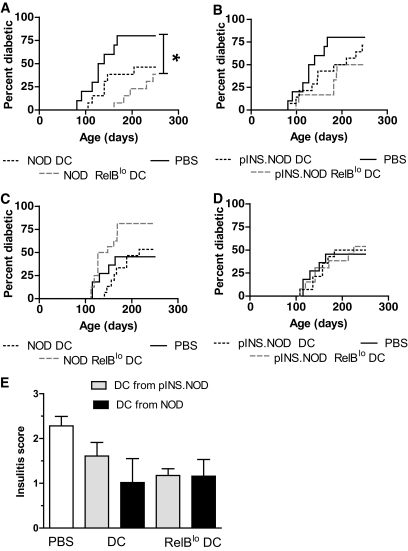
To investigate the consequences of administration of RelBlo DCs to young or insulitic NOD mice, we generated RelBlo DCs from NOD mice by incubation of bone marrow cells in the presence of GM-CSF, IL-4, and Bay11-7082 (an irreversible inhibitor of NF-κB and inflammasome, but not p38 mitogen-activated protein kinase [40,41]) as previously described (33,35). CD40 and MHC class II expression were reduced by addition of Bay11-7082 to NOD DC cultures compared with control DCs without Bay11-7082, as expected (supplementary Fig. 1 in the online appendix, available at http://diabetes.diabetesjournals.org/cgi/content/full/db10-0104/DC1). The concentration of inhibitor required to inhibit expression of CD40 and MHC class II in NOD bone marrow cell cultures was generally 50% of that required for C57BL/6 RelBlo DC cultures, consistent with the constitutively higher levels of NF-κB expression in NOD cells (11). To determine the potential of RelBlo DCs to suppress disease generation in an antigen-specific manner, we also generated RelBlo DCs from NOD mice where proinsulin is driven as a transgene by the MHC class II promoter (pINS.NOD) (42). Mean survival without diabetes was increased from 161 days (95% CI 132–191) if mice were untreated to 232 days (215–248) in mice administered 5 × 10⁵ RelBlo DCs subcutaneously, generated from NOD.Lt mice (P = 0.002). The smaller increases in survival time in mice administered pINS.NOD DCs, or control DCs, at 28 days of age were not statistically significant (Fig. 1). There was a trend for reduction in insulitis at 85 days in mice treated with NOD.Lt or pINS.NOD RelBlo DCs compared with PBS, but because islets from only four mice per group were examined, it was not meaningful to analyze the data statistically (Fig. 1E). In contrast, diabetes incidence was not reduced relative to saline-treated controls by transfer of RelBlo or control NOD or pINS.NOD DCs at 100 days of age and if anything, NOD RelBlo DC reduced the survival time without diabetes at this age (Fig. 1C and D). Thus, as expected, administration of RelBlo DCs prevented development of diabetes when administered to 28- but not 100-day-old NOD mice. This was not dependent on transgenic expression of proinsulin autoantigen by the DCs.
FIG. 1.
RelBlo tolerizing DCs inhibit diabetes when administered to young, but not insulitic, NOD mice. RelBlo DCs were generated from the bone marrow of NOD or pINS.NOD transgenic mice in the presence of GM-CSF, IL-4, and Bay11-7082. Control DCs were generated from the same mice in the presence of cytokines and the absence of Bay11-7082. Twenty-eight-day-old (A and B) or 100-day-old female NOD (C and D) mice were injected subcutaneously with 5 × 10⁵ DCs. Mice were screened weekly for diabetes until 250 days of age. Diabetes incidence curves are shown for groups each containing 12 mice. *P < 0.05 (Kaplan-Meier survival analysis with Bonferroni correction for multiple groups). Insulitis was assessed at 12 weeks (E) in four female NOD mice per group treated at 4 weeks of age with DCs as shown. Data represent means ± SD.
IL-1β is overexpressed as islet inflammation develops, in response to self-antigen presentation.
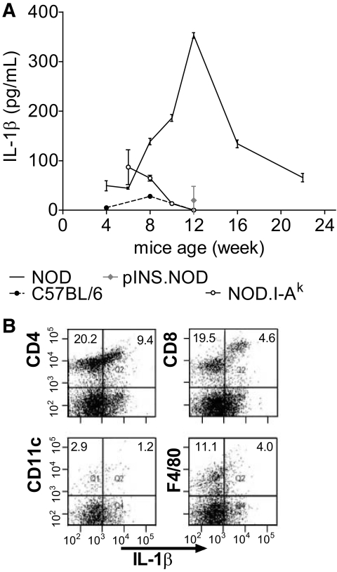
We showed previously that IL-1β overproduction in NOD mice is systemic and can be measured by stimulation of splenocytes with anti-CD3; IL-1 production in humans is also systemic and measurable in PBMCs (22). IL-1 dysregulates Treg cell function, and RelBlo DCs suppress inflammation through Treg cells. We therefore determined the timing and mechanism of systemic IL-1β overproduction prior to the onset of diabetes relative to the timing of administration of RelBlo DCs. We first analyzed supernatants from splenocytes stimulated with anti-CD3 every 4 weeks from weaning till diabetes. IL-1β secretion by NOD splenocytes was increased relative to C57BL/6 controls, between weeks 6 and 16, peaking at ∼12 weeks of age (Fig. 2A), as insulitis develops (43). IL-1β was not overexpressed by splenocytes from NOD.I-Ak congenic mice, which do not express the MHC class II proinsulin antigen restriction element, I-Ag7, or from pINS.NOD transgenic mice. These mice express mouse proinsulin II under control of the I-Eαk MHC class II promoter, are tolerant to proinsulin, and do not develop diabetes (but remain susceptible to other autoimmune diseases) (44,45). When anti-CD3–stimulated splenocytes were analyzed with intracellular staining and flow cytometry, we found that T-cells including both CD4+ and CD8+ subsets, and APCs including CD11c+ DC and F4/80+ macrophages, produced IL-1β (Fig. 2B). Together, the data indicate that IL-1β is overexpressed by innate immune cells and T-cells, and, unexpectedly, that IL-1β overproduction during the development of islet inflammation is driven or accelerated by islet autoantigen presentation in the context of the NOD genetic background.
FIG. 2.
Secretion of IL-1β by NOD splenocytes in response to anti-CD3ε antibody during the phase of insulitis does not occur in the absence of autoantigen presentation and responding T-cells. A: Splenocytes from NOD.Lt, C57BL/6, NOD.I-Ak, or pINS.NOD mice of different ages were incubated for 24 h in presence of anti-CD3ε antibody, and IL-1β was assayed in the supernatants of three mice per group by ELISA. B: NOD.Lt splenocytes were incubated with anti-CD3ε antibody for 24 h, the last 4 h in presence of brefeldin A 5 μg/ml, before staining for surface markers as shown and intracellular IL-1β. Representative of four experiments.
Because sera from patients with or at risk of type 1 diabetes, when incubated with healthy PBMCs, promote a microarray signature dominated by IL-1β (15), we assessed whether a factor in mouse serum similarly promotes IL-1β mRNA expression when incubated with splenocytes from either C57BL/6 or 6-week-old NOD mice. Although serum from diabetic NOD mice induced expression of IL-1β by healthy splenocytes, serum derived from NOD mice during the insulitis phase did not (supplementary Fig. 2). Glucose at concentrations similar to those in diabetic mice or humans promote IL-1 secretion by monocytes in vitro (46). Thus, our data are consistent with the conclusion that islet autoreactivity and not metabolic factors, such as hyperglycemia, drive IL-1β overexpression during the insulitis phase in NOD mice.
IL-1β blocks Treg function during the insulitis phase in NOD mice.
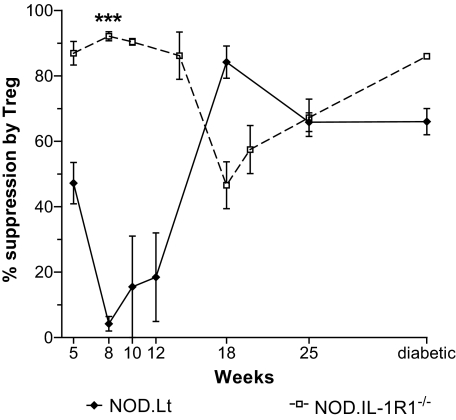
We next determined the effects of IL-1β in NOD mice. We first tracked the capacity of NOD effector T-cells (Teff) to be suppressed by regulatory (Treg) T-cells, in parallel to IL-1β production, at different ages in NOD.Lt mice. We incubated syngeneic splenic DCs, CD4+CD25− Teff, CD4+CD25+ Treg, and anti-CD3 and plotted the percent suppression of Teff by Treg over time (Fig. 3). Splenic Treg suppressed proliferation of Teff in vitro before 6 weeks of age to 40% maximal. However, Treg were unable to suppress Teff by 8 weeks of age, and this function only returned by 18 weeks, associated with the fall in splenocyte IL-1 secretion (Fig. 2A). The same experiments were carried out using cells from NOD mice lacking the IL-1R1 receptor (NOD.IL-1R1−/−). The capacity of NOD.Lt Treg to suppress NOD.Lt Teff was significantly different from the capacity of NOD.IL-1R1−/− Treg to suppress NOD.IL-1R1−/− Teff over the same time period (P < 0.001 by two-way ANOVA). Thus, the reduced capacity of NOD.Lt splenic CD4+CD25+ T-cells to suppress Teff is IL-1β and time dependent. The results are consistent with reduction in Treg function in NOD mice in vivo after 4–6 weeks of age (7).
FIG. 3.
Transient inhibition of Treg cell function is IL-1β dependent, corresponding to the peak of insulitis. CD11c+, CD4+CD25+, and CD4+CD25− cells were purified from spleens and lymph nodes of NOD mice of different ages (four mice pooled per group), using immunomagnetic separation. A total of 100,000 CD11c+ DCs, 105 CD4+CD25− T-cells, and 7.5 × 10⁴ CD4+CD25+ T-cells were incubated for 3 days in presence of anti-CD3ε antibody before determination of the T-cell proliferative response by [3H]thymidine uptake. The regulatory capacity is expressed as the percentage suppression of T-cell proliferation in presence of CD4+CD25+ T-cells relative to the maximum proliferation observed in presence of CD4+CD25− T-cells and DCs alone. Data from three separate experiments are expressed as means ± SE percent suppression. ***P < 0.0001 analyzed by two-way ANOVA comparing NOD and NOD.IL-1R1−/− over time.
An inflammatory profile associated with IL-1β.
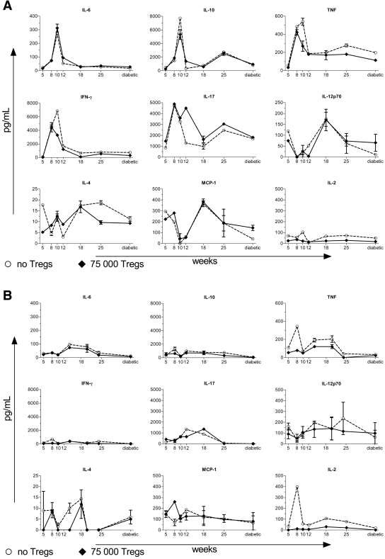
Given the marked change in proinflammatory IL-1β secretion associated with altered Treg function during the insulitis phase in NOD mice, we extended our analysis to determine whether other proinflammatory cytokines were produced simultaneously and whether these could be suppressed by Treg cells. IL-6, tumor necrosis factor (TNF), IL-17, IFN-γ, and IL-10 were overexpressed at the same time as IL-1β (8–12 weeks). Treg did not suppress these cytokines when added to Teff (Fig. 4A). We did not observe similar increases in IL-6, IL-17, IFN-γ, or IL-10 when analyzing cells from NOD.IL-1R1−/− mice (Fig. 4B). TNF was overexpressed in NOD.IL-1R1−/− mice between weeks 5 and 10 but was suppressed by the addition of Treg. IL-23 was undetectable in these supernatants (data not shown).
FIG. 4.
IL-1β drives IL-6, TNF, IL-10, and IL-17, which are not subject to Treg suppression, as insulitis develops in NOD mice. Cytokines were assayed in supernatants from the T-cell proliferation assays (Fig. 3) from mice of the ages indicated using CBA kits and IL-17 ELISA. ○, cytokine production by CD4+CD25− T-cells in the absence of CD4+CD25+ Treg; ♦, cytokine production by CD4+CD25− T-cells in the presence of CD4+CD25+ Treg. A: Wild-type mice. B: NOD.IL-1R1−/− mice. Data represent mean ± SEM.
IL-17 production increased again in supernatants from NOD mice at ∼25 weeks and in IL-1R1−/−.NOD mice at 18 weeks, concomitant with reduction in Treg function (Fig. 4) and heralding the onset of diabetes. Levels of the IL-17 regulatory cytokine, IFN-γ, dropped after 12 weeks, whereas IL-17 levels increased. The data suggest that in the presence of high levels of IL-1β, Treg are unable to contain the expression of a set of proinflammatory cytokines by effector T-cells or APCs.
Spontaneous pancreatic islet cytokine production is time dependent and reflects production by stimulated splenocytes.
To determine the relevance of cytokine production stimulated from splenocytes, we isolated pancreatic islets from NOD mice between the ages of 4 and 15 weeks, extracted RNA from whole islet tissue and quantitated IL-1β, TNF, IL-6, and IFN-γ RNA by Taqman real-time PCR. Islet IL-1β and IL-6 mRNA expression increased between 4 and 15 weeks (P < 0.05, P < 0.0001, respectively, one-way ANOVA with post hoc analysis for linear trend), reflecting the time course of stimulated splenocytes (Fig. 5). IL-17 signal was too low to amplify from whole islets, likely because of a small proportion of infiltrating Th17, as IL-17 was amplified from stimulated NOD.Lt splenic T-cells (not shown). Thus, the Th17-promoting cytokines, IL-1β, and IL-6 are also expressed by inflamed islets.
FIG. 5.
Age-dependent expression of proinflammatory cytokines by pancreatic islets. Pancreatic islets were purified from NOD.Lt mice (n = 5 per group) of the ages shown, and RNA was analyzed by Taqman PCR for relative expression of IL-1β, IL-6, TNF, and IFN-γ. *P < 0.05; ***P < 0.0001 (one-way ANOVA with post hoc analysis for linear trend).
IL-17 production is promoted by addition of Tregs to Teff in an IL-1β–dependent manner at the time of peak insulitis.
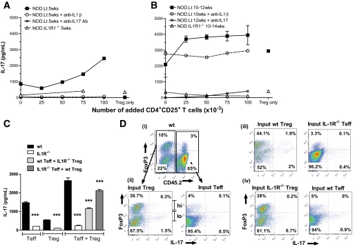
We therefore tested the relationship between NOD mouse age, Treg, Teff, IL-1β, and IL-17 production by adding Treg from NOD or NOD IL-1R1−/− mice to syngeneic Teff stimulated by DCs and anti-CD3 (Fig. 6). IL-17 increased approximately threefold with the addition of Treg cells to Teff from 5-week NOD.Lt mice (Fig. 6A). IL-17 secretion was dependent on IL-1β signaling in vitro and in vivo, as anti–IL-1β mAb blocked the secretion of IL-17 in vitro, and NOD.IL1R1−/− T-cells did not secrete IL-17. By 10 weeks of age, both Teff and Treg secreted IL-17 and this was no longer dependent on IL-1β in vitro (Fig. 6B). However, because T-cells isolated from 10-week NOD.IL1R1−/− mice did not secrete IL-17 ex vivo, the data suggest that IL-1β (and concomitant IL-6) produced in response to antigen presentation promotes the development of Th17 from a young age in NOD.Lt mice through FoxP3+ Treg cell conversion to Th17 (47). To assess this in vitro, Teff and Treg purified from 6-week CD45.1 NOD.Lt, CD45.2.NOD, and CD45.1 NOD.IL-1R1−/− mice were stimulated with DCs and anti-CD3. Purified CD45.2 CD25− Teff included 4% CD4+FoxP3+ cells (of which <1% were CD25+), and purified CD45.1 CD25+ Treg comprised 86% FoxP3hi and 14% FoxP3lo cells. After 72 h, IL-17 was measured in supernatants, and PMA-restimulated cells were stained for CD4, CD45.2, FoxP3, and IL-17. Teff and Treg secreted IL-17 in an IL-1–dependent manner. When Teff were mixed with Treg, IL-17 was secreted if either Teff or Treg but not both lacked IL-1R1 (Fig. 6C). The proportion of CD45.1+ FoxP3hi cells fell to 30% in culture (Fig. 6D, i). Approximately 0.6% of the input CD45.2+ Teff and 1.8% of input CD45.1+ Treg expressed IL-17 (Fig. 6D, ii). Of the IL-17+ Treg, the majority were FoxP3lo. When all cells were NOD.IL-1R1−/−, 0.3% of Teff and 0.1% of Treg cells expressed IL-17 in culture (data not shown). However, if NOD.IL-1R1−/− Teff were mixed with wild-type Treg, 4% of input wild-type FoxP3lo and FoxP3hi Treg expressed IL-17 (Fig. 6D, iii), and if NOD.IL-1R1−/− Treg were mixed with wild-type Teff, 0.9% of Teff expressed IL-17 (Fig. 6D, iv). Thus, NOD.IL-1R1−/− Treg were unable to prevent low level of IL-17 production by wild-type Teff. Moreover, FoxP3 expression is unstable after activation in the presence of effector cells, and conversion of FoxP3hi and FoxP3lo Th17 is IL-1 dependent.
FIG. 6.
IL-17 is produced by Teff and reprogrammed Tregs in an IL-1β–dependent manner during insulitis. IL-17 was assayed at 5 (A) or 10–14 weeks (B) in supernatants from the T-cell proliferation assay by ELISA from NOD.Lt cells with or without anti–IL-1 or anti–IL-17 mAb (10 μg/ml) or from NOD.IL-1R1−/− cells. C: CD4+CD25− Teff and CD4+CD25+ Treg purified from 6-week CD45.1 NOD.Lt, CD45.2.NOD, and CD45.1 NOD.IL-1R1−/− mice were stimulated with DCs and anti-CD3. After 72 h, IL-17 was measured in supernatants. ***P < 0.0001 (one-way ANOVA). D: Cells from the same experiment were restimulated with PMA in the presence of brefeldin A and stained for CD4, CD45.2, FoxP3, and IL-17. Cells are gated on CD4 and the relevant congenic marker to analyze Teff and Treg FoxP3 and IL-17 expression individually.
In the IL-1–rich insulitic environment, RelBlo DCs reduce suppressive capacity of Treg cells at the expense of Th17 cells.
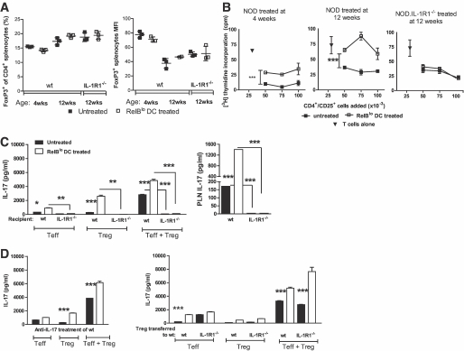
Given the IL-1–dependent dysregulation of Treg, coincident with the failure of RelBlo DCs to prevent type 1 diabetes, we analyzed the relationship between RelBlo DC administration and Treg number and function. As NOD mice age, Treg lose expression of FoxP3 (48). The frequency of FoxP3+ Treg in spleen and intensity of FoxP3 expression 4 weeks later were not affected by RelBlo DC administration to 4- or 12-week mice (Fig. 7A). Treg cells purified from untreated 8-week-old NOD mice suppressed Teff purified from naïve 6-week-old NOD mice, but this was significantly reduced by administration of RelBlo DC 4 weeks previously (Fig. 7B). Suppressive capacity of Treg cells purified from 16-week-old NOD mice was reduced relative to the activity of Treg cells purified from 8-week-old mice and significantly reduced by administration of RelBlo DCs 4 weeks previously (Fig. 7B). In contrast, suppressive activity of Treg cells isolated from 16-week-old NOD.IL-1R1−/− recipients of RelBlo DCs administered 4 weeks earlier and untreated NOD.IL-1R1−/− mice was equivalent (Fig. 7B). The data demonstrate that RelBlo DCs exacerbate the age-dependent decline in Treg cell function. Similarly, when 6-week wild-type or NOD.IL-1R1−/− mice were untreated or injected with RelBlo DCs, IL-17 secretion by splenic Teff and Treg was promoted by RelBlo DCs in an IL-1–dependent manner 4 weeks later (Fig. 7C). IL-1–dependent IL-17 secretion by anti-CD3–stimulated pancreatic draining lymph node cells from these mice was greater after treatment with RelBlo DCs (Fig. 7C). Consistent with this IL-1 dependence, promotion of IL-17 production by RelBlo DCs was not altered by treatment of wild-type recipients with anti–IL-17 mAb (Fig. 7D). Consistent with the inability of IL-1R1−/− Treg to suppress IL-17 production by wild-type cells in vitro (Fig. 6), adoptive transfer of wild-type or NOD.IL-1R1−/− Treg purified from 4-week-old donors to 6-week-old recipients did not impact IL-17 secretion in the presence of RelBlo DCs (Fig. 7D). By staining, IL-17 was almost all produced by host FoxP3lo Treg (data not shown). The data demonstrate that RelBlo DCs exacerbate the age-dependent decline in Treg cell function at the expense of conversion to Th17 in NOD mice including in pancreatic draining lymph node, in an IL-1–dependent manner.
FIG. 7.
In the IL-1–rich insulitic environment, tolerizing DCs reduce suppressor function at the expense of IL-17. Four-week-old, 12-week-old NOD mice, and 12-week-old NOD.IL1R−/− mice were injected subcutaneously with RelBlo DCs or saline. A: The percentage of CD4+ cells expressing FoxP3 in spleen and the mean fluorescence intensity (MFI) of FoxP3 expressed by CD4+ T-cells were enumerated. B: Four weeks later, CD4+CD25+ T-cells were isolated from spleen and various numbers were added to 1 × 10⁵ CD11c+ splenic DCs and 1 × 10⁵ CD4+CD25− T-cells purified from naïve 6-week NOD mice and stimulated with 0.5 μg/ml anti-CD3. Proliferation was assessed by incorporation of [3H] thymidine. Data represent the mean of triplicate wells ± SE. A total of 6–9 mice were pooled in each of two separate experiments. ***P < 0.0001 (two-way ANOVA). C and D: Six-week wild-type or NOD.IL-1R1−/− mice were untreated or injected with RelBlo DCs. As DCs were administered, some groups of wild-type mice were administered anti–IL-17 mAb intraperitoneally on alternate days for 10 days or CD4+CD25+ Treg purified from either wild-type or NOD.IL-1R1−/− mice intravenously once. After 4 weeks, Teff and Treg were purified from each group and incubated with DCs in the presence of anti-CD3. IL-17 levels were measured in supernatants by ELISA (C) and cells restimulated with PMA in the presence of brefeldin A were stained for CD4, CD45.2, FoxP3, and IL-17 (D). *P < 0.05; **P < 0.001; ***P < 0.0001 (one-way ANOVA) (C and D).
IL-17 impairs Treg function systemically.
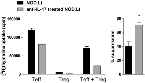
Although IL-1 impairs the suppressive function of Treg (Fig. 4) (17), a recent publication (49) showed that inhibition of IL-17 for 10 days in 10-week NOD mice was sufficient to impair progression to diabetes, associated with increased Treg infiltration of islets. When we treated 6-week NOD with anti–IL-17, Teff were suppressed by splenic Treg significantly better than those of untreated mice (Fig. 8). The data support the conclusion that in addition to the broad impact of IL-1, IL-17 itself impacts suppression by FoxP3+ Treg.
FIG. 8.
Enhanced Treg suppressive capacity in response to anti–IL-17. CD4+CD25+ and CD4+CD25− T-cells purified from spleens of wild-type mice administered anti–IL-17 mAb intraperitoneally on alternate days for 10 days 4 weeks prior at age 6 weeks were incubated alone or in a 1:1 ratio with DCs and anti-CD3. Proliferation was assessed by incorporation of [3H] thymidine and means ± SE; percent suppression was calculated. Three mice were pooled in each of two separate experiments. *P < 0.05 (t test).
DISCUSSION
Many newly diagnosed type 1 diabetic patients exhibit high levels of IL-1β or a microarray signature, including IL-1β (22,50). Although metabolic disturbance is implicated in the IL-1β expression by such patients, there are suggestions of IL-1 overexpression earlier in the disease course. For example, sera from at-risk first-degree relatives induced an IL-1–related gene expression signature, when incubated with healthy PBMCs (15). We show here that as NOD mice age, IL-1 is produced systemically, including in the pancreatic islet, and is implicated in the immune dysregulation that occurs as islet autoantigen is presented in at-risk mice, by virtue of their predisposing MHC class II alleles. Tolerant pINS.NOD mice also fail to upregulate IL-1. These data suggest that IL-1 production by self-reactive peripheral blood T-cells could be a useful early prognostic indicator in children with high-risk MHC alleles, even before development of islet autoantibodies. Not all type 1 diabetic patients show elevated IL-1β levels or gene signature. Delayed diabetes progression in NOD.IL-1R1−/− mice suggests that IL-1β is not necessary for this form of diabetes; however, high IL-1 might predict a more aggressive disease course associated with high-risk MHC alleles.
In NOD mice, early IL-1 overproduction has functional consequences: systemic (including islet) induction of IL-6 and IFN-γ, reduction in Treg suppressor function, FoxP3 instability, reprogramming of Treg to Th17, and dysregulation of the mechanisms by which tolerizing DCs suppress autoimmunity. Similar to NOD mice, IL-6 downregulated FoxP3 expression and (with IL-1 and transforming growth factor-β) reprogrammed Treg to Th17 (47,51–53). Reprogramming is of particular concern in the context of autoimmunity, because FoxP3+Treg are selected in the thymus for their reactivity toward self-antigens, and FoxP3+Th17 could contribute to pathogenic self-reactivity (54). In experimental allergic encephalomyelitis, ∼10% of splenic FoxP3+ cells produced IL-17 (47). Th17 differentiation is initiated but not maintained by IL-1 in NOD mice and can be initiated by other factors in NOD.IL-1R1−/− mice. Thus, IL-1β accelerates diabetes by reprogramming Treg to Th17 at the expense of suppressor function, between 6 and 18 weeks in NOD mice. Moreover, IL-1 enhances T-cell IFN-γ, which drives insulitis, and must be contained by FoxP3+Treg (6,17).
Many immunotherapeutic strategies, which induce Treg, prevent diabetes when administered to young NOD mice but not during insulitis (25,55–58). This is likely reflected in human type 1 diabetes, where the outcome of preventive trials of antigen-specific tolerizing immunotherapy has varied (59,60). Nasal insulin, which was ineffective, was administered to very high-risk children in the late preclinical phase (60) when IL-1 was likely already expressed. Similar to previous studies, RelBlo DC immunotherapy in young NOD mice did not depend on presentation of proinsulin antigen (25). This could be because nontransgenic RelBlo DCs present a range of self-antigens that tolerize relevant autoreactive T-cells, or because of nonspecific Th2 immune deviation by DCs cultured in FCS, presenting FCS-derived epitopes (61,62). The latter is less likely because DCs cultured without NF-κB inhibitor were ineffective, and neither Teff nor Treg secreted more IL-4 after administration of RelBlo DCs (not shown) (61,62).
In a recent study (49), anti–IL-17 mAb administered to 10-week but not 4- to 5-week NOD mice prevented diabetes. The current studies demonstrate that 10 weeks is ideal and 4 weeks is too early to block IL-17. Furthermore, IL-17 may impact the capacity of Teff to be suppressed by Treg. This may be indirect, as anti–IL-17 reduces DC differentiation and promotes IL-10 production (63,64).
Tolerizing DCs delivered to young mice likely regulate autoantigen presentation, preventing expression of proinflammatory triggers for Th17. RelBlo DCs, which suppress inflammatory arthritis, also prevent diabetes in NOD mice when administered at 4 weeks but not 14 weeks. At 10–14 weeks, RelBlo DCs exacerbated the IL-1–dependent decline in Treg cell function and conversion to Th17. When IL-1β and IL-6 are overexpressed by NOD mice, reprogramming Th17, the ground is no longer fertile for therapies which induce Treg. In contrast, GAD-specific Ig-GAD immunotherapy, which promotes IFN-γ and blocks IL-17, was only effective when delivered after 8 weeks of age (65). Our data in NOD mice have important implications for appropriate timing of immunotherapy in humans at risk of type 1 diabetes. IL-1 and IL-17 would be interesting early biomarkers in children at risk of type 1 diabetes to identify similar pathogenetic stages and to stratify treatment to tolerizing immunotherapy or cytokine blockade (66).
Supplementary Material
ACKNOWLEDGMENTS
This work was supported by National Health and Medical Research Council (NHMRC) grants 351439 and 569938 and Juvenile Diabetes Research Foundation grants 1-2006-149 and 5-2003-269. R.T. was supported by Arthritis Queensland and an Australian Research Council Future Fellowship. R.S. was supported by an NHMRC Career Development Award. B.O. was supported by a Queensland Smart State Fellowship.
No potential conflicts of interest relevant to this article were reported.
S.B.-M., D.P., S.B., E.D., S.P., and R.S. researched data. S.B.-M., D.P., S.P., R.S., and R.T. wrote the manuscript. B.O., S.P., L.C.H., and T.W.H.K. contributed to the discussion and reviewed/edited the manuscript.
Footnotes
The costs of publication of this article were defrayed in part by the payment of page charges. This article must therefore be hereby marked “advertisement” in accordance with 18 U.S.C. Section 1734 solely to indicate this fact.
REFERENCES
- 1.Dahlen E, Dawe K, Ohlsson L, Hedlund G: Dendritic cells and macrophages are the first and major producers of TNF-alpha in pancreatic islets in the nonobese diabetic mouse. J Immunol 1998;160:3585–3593 [PubMed] [Google Scholar]
- 2.Jansen A, Homo-Delarche F, Hooijkaas H, Leenen PJ, Dardenne M, Drexhage HA: Immunohistochemical characterization of monocytes-macrophages and dendritic cells involved in the initiation of the insulitis and beta-cell destruction in NOD mice. Diabetes 1994;43:667–675 [DOI] [PubMed] [Google Scholar]
- 3.Serreze DV, Leiter EH: Genes and cellular requirements for autoimmune diabetes susceptibility in nonobese diabetic mice. Curr Dir Autoimmun 2001;4:31–67 [DOI] [PubMed] [Google Scholar]
- 4.Krishnamurthy B, Dudek NL, McKenzie MD, Purcell AW, Brooks AG, Gellert S, Colman PG, Harrison LC, Lew AM, Thomas HE, Kay TW: Responses against islet antigens in NOD mice are prevented by tolerance to proinsulin but not IGRP. J Clin Invest 2006;116:3258–3265 [DOI] [PMC free article] [PubMed] [Google Scholar]
- 5.Marino E, Grey ST: A new role for an old player: do B cells unleash the self-reactive CD8+ T cell storm necessary for the development of type 1 diabetes? J Autoimmun 2008;31:301–305 [DOI] [PubMed] [Google Scholar]
- 6.Feuerer M, Shen Y, Littman DR, Benoist C, Mathis D: How punctual ablation of regulatory T cells unleashes an autoimmune lesion within the pancreatic islets. Immunity 2009;31:654–664 [DOI] [PMC free article] [PubMed] [Google Scholar]
- 7.Tritt M, Sgouroudis E, d'Hennezel E, Albanese A, Piccirillo CA: Functional waning of naturally occurring CD4+ regulatory T-cells contributes to the onset of autoimmune diabetes. Diabetes 2008;57:113–123 [DOI] [PubMed] [Google Scholar]
- 8.Tang Q, Adams JY, Penaranda C, Melli K, Piaggio E, Sgouroudis E, Piccirillo CA, Salomon BL, Bluestone JA: Central role of defective interleukin-2 production in the triggering of islet autoimmune destruction. Immunity 2008;28:687–697 [DOI] [PMC free article] [PubMed] [Google Scholar]
- 9.Mollah ZUA, Pai S, Moore C, O'Sullivan BJ, Harrison MJ, Peng J, Phillips K, Prins JB, Cardinal J, Thomas R: Abnormal NF-kappa B function characterizes human type 1 diabetes dendritic cells and monocytes. J Immunol 2008;180:3166–3175 [DOI] [PubMed] [Google Scholar]
- 10.Weaver DJ, Jr, Poligone B, Bui T, Abdel-Motal UM, Baldwin AS, Jr, Tisch R: Dendritic cells from nonobese diabetic mice exhibit a defect in NF- kappa B regulation due to a hyperactive I kappa B kinase. J Immunol 2001;167:1461–1468 [DOI] [PubMed] [Google Scholar]
- 11.Poligone B, Weaver DJ, Jr, Sen P, Baldwin AS, Jr, Tisch R: Elevated NF-kappaB activation in nonobese diabetic mouse dendritic cells results in enhanced APC function. J Immunol 2002;168:188–196 [DOI] [PubMed] [Google Scholar]
- 12.Wheat W, Kupfer R, Gutches DG, Rayat GR, Beilke J, Scheinman RI, Wegmann DR: Increased NF-kappa B activity in B cells and bone marrow-derived dendritic cells from NOD mice. Eur J Immunol 2004;34:1395–1404 [DOI] [PubMed] [Google Scholar]
- 13.Devaraj S, Dasu MR, Rockwood J, Winter W, Griffen SC, Jialal I: Increased toll-like receptor (TLR) 2 and TLR4 expression in monocytes from patients with type 1 diabetes: further evidence of a proinflammatory state. J Clin Endocrinol Metab 2008;93:578–583 [DOI] [PMC free article] [PubMed] [Google Scholar]
- 14.Devaraj S, Glaser N, Griffen S, Wang-Polagruto J, Miguelino E, Jialal I: Increased monocytic activity and biomarkers of inflammation in patients with type 1 diabetes. Diabetes 2006;55:774–779 [DOI] [PubMed] [Google Scholar]
- 15.Wang X, Jia S, Geoffrey R, Alemzadeh R, Ghosh S, Hessner MJ: Identification of a molecular signature in human type 1 diabetes mellitus using serum and functional genomics. J Immunol 2008;180:1929–1937 [DOI] [PubMed] [Google Scholar]
- 16.Pfleger C, Mortensen HB, Hansen L, Herder C, Roep BO, Hoey H, Aanstoot HJ, Kocova M, Schloot NC: Association of IL-1ra and adiponectin with C-peptide and remission in patients with type 1 diabetes. Diabetes 2008;57:929–937 [DOI] [PubMed] [Google Scholar]
- 17.O'Sullivan B, Thomas HE, Pai S, Santamaria P, Iwakura Y, Steptoe RJ, Kay TW, Thomas R: IL-1 breaks tolerance through expansion of CD25+ effector T cells. J Immunol 2006;176:7278–7287 [DOI] [PubMed] [Google Scholar]
- 18.Mandrup-Poulsen T, Spinas GA, Prowse SJ, Hansen BS, Jorgensen DW, Bendtzen K, Nielsen JH, Nerup J: Islet cytotoxicity of interleukin 1: influence of culture conditions and islet donor characteristics. Diabetes 1987;36:641–647 [DOI] [PubMed] [Google Scholar]
- 19.Eriksson U, Kurrer MO, Sonderegger I, Iezzi G, Tafuri A, Hunziker L, Suzuki S, Bachmaier K, Bingisser RM, Penninger JM, Kopf M: Activation of dendritic cells through the interleukin 1 receptor 1 is critical for the induction of autoimmune myocarditis. J Exp Med 2003;197:323–331 [DOI] [PMC free article] [PubMed] [Google Scholar]
- 20.Yaacob NS, Kaderi MA, Norazmi MN: The expression of cytokine genes in the peritoneal macrophages and splenic CD4- and CD8-positive lymphocytes of the nonobese diabetic mice. J Clin Immunol 2004;24:177–184 [DOI] [PubMed] [Google Scholar]
- 21.Thomas HE, Irawaty W, Darwiche R, Brodnicki TC, Santamaria P, Allison J, Kay TW: IL-1 receptor deficiency slows progression to diabetes in the NOD mouse. Diabetes 2004;53:113–121 [DOI] [PubMed] [Google Scholar]
- 22.Kaizer EC, Glaser CL, Chaussabel D, Banchereau J, Pascual V, White PC: Gene expression in peripheral blood mononuclear cells from children with diabetes. J Clin Endocrinol Metab 2007;92:3705–3711 [DOI] [PubMed] [Google Scholar]
- 23.Balasa B, Krahl T, Patstone G, Lee J, Tisch R, McDevitt HO, Sarvetnick N: CD40 ligand-CD40 interactions are necessary for the initiation of insulitis and diabetes in nonobese diabetic mice. J Immunol 1997;159:4620–4627 [PubMed] [Google Scholar]
- 24.Feili-Hariri M, Falkner DH, Gambotto A, Papworth GD, Watkins SC, Robbins PD, Morel PA: Dendritic cells transduced to express interleukin-4 prevent diabetes in nonobese diabetic mice with advanced insulitis. Human Gene Ther 2003;14:13–23 [DOI] [PubMed] [Google Scholar]
- 25.Ma L, Qian S, Liang X, Wang L, Woodward JE, Giannoukakis N, Robbins PD, Bertera S, Trucco M, Fung JJ, Lu L: Prevention of diabetes in NOD mice by administration of dendritic cells deficient in nuclear transcription factor-κB activity. Diabetes 2003;52:1976–1985 [DOI] [PubMed] [Google Scholar]
- 26.Nitta Y, Tashiro F, Tokui M, Shimada A, Takei I, Tabayashi K, Miyazaki J: Systemic delivery of interleukin 10 by intramuscular injection of expression plasmid DNA prevents autoimmune diabetes in nonobese diabetic mice. Human Gene Ther 1998;9:1701–1707 [DOI] [PubMed] [Google Scholar]
- 27.O'Sullivan BJ, Thomas R: CD40 ligation conditions dendritic cell antigen-presenting function through sustained activation of NF-kappaB. J Immunol 2002;168:5491–5498 [DOI] [PubMed] [Google Scholar]
- 28.O'Sullivan BJ, Thomas R: CD40 ligation conditions dendritic cell antigen-presenting function through sustained activation of NF-kappaB. J Immunol 2002;168:5491–5498 [DOI] [PubMed] [Google Scholar]
- 29.Pettit AR, MacDonald KPA, O'Sullivan B, Thomas R: Differentiated dendritic cells expressing nuclear RelB are predominantly located in rheumatoid synovial tissue perivascular mononuclear cell aggregates. Arthritis Rheum 2000;43:791–800 [DOI] [PubMed] [Google Scholar]
- 30.Kobayashi T, Walsh PT, Walsh MC, Speirs KM, Chiffoleau E, King CG, Hancock WW, Caamano JH, Hunter CA, Scott P, Turka LA, Choi Y: TRAF6 is a critical factor for dendritic cell maturation and development. Immunity 2003;19:353–363 [DOI] [PubMed] [Google Scholar]
- 31.Martin E, O'Sullivan B, Low P, Thomas R: Antigen-specific suppression of a primed immune response by dendritic cells mediated by regulatory T cells secreting interleukin-10. Immunity 2003;18:155–167 [DOI] [PubMed] [Google Scholar]
- 32.Li M, Zhang X, Zheng X, Lian D, Zhang ZX, Ge W, Yang J, Vladau C, Suzuki M, Chen D, Zhong R, Garcia B, Jevnikar AM, Min WP: Immune modulation and tolerance induction by RelB-silenced dendritic cells through RNA interference. J Immunol 2007;178:5480–5487 [DOI] [PubMed] [Google Scholar]
- 33.Martin E, Capini C, Duggan E, Lutzky VP, Stumbles P, Pettit AR, O'Sullivan B, Thomas R: Antigen-specific suppression of established arthritis in mice by dendritic cells deficient in NF-kappaB. Arthritis Rheum 2007;56:2255–2266 [DOI] [PubMed] [Google Scholar]
- 34.Zheng SG, Wang JH, Gray JD, Soucier H, Horwitz DA: Natural and induced CD4+CD25+ cells educate CD4+CD25- cells to develop suppressive activity: the role of IL-2, TGF-beta, and IL-10. J Immunol 2004;172:5213–5221 [DOI] [PubMed] [Google Scholar]
- 35.Capini C, Jaturanpinyo M, Chang HI, Mutalik S, McNally A, Street S, Steptoe R, O'Sullivan B, Davies N, Thomas R: Antigen-specific suppression of inflammatory arthritis using liposomes. J Immunol 2009;182:3556–3565 [DOI] [PubMed] [Google Scholar]
- 36.Steptoe RJ, Stankovic S, Lopaticki S, Jones LK, Harrison LC, Morahan G: Persistence of recipient lymphocytes in NOD mice after irradiation and bone marrow transplantation. J Autoimmun 2004;22:131–138 [DOI] [PubMed] [Google Scholar]
- 37.French MB, Allison J, Cram DS, Thomas HE, Dempsey-Collier M, Silva A, Georgiou HM, Kay TW, Harrison LC, Lew AM: Transgenic expression of mouse proinsulin II prevents diabetes in nonobese diabetic mice. Diabetes 1997;46:34–39 [DOI] [PubMed] [Google Scholar]
- 38.Leiter EH: Multiple low-dose streptozotocin-induced hyperglycemia and insulitis in C57BL mice: influence of inbred background, sex and thymus. Proc Natl Acad Sci U S A 1982;79:630–634 [DOI] [PMC free article] [PubMed] [Google Scholar]
- 39.Liu M, Shapiro ME: A new method for isolation of murine islets with markedly improved yields. Transplant Proc 1995;27:3208–3210 [PubMed] [Google Scholar]
- 40.Pierce JW, Schoenleber R, Jesmok G, Best J, Moore SA, Collins T, Gerritsen ME: Novel inhibitors of cytokine-induced IkappaBalpha phosphorylation and endothelial cell adhesion molecule expression show anti-inflammatory effects in vivo. J Biol Chem 1997;272:21096–21103 [DOI] [PubMed] [Google Scholar]
- 41.Juliana C, Fernandes-Alnemri T, Wu J, Datta P, Solorzano L, Yu JW, Meng R, Quong AA, Latz E, Scott CP, Alnemri ES: Anti-inflammatory compounds parthenolide and Bay 11–7082 are direct inhibitors of the inflammasome. J Biol Chem 2010;285:9792–9802 [DOI] [PMC free article] [PubMed] [Google Scholar]
- 42.Steptoe RJ, Ritchie JM, Jones LK, Harrison LC: Autoimmune diabetes is suppressed by transfer of proinsulin-encoding Gr-1+ myeloid progenitor cells that differentiate in vivo into resting dendritic cells. Diabetes 2005;54:434–442 [DOI] [PubMed] [Google Scholar]
- 43.Fox CJ, Danska JS: IL-4 expression at the onset of islet inflammation predicts nondestructive insulitis in nonobese diabetic mice. J Immunol 1997;158:2414–2424 [PubMed] [Google Scholar]
- 44.Slattery RM, Kjer-Nielsen L, Allison J, Charlton B, Mandel TE, Miller JF: Prevention of diabetes in non-obese diabetic I-Ak transgenic mice. Nature 1990;345:724–726 [DOI] [PubMed] [Google Scholar]
- 45.French MB, Allison J, Cram DS, Thomas HE, Dempsey-Collier M, Silva A, Georgiou HM, Kay TW, Harrison LC, Lew AM: Transgenic expression of mouse proinsulin II prevents diabetes in nonobese diabetic mice. Diabetes 1997;46:34–39 [DOI] [PubMed] [Google Scholar]
- 46.Dasu MR, Devaraj S, Jialal I: High glucose induces IL-1beta expression in human monocytes: mechanistic insights. Am J Physiol Endocrinol Metab 2007;293:E337–E346 [DOI] [PMC free article] [PubMed] [Google Scholar]
- 47.Chung Y, Chang SH, Martinez GJ, Yang XO, Nurieva R, Kang HS, Ma L, Watowich SS, Jetten AM, Tian Q, Dong C: Critical regulation of early Th17 cell differentiation by interleukin-1 signaling. Immunity 2009;30:576–587 [DOI] [PMC free article] [PubMed] [Google Scholar]
- 48.Pop SM, Wong CP, Culton DA, Clarke SH, Tisch R: Single cell analysis shows decreasing FoxP3 and TGFbeta1 coexpressing CD4+CD25+ regulatory T cells during autoimmune diabetes. J Exp Med 2005;201:1333–1346 [DOI] [PMC free article] [PubMed] [Google Scholar]
- 49.Emamaullee JA, Davis J, Merani S, Toso C, Elliott JF, Thiesen A, Shapiro AM: Inhibition of Th17 cells regulates autoimmune diabetes in NOD mice. Diabetes 2009;58:1302–1311 [DOI] [PMC free article] [PubMed] [Google Scholar]
- 50.Netea MG, Hancu N, Blok WL, Grigorescu-Sido P, Popa L, Popa V, van der Meer JW: Interleukin 1 beta, tumour necrosis factor-alpha and interleukin 1 receptor antagonist in newly diagnosed insulin-dependent diabetes mellitus: comparison to long-standing diabetes and healthy individuals. Cytokine 1997;9:284–287 [DOI] [PubMed] [Google Scholar]
- 51.Yang XO, Nurieva R, Martinez GJ, Kang HS, Chung Y, Pappu BP, Shah B, Chang SH, Schluns KS, Watowich SS, Feng XH, Jetten AM, Dong C: Molecular antagonism and plasticity of regulatory and inflammatory T cell programs. Immunity 2008;29:44–56 [DOI] [PMC free article] [PubMed] [Google Scholar]
- 52.Deknuydt F, Bioley G, Valmori D, Ayyoub M: IL-1beta and IL-2 convert human Treg into T(H)17 cells. Clin Immunol 2009;131:298–307 [DOI] [PubMed] [Google Scholar]
- 53.Osorio F, LeibundGut-Landmann S, Lochner M, Lahl K, Sparwasser T, Eberl G, Reis e Sousa C: DC activated via dectin-1 convert Treg into IL-17 producers. Eur J Immunol 2008;38:3274–3281 [DOI] [PMC free article] [PubMed] [Google Scholar]
- 54.Hsieh CS, Liang Y, Tyznik AJ, Self SG, Liggitt D, Rudensky AY: Recognition of the peripheral self by naturally arising CD25+ CD4+ T cell receptors. Immunity 2004;21:267–277 [DOI] [PubMed] [Google Scholar]
- 55.Shoda LK, Young DL, Ramanujan S, Whiting CC, Atkinson MA, Bluestone JA, Eisenbarth GS, Mathis D, Rossini AA, Campbell SE, Kahn R, Kreuwel HT: A comprehensive review of interventions in the NOD mouse and implications for translation. Immunity 2005;23:115–126 [DOI] [PubMed] [Google Scholar]
- 56.Goudy KS, Burkhardt BR, Wasserfall C, Song S, Campbell-Thompson ML, Brusko T, Powers MA, Clare-Salzler MJ, Sobel ES, Ellis TM, Flotte TR, Atkinson MA: Systemic overexpression of IL-10 induces CD4+CD25+ cell populations in vivo and ameliorates type 1 diabetes in nonobese diabetic mice in a dose-dependent fashion. J Immunol 2003;171:2270–2278 [DOI] [PubMed] [Google Scholar]
- 57.Gadkar KG, Shoda LK, Kreuwel HT, Ramanujan S, Zheng Y, Whiting CC, Young DL: Dosing and timing effects of anti-CD40L therapy: predictions from a mathematical model of type 1 diabetes. Ann N Y Acad Sci 2007;1103:63–68 [DOI] [PubMed] [Google Scholar]
- 58.Zhang ZJ, Davidson L, Eisenbarth G, Weiner HL: Suppression of diabetes in nonobese diabetic mice by oral administration of porcine insulin. Proc Natl Acad Sci U S A 1991;88:10252–10256 [DOI] [PMC free article] [PubMed] [Google Scholar]
- 59.Skyler JS, Krischer JP, Wolfsdorf J, Cowie C, Palmer JP, Greenbaum C, Cuthbertson D, Rafkin-Mervis LE, Chase HP, Leschek E: Effects of oral insulin in relatives of patients with type 1 diabetes: the Diabetes Prevention Trial–Type 1. Diabetes Care 2005;28:1068–1076 [DOI] [PubMed] [Google Scholar]
- 60.Nanto-Salonen K, Kupila A, Simell S, Siljander H, Salonsaari T, Hekkala A, Korhonen S, Erkkola R, Sipila JI, Haavisto L, Siltala M, Tuominen J, Hakalax J, Hyoty H, Ilonen J, Veijola R, Simell T, Knip M, Simell O: Nasal insulin to prevent type 1 diabetes in children with HLA genotypes and autoantibodies conferring increased risk of disease: a double-blind, randomised controlled trial. Lancet 2008;372:1746–1755 [DOI] [PubMed] [Google Scholar]
- 61.Haase C, Ejrnaes M, Juedes AE, Wolfe T, Markholst H, von Herrath MG: Immunomodulatory dendritic cells require autologous serum to circumvent nonspecific immunosuppressive activity in vivo. Blood 2005;106:4225–4233 [DOI] [PMC free article] [PubMed] [Google Scholar]
- 62.Kadri N, Potiron N, Ouary M, Jegou D, Gouin E, Bach JM, Lieubeau B: Fetal calf serum-primed dendritic cells induce a strong anti-fetal calf serum immune response and diabetes protection in the non-obese diabetic mouse. Immunol Lett 2007;108:129–136 [DOI] [PubMed] [Google Scholar]
- 63.Bai H, Cheng J, Gao X, Joyee AG, Fan Y, Wang S, Jiao L, Yao Z, Yang X: IL-17/Th17 promotes type 1 T cell immunity against pulmonary intracellular bacterial infection through modulating dendritic cell function. J Immunol 2009;183:5886–5895 [DOI] [PubMed] [Google Scholar]
- 64.Higgins SC, Jarnicki AG, Lavelle EC, Mills KH: TLR4 mediates vaccine-induced protective cellular immunity to Bordetella pertussis: role of IL-17-producing T cells. J Immunol 2006;177:7980–7989 [DOI] [PubMed] [Google Scholar]
- 65.Jain R, Tartar DM, Gregg RK, Divekar RD, Bell JJ, Lee HH, Yu P, Ellis JS, Hoeman CM, Franklin CL, Zaghouani H: Innocuous IFNgamma induced by adjuvant-free antigen restores normoglycemia in NOD mice through inhibition of IL-17 production. J Exp Med 2008;205:207–218 [DOI] [PMC free article] [PubMed] [Google Scholar]
- 66.Pickersgill LM, Mandrup-Poulsen TR: The anti-interleukin-1 in type 1 diabetes action trial: background and rationale. Diabetes Metab Res Rev 2009;25:321–324 [DOI] [PubMed] [Google Scholar]
Associated Data
This section collects any data citations, data availability statements, or supplementary materials included in this article.


