Abstract
Autophagy degrades pathogens in vitro. The autophagy gene Atg5 has been reported to be required for IFN-γ-dependent host protection in vivo. However, these protective effects occur independently of autophagosome formation. Thus, the in vivo role of classic autophagy in protection conferred by adaptive immunity and how adaptive immunity triggers autophagy are incompletely understood. Employing biochemical, genetic and morphological studies, we found that CD40 upregulates the autophagy molecule Beclin 1 in microglia and triggers killing of Toxoplasma gondii dependent on the autophagy machinery. Infected CD40−/− mice failed to upregulate Beclin 1 in microglia/macrophages in vivo. Autophagy-deficient Beclin 1+/− mice, mice with deficiency of the autophagy protein Atg7 targeted to microglia/macrophages as well as CD40−/− mice exhibited impaired killing of T. gondii and were susceptible to cerebral and ocular toxoplasmosis. Susceptibility to toxoplasmosis occurred despite upregulation of IFN-γ, TNF-α and NOS2, preservation of IFN-γ-induced microglia/macrophage anti-T. gondii activity and the generation of anti-T. gondii T cell immunity. CD40 upregulated Beclin 1 and triggered killing of T. gondii by decreasing protein levels of p21, a molecule that degrades Beclin 1. These studies identified CD40-p21-Beclin 1 as a pathway by which adaptive immunity stimulates autophagy. In addition, they support that autophagy is a mechanism through which CD40-dependent immunity mediates in vivo protection and that the CD40-autophagic machinery is needed for host resistance despite IFN-γ.
Introduction
The lysosomal system is an effector of microbial degradation. Unfortunately, many pathogens including the intracellular protozoan Toxoplasma gondii have developed various strategies to avoid lysosomal degradation. Autophagy is a mechanism that can re-route pathogens to lysosomes. Autophagy is a process where an isolation membrane encircles portions of cytosol and organelles leading to the formation of autophagosomes [1], [2]. Fusion between autophagosomes and endosomes-lysosomes culminates in the formation of autolysosomes and degradation of their contents. Autophagy can mediate in vitro anti-microbial activity against various pathogens [3], [4], [5], [6], [7]. In the case of T. gondii infection, the CD40 – CD154 pathway triggers killing of the parasite within macrophages that is dependent on the autophagy pathway [6], [8]. CD40 is a member of the TNF receptor superfamily expressed on antigen presenting cells as well as on some non-hematopoietic cells, while CD154 (CD40 ligand) is expressed primarily on activated CD4+ T cells. The interaction between T. gondii-reactive T cells and infected macrophages results in CD40-dependent killing of the parasite through the autophagy pathway [6].
Innate immunity can activate autophagy to mediate host protection in vivo. Autophagy protects Drosophila against Listeria monocytogenes and Vesicular stomatitis virus [9], [10]. In the case of Herpes simplex virus 1 (HSV-1), the virus prevents autophagy by producing the neurovirulence factor ICP34.5 that binds and blocks the effect of the autophagy protein Beclin 1 [11]. A strain of HSV-1 deficient in ICP34.5 does not cause encephalitis in wild type mice but causes disease in mice deficient in PKR, a signaling molecule linked to autophagy [11]. Similarly, in a model of Salmonella infection in Caenorhabditis elegans and Dictyostelium discoideum, autophagy gene inactivation results in increased bacterial replication and decreased animal lifespan [12].
The autophagy gene ATG5 mediates autophagosome-independent host protection. Studies in mice deficient in ATG5 in phagocytes revealed that this autophagy gene was required for IFN-γ-mediated in vivo host protection likely because ATG5 was required for the induction of IFN-γ-dependent anti-microbial activity in macrophages [13]. ATG5 was required for recruitment of the Immunity- Related GTPase (IRG) Irga6 to the parasitophorous vacuole, the damage to this structure and clearance of the parasite [13]. This process occurred independently of classical autophagosome formation. In addition, mice with inactivation of ATG5 in dendritic cells revealed that this gene promoted the induction of a Th1 response and protection against HSV-1 [14]. The role of ATG5 in dendritic cells was to enhance MHC II processing of phagocytosed antigens that contain TLR agonists [14]. Processing of antigens for MHC II did not appear to be dependent on canonical autophagy [14]. These studies indicate that ATG5 regulates IFN-γ-dependent host protection and it does so through a manner that is does not rely on classical autophagy. Thus, the in vivo role of autophagy in mediating protection conferred by adaptive immunity is still not completely understood. In addition, it is unclear how adaptive immunity activates autophagy.
T. gondii can cause disease that manifests as cerebral and/or ocular toxoplasmosis. IFN-γ, TNF-α and their downstream effector molecule NOS2 are considered the mediators of resistance against ocular and cerebral toxoplasmosis [15], [16], [17], [18], [19], [20]. We report herein that despite the induction of pathogen-reactive T cells, IFN-γ, TNF-α, NOS2, and the preservation of IFN-γ-induced antimicrobial activity in microglia/macrophages, adaptive immunity still required the autophagic pathway to confer in vivo protection against cerebral and ocular toxoplasmosis. CD40 upregulates Beclin 1, triggers autophagy and killing of T. gondii in microglia independently of IFN-γ, nitric oxide and Irga6. This pathway is required for host protection since mice deficient in Beclin 1, mice with defective expression of Atg7 in microglia/macrophages and CD40−/− mice exhibit impaired autophagy protein-dependent killing of T. gondii and are susceptible to cerebral and ocular toxoplasmosis despite induction of T. gondii-specific T cell response, upregulation of IFN-γ, TNF-α and NOS2 and unimpaired IFN-γ-induced anti-microbial activity in microglia/macrophages. We also identified a pathway whereby adaptive immunity enhances autophagy. CD40 down-regulates p21 leading to increased levels of Beclin 1 and enhanced autophagy.
Results
CD40−/− mice are susceptible to ocular toxoplasmosis and toxoplasmic encephalitis despite upregulation of IFN-γ, TNF-α and NOS2
B6 and CD40−/− mice were infected with T. gondii. As shown in Figure 1A, CD40−/− died during the chronic phase of infection between weeks 4 to 6 while B6 mice survived at least 11 weeks (p<0.001). We conducted histopathologic studies of the eye and brain. The histological features of the choroid and retina from uninfected B6 and CD40−/− mice were similar. At 4 weeks post-infection, infected B6 mice revealed mild histopathologic changes in the retina and choroid (Figure 1B). In contrast, CD40−/− mice revealed more pronounced histopathology (p<0.01) that included distortion of the retinal architecture, hypertrophy of retinal pigmented epithelial cells, invasion of the retina by these cells, inflammatory infiltrates as well as occasional tissue cysts (Figure 1C–E). Next, we examined the parasite load to determine if worse histopathology in CD40−/− mice was linked to inability to control the parasite. Compared to B6 mice, CD40−/− mice exhibited a significantly higher parasite load in the eye as assessed by quantification of parasite DNA (Figure 1F) (p<0.02).
Figure 1. CD40−/− mice are susceptible to the chronic phase of T. gondii infection.
B6 and CD40−/− mice were infected with tissue cysts of the ME49 strain of T. gondii. A, Survival was monitored daily. Data shown are from groups containing 8 mice. B–I, B6 and CD40−/− mice were euthanized at 4 weeks post-infection. B, Eyes from infected B6 mice revealed mild histopathology. C–E, Eyes from infected CD40−/− mice revealed disruption of retinal architecture, more prominent inflammation (C), invasion of the retina by retinal pigmented epithelial cells (D) and occasional tissue cysts (E). Periodic Acid Schiff-Hematoxylin (PASH); B, C X200; D X400; E X1,000. F, CD40−/− mice exhibit higher parasite load in the eye than B6 mice. DNA was isolated from eyes. Levels of the T. gondii B1 gene were examined using quantitative PCR. Each group contained 5 mice. Results are shown as the mean ± SEM. G, Brains from infected B6 mice showed slight inflammation. H, Brains from infected CD40−/− mice show prominent areas of inflammation (arrow) and numerous tissue cysts (arrow head). PASH X100. I, CD40−/− mice exhibit higher numbers of tissue cysts in the brain than B6 mice. Each group contained 5 mice. Results are representative of 4 independent experiments. *≤0.05; ***p≤0.001.
Histopathologic studies of the brain at 4 weeks post-infection showed that CD40−/− mice had more extensive parenchymal inflammatory foci, perivascular cuffing and more numerous tissue cysts than infected B6 mice (Figure 1G, H). Histopathologic changes were more pronounced in CD40−/− mice than in B6 mice (p<0.01). A significantly higher parasite load in the brain as assessed by the number of tissue cysts accompanied worsened histopathology in CD40−/− mice (Figure 1I) (p<0.001). CD40−/−mice also exhibited increased parasite load and more pronounced histopathology at 2 weeks post-infection (not shown). Thus, CD40 is critical for control of ocular and cerebral toxoplasmosis. These results are similar to those obtained with CD154−/− mice [21] and indicate that in contrast to studies with Mycobacterium tuberculosis [22], both components of the CD40 – CD154 pathway are critical for resistance against the chronic phase of T. gondii infection.
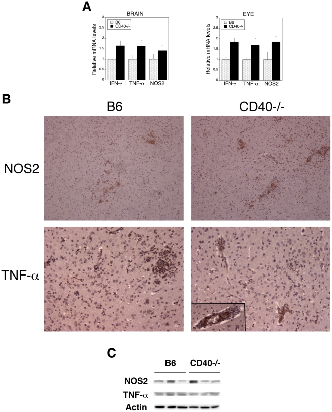
Next, we examined whether susceptibility to toxoplasmosis in CD40−/− mice could be explained by impaired expression of IFN-γ, TNF-α and NOS2. Compared to uninfected mice, T. gondii infected animals exhibited at least a 2-log increase in mRNA levels of these genes (not shown) and eyes and brains from both infected CD40−/− and infected B6 mice exhibited upregulation of mRNA of these genes at 2 and 4 weeks post-infection (Figure 2A and not shown). Immunohistochemical studies of the brain and immunoblot of brain lysates at 4 weeks post-infection revealed protein expression of NOS2 and TNF-α in both CD40−/− and control mice (Figure 2B and 2C). Thus, CD40−/− mice are susceptible to ocular and cerebral toxoplasmosis despite upregulation of key mediators of host protection: IFN-γ, TNF-α and NOS2.
Figure 2. T. gondii-infected CD40−/− mice exhibit upregulation of IFN-γ, TNF-α and NOS2 in the eye and brain.
B6 and CD40−/− mice were euthanized 4 weeks after infection with tissue cysts of the ME49 strain of T. gondii. A, Levels of IFN-γ, TNF-α and NOS2 mRNA in eyes and brains were examined using quantitative PCR and normalized against the levels of 18s rRNA. Each group contained 5 mice. Results are shown as the mean ± SEM. B, Brains were subjected to immunohistochemistry for NOS2 (X100) and TNF-α (X200). TNF-α was detected not only in the brain parenchyma but also within blood vessel walls in both B6 and CD40−/− mice (inset). C, Brains were lysed and subjected to immunoblot for NOS2, TNF-α and actin. Each lane represents one mouse. Results are representative of 3 to 4 independent experiments.
CD40 upregulates autophagy proteins and stimulates autophagy
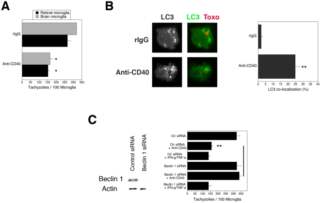
Microglia are considered central mediators of resistance against T. gondii in neural tissue [16]. Microglia were incubated with a stimulatory anti-CD40 or control mAb. Primary brain or retinal microglia treated with a stimulatory anti-CD40 mAb exhibited a significant lower parasite load than control microglia (p<0.02) (Figure 3A). Similar results were obtained with 2 mouse microglia cell lines (BV-2 and N9) or by treating cells with mouse CD154 (not shown). Anti-T. gondii activity induced by CD40 was not impaired by a neutralizing anti-IFN-γ mAb, or NMA, an inhibitor of NOS2 (p>0.5) (Figure S1A and S1B). In contrast, anti-IFN-γ mAb or NMA blocked the anti-T. gondii activity induced by IFN-γ/TNF-α (p<0.001). The autophagy pathway was required for CD40-induced anti-T. gondii activity in microglia because CD40 stimulation caused recruitment of the autophagy marker LC3 around the parasite (Figure 3B) and silencing of Beclin 1 using siRNA ablated CD40-induced anti-T. gondii activity (p<0.01) (Figure 3C). In contrast, silencing of Beclin 1 had no effect on IFN-γ/TNF-α induced anti-T. gondii activity. The role of the autophagy pathway was also studied in primary microglia and macrophages. Treatment of microglia with the autophagy inhibitor 3-methyl adenine (3-MA) inhibited toxoplasmacidal activity induced by CD40 stimulation (p<0.01) (Figure S2A). Silencing of Atg5 prevented CD40 stimulation from decreasing the parasite load in primary macrophages and microglia (p<0.01) (Figures S2B and S2C). Similarly, silencing of Atg5 in BV-2 cells ablated toxoplasmacidal activity induced by CD40 (p<0.01) (Figure S2D).
Figure 3. Autophagy mediates anti-T. gondii activity induced by CD40 in microglia.
A, Primary retinal and brain microglia obtained from B6 mice were incubated with a stimulatory anti-CD40 or control mAb, or with IFN-γ/TNF-α followed by infection with tachyzoites of RH T. gondii. The numbers of tachyzoites per 100 microglia were determined at 18 h post-infection. B, BV-2 cells were transfected with LC3-EGFP followed by incubation with anti-CD40 or control mAb. Cells were infected with RFP-T. gondii. Cells were examined at 5 h to determine the percentages of cells where LC3 accumulated around the parasites (arrow head). C, BV-2 cells were transfected with control siRNA or siRNA against Beclin 1. Protein expression of Beclin 1 and actin were examined by immunoblot. BV-2 cells transfected with control siRNA or siRNA against Beclin 1 were incubated with a stimulatory anti-CD40 mAb, control mAb or IFN-γ/TNF-α followed by RH T. gondii infection. Results are representative of 3 independent experiments. *≤0.05; **p≤0.01.
Atg5 mediates anti-T. gondii activity in IFN-γ-activated macrophages independent of autophagosome formation [13]. IFN-γ induces killing of type II strains of the parasite through Atg5-dependent recruitment of Irga6 to the membrane of the parasitophorous vacuole [13]. We further determined whether the mechanism of anti-microbial activity triggered by CD40 is distinct from that utilized by IFN-γ. Figure S3A shows that CD40 induced anti-microbial activity against not only a type I strain of T. gondii (RH) but also against a type II strain (ME49). Knockdown of Beclin 1 ablated the anti-T. gondii activity against a type II strain of the parasite induced by CD40 but had no effect on anti-T. gondii activity mediated by IFN-γ (Figure S3B). Importantly, knockdown of Irga6 did not impair CD40-induced killing of T. gondii while it inhibited the effect of IFN-γ (Figure S3C). Taken together, these studies indicate that CD40 induces entrapment of T. gondii by autophagosomes and triggers anti-T. gondii activity in retinal and brain microglia through the autophagy pathway, a process that is distinct from that utilized by IFN-γ since CD40 not only acts independently of this cytokine but also independently of Irga6 and nitric oxide. Moreover, CD40 but not IFN-γ requires Beclin 1, an autophagy protein that controls autophagosome formation [23].
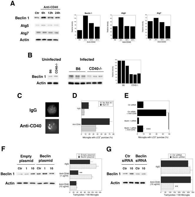
The level of Beclin 1 expression has been linked to autophagic activity [24], [25]. We examined whether CD40 modulates Beclin 1 expression. As shown in Figure 4A, CD40 stimulation caused a rapid increase in Beclin 1 protein expression in primary microglia (2.8-fold increase on average; n = 4). CD40 stimulation also caused an apparently less pronounced upregulation of the Atg5 expression (average 1.7-fold) but did not appear to affect expression of Atg7 (Figure 4A). We examined whether CD40 affects the in vivo levels of Beclin 1 during T. gondii infection. While we did not detect differences in Beclin 1 protein expression between brain microglia from uninfected B6 and CD40−/− mice, Beclin 1 expression in microglia/macrophages from T. gondii-infected B6 mice was higher than in microglia/macrophages from infected CD40−/− mice (Figure 4B).
Figure 4. CD40 upregulates autophagy protein expression and leads to increased autophagy.
A, Primary brain microglia from B6 mice were incubated with anti-CD40 mAb or control mAb. Beclin 1, Atg5 and Atg7 protein expression were normalized against actin. B, Beclin 1 and actin protein expression in purified brain microglia from uninfected B6 and CD40−/− mice or purified brain microglia/macrophages from B6 or CD40−/− mice at 4 weeks post-infection. Each lane represents a pool of 2 mice. C, D, BV-2 cells transfected with LC3-EGFP were incubated with anti-CD40 mAb or control mAb in the presence or absence of bafilomycin A1 (BafA1). E, BV-2 cells were transfected with control or Beclin 1 siRNA. After 2 days, cells were transfected with LC3-EGFP followed by incubation with anti-CD40 or control mAb. F, BV-2 cells were transfected with Beclin 1-encoding or empty plasmids. Expression of Beclin 1 and actin were examined by immunoblot. Cells were incubated with anti-CD40 mAb (1 or 10 µg/ml) or control mAb followed by infection with tachyzoites of RH T. gondii. G, BV-2 cells transfected with a suboptimal concentration of Beclin 1 siRNA or control were incubated with anti-CD40 mAb (10 µg/ml) or control mAb followed by RH T. gondii infection. Results are representative of 3 to 4 independent experiments. **p≤0.01; ***p≤0.001.
We determined if CD40 not only increased expression of autophagy protein but also enhanced basal levels of autophagy. BV-2 cells were transfected with LC3-EGFP and were incubated with or without anti-CD40 mAb. CD40 stimulation increased the percentage of microglia with LC3+ punctae (Figure 4C, D). An increase in these structures could be due to increased autophagosome formation or decreased degradation. Microglia were treated with bafilomycin A1, an inhibitor of vacuolar ATPase, which prevents autophagosome degradation [26]. CD40 stimulation still upregulated LC3+ punctae when autophagosome degradation was prevented. CD40 stimulated autophagy through Beclin 1 because knock-down of this autophagy protein inhibited upregulation of LC3+ punctae (Figure 4E) (p<0.001). Together, CD40 increases expression of Beclin 1 and Atg5 in microglia and enhances autophagy.
Next, we tested whether changes in the level of autophagy protein expression affect toxoplasmacidal activity induced by CD40. We centered on Beclin 1 given that levels of expression of this protein can affect autophagy. BV-2 cells were transfected with a Beclin 1-encoding plasmid or with empty plasmid (Figure 4F). A suboptimal concentration of anti-CD40 mAb (1 µg/ml) did not affect Beclin 1 expression and did not induce anti-T. gondii activity in BV-2 cells transfected with the empty plasmid. In contrast, Beclin 1-transfected BV-2 cells exhibited on average a 2.4-fold increase in Beclin 1 protein expression that was accompanied by anti-T. gondii activity even if stimulated with 1 µg/ml of anti-CD40 mAb (Figure 4F). Conversely, BV-2 cells transfected with a suboptimal concentration of Beclin 1 siRNA exhibited on average a 53% reduction in Beclin 1 protein levels and ablated CD40-induced anti-T. gondii activity despite treatment with a fully activating dose of anti-CD40 mAb (10 µg/ml) (Figure 4G). Thus, relatively modest changes in Beclin 1 expression modulate CD40-induced anti-T. gondii activity.
CD40 stimulation diminishes p21 expression leading to Beclin 1 upregulation
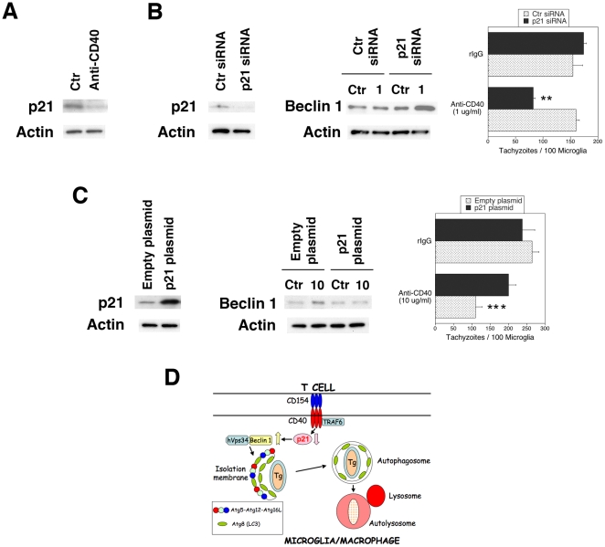
p21 has been reported to decrease Beclin 1 expression through protein degradation [27]. CD40 upregulates Beclin 1 without affecting Beclin 1 mRNA levels as assessed by real time PCR (Figure S4). Thus, we examined the role of p21 in Beclin 1 upregulation caused by CD40. CD40 stimulation of primary microglia and BV-2 cells caused a rapid decrease in p21 levels (Figure 5A). In addition, knockdown of p21 by transfection with siRNA induced Beclin 1 upregulation in BV-2 cells incubated with a sub-optimal concentration of anti-CD40 mAb (Figure 5B). In contrast, p21 overexpression by transfection with a p21-encoding plasmid prevented CD40 from increasing Beclin 1 levels (Figure 5C). The effects of p21 were of functional relevance because knockdown of p21 promoted anti-T. gondii activity in microglia stimulated with a suboptimal concentration of anti-CD40 mAb (Figure 5B) while overexpression of p21 inhibited anti-T. gondii activity induced by CD40 (Figure 5C) (p<0.004). These results indicate that p21 regulates Beclin 1 expression and autophagy protein-dependent killing of T. gondii in CD40-stimulated microglia (Figure 5D).
Figure 5. CD40 downregulates p21, increases Beclin 1 expression and autophagic killing of T. gondii.
A, Primary brain microglia from B6 mice were incubated with anti-CD40 mAb or control mAb for 1 h followed by assessment of p21 and actin expression. B, BV-2 cells were transfected with control or p21 siRNA. Cells were incubated with a suboptimal concentration of anti-CD40 (1 µg/ml) or control mAb and followed by assessment of Beclin 1 expression. In addition, cells were challenged with RH T. gondii and the numbers of tachyzoites per 100 microglia were determined. C, BV-2 cells were transfected with p21-encoding or empty plasmids. Cells were incubated with an optimal concentration of anti-CD40 (10 µg/ml) or control mAb and followed by assessment of Beclin 1 expression. Cells were challenged with RH T. gondii and the numbers of tachyzoites per 100 microglia were determined. Results are shown as the mean ± SEM and are representative of 3 independent experiments. D, Proposed model of the role of p21 in modulation of Beclin 1 expression and activation of T. gondii killing induced by CD40. We previously reported that CD40 induces autophagy and killing of T. gondii through the adaptor protein TRAF6 [6], [8]. This process would be dependent on down-regulation of p21 that would result in increased levels of Beclin 1 and enhanced autophagy. **p≤0.01; ***p≤0.001.
Autophagy-deficient mice are susceptible to ocular toxoplasmosis and toxoplasmic encephalitis
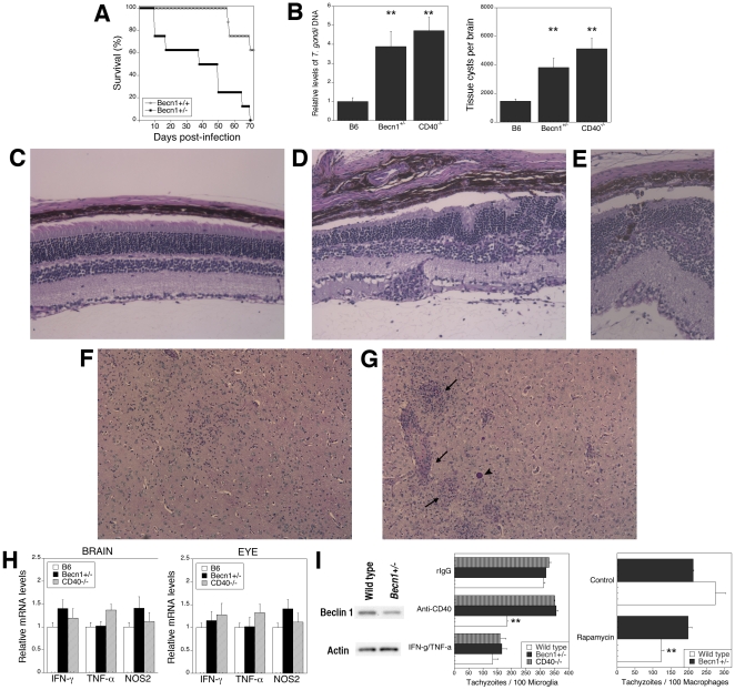
Our in vitro studies indicate that the levels of Beclin 1 modulate killing of T. gondii. Based on these findings, we hypothesized that mice with a partial defect in Beclin 1 would exhibit impaired killing of T. gondii and susceptibility to ocular and cerebral toxoplasmosis. We used mice with heterozygous deletion of Beclin 1 (Becn1 +/− mice) to begin to test the in vivo relevance of the autophagic pathway in protection against cerebral and ocular toxoplasmosis. Becn1 +/− mice exhibit diminished Beclin 1 protein levels and reduced autophagy in vivo [24]. Becn1 +/− mice infected with T. gondii exhibited decreased survival compared to littermate controls (Becn1+/+) (Figure 6A) (p<0.001). Moreover, infected Becn1 +/− mice exhibited higher parasite loads in the brain and eye (Figure 6B) (p<0.01). Similar to CD40−/− animals, Becn1 +/− mice exhibited more pronounced retinal histopathology (p<0.01) that included distortion of the retinal architecture, hypertrophy of retinal pigmented epithelial cells, retinal invasion by these cells and inflammatory infiltrates (Figure 6C–E). In the brain, Becn1 +/− mice had more extensive parenchymal inflammatory foci, perivascular cuffing as well as more numerous tissue cysts than control mice (Figure 6F–G) (p<0.01). This increased susceptibility to toxoplasmosis was not explained by diminished expression of IFN-γ, NOS2 and TNF-α in the brain and eye (Figure 6H). Mice with Atg5−/− hematopoietic cells exhibit defects in T cell homeostasis [28]. However, Becn1 +/− mice have a partial defect in Beclin 1 expression. Indeed, the phenotypic composition of lymphoid tissue in these mice as well as in CD40−/− mice was similar to that of control mice (Figure S5A). Moreover, these groups of mice exhibited similar percentages of T. gondii-reactive IFN-γ- or IL-4-producing CD4+ and CD8+ T cells as well as equivalent in vitro secretion of IFN-γ after stimulation with T. gondii lysate antigen (TLA) (Figure S5B and S5C). Next, we studied microglial anti-microbial activity. Microglia from Becn1 +/− mice had diminished Beclin 1 protein levels compared to control mice (Figure 6I). In marked contrast to the cytokine studies, microglia from Becn1 +/− mice had a defect in killing of T. gondii induced by CD40 but not in anti-T. gondii activity triggered by IFN-γ/TNF-α (Figure 6I). This defect occurred despite normal expression of CD40 on microglia from Becn1 +/− mice (cMFI: Control = 103±10; Becn1 +/− = 112±15). To further confirm that Becn1+/− mice exhibit defective autophagic killing of T. gondii, macrophages from controls and Becn1+/− mice were incubated with rapamycin, an agent that induces anti-T. gondii activity via autophagy [6]. Macrophages from Becn1+/− mice exhibited impaired killing induced by rapamycin (Figure 6I). Thus, in spite of upregulation of IFN-γ, TNF-α and NOS2, and the development of T cell-mediated immunity against T. gondii, mice with defective autophagy and with impaired autophagy protein-dependent killing of the parasite are susceptible to toxoplasmosis.
Figure 6. Autophagy-deficient mice are susceptible to cerebral and ocular toxoplasmosis.
Becn1 +/− and littermate control mice (B6) were infected with ME49 T. gondii. A, Survival was monitored daily. Data shown are from groups containing 8 mice. B, Mice (5 mice per group) were euthanized at 4 weeks post-infection. DNA was isolated from eyes and the levels of the T. gondii B1 gene were examined using real time quantitative PCR. Tissue cysts were counted in brain homogenates. C, Eyes from infected control mice revealed mild histopathology. D, E, Eyes from infected Becn1 +/− mice revealed disruption of retinal architecture, more prominent inflammation and invasion of the retina by retinal pigmented epithelial cells (E). PASH; X200. F, Brains from infected control mice showed slight inflammation. G, Brains from infected Becn1 +/− mice show prominent areas of inflammation (arrow) and frequent tissue cysts (arrow head). PASH X100. H, Levels of IFN-γ, TNF-α and NOS2 mRNA in eyes and brains were examined using real time quantitative PCR and normalized against the levels of 18s rRNA. Each group contained 5 mice. I, Beclin 1 and actin protein expression in primary microglia from control and Becn1 +/− mice. Primary brain microglia from control and Becn1 +/− mice and were incubated with anti-CD40 or control mAb, or with IFN-γ/TNF-α followed by infection with tachyzoites of RH T. gondii. Primary bone marrow derived macrophages were obtained from control and Becn1 +/− mice and were incubated with or without rapamycin (1 µM) 2 h after infection with tachyzoites of RH T. gondii. Results are shown as the mean ± SEM and are representative of 3 independent experiments. **p≤0.01.
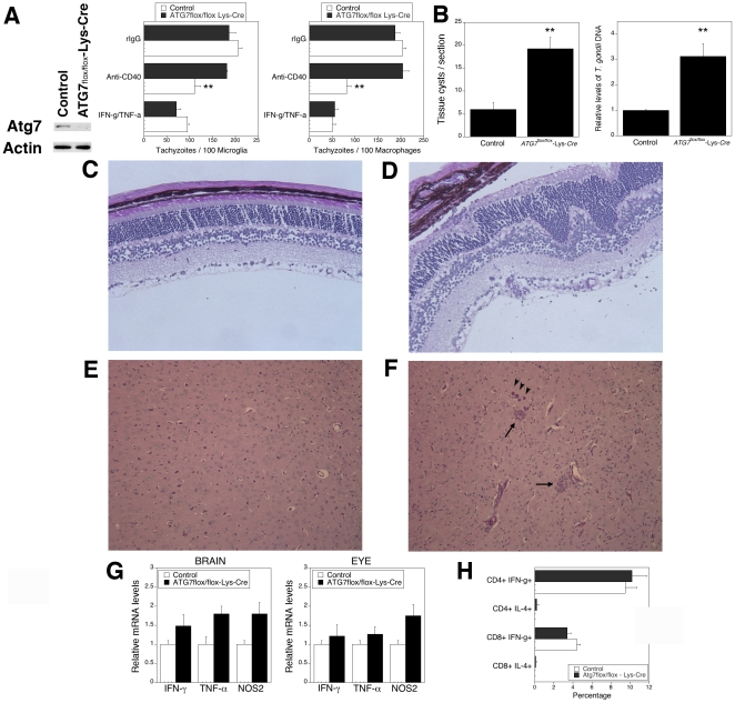
To further evaluate the role of autophagy for in vivo control of T. gondii in the brain and eye, we conducted experiments using mice where another autophagy gene, ATG7, was inactivated in macrophages/microglia. ATG7 was inactivated in myelomonocytic cells by breeding of ATG7flox/flox mice [29] with mice that express the Cre recombinase from endogenous lysozyme M locus (Lyz-Cre). Indeed, microglia and macrophages from ATG7flox/flox-Lyz-Cre mice had impaired expression of Atg7 (Figure 7A and not shown). Moreover, microglia as well as macrophages from these mice exhibited defective killing of T. gondii induced by CD40 but unimpaired parasite killing in response to IFN-γ/TNF-α (Figure 7A). Compared to control mice (ATG7 flox/flox), ATG7flox/flox-Lyz-Cre mice infected with T. gondii were unable to control the parasite in the brain and eye (Figure 7B) (p<0.01) and exhibited more prominent histopathology in these organs (Figure 7C–F) (p<0.01). Similar to the studies with Becn1+/− mice, ATG7flox/flox-Lyz-Cre mice were susceptible to toxoplasmosis despite upregulation of IFN-γ, TNF-α and NOS2 mRNA (Figure 7G) and the generation of T. gondii-specific T cell response (Figure 7H). Thus, mice with defective expression of Atg7 targeted to microglia/macrophages and impaired killing of T. gondii exhibit defective in vivo control of T. gondii in neural tissue.
Figure 7. Mice deficient in autophagy in macrophages/microglia are susceptible to toxoplasmosis.
A, Primary microglia and macrophages were obtained from control and ATG7flox/flox-Lyz-Cre mice. Atg7 and actin expression were assessed by immunoblot. Results shown are those obtained with microglia. Similar findings were observed in primary macrophages. Microglia and macrophages were incubated with a stimulatory anti-CD40 or control mAb, or with IFN-γ/TNF-α followed by infection with tachyzoites of RH T. gondii. B–F, ATG7flox/flox-Lyz-Cre and control mice were infected with ME49 T. gondii. Mice (4–5 mice per group) were euthanized at 3 weeks post-infection. B, ATG7flox/flox-Lyz-Cre mice exhibit higher parasite load in the eye and brain than control mice. Numbers of tissue cysts were determined in 100 high power fields. C, D, Compared to control mice (C), infected ATG7flox/flox-Lyz-Cre mice (D) revealed more prominent disruption of retinal architecture and inflammation. PASH; X200. E, F, Compared to control mice (E), infected ATG7flox/flox-Lyz-Cre mice (F) show more prominent areas of inflammation (arrow) and frequent tissue cysts (arrow head) in the brain. PASH X100. G, Eyes and brains were obtained at 3 weeks post-infection and used to isolate RNA. Levels of IFN-γ, TNF-α and NOS2 mRNA were examined using real time quantitative PCR and normalized against the levels of 18s rRNA. Each group contained 5 mice. H, Splenocytes were obtained from control and ATG7flox/flox-Lyz-Cre mice 3 weeks post-infection with ME49 T. gondii and were incubated with TLA as described in Materials and Methods. The percentages of CD3+ CD4+ or CD3+ CD8+ T cells that became IFN-γ+ or IL-4+ cells were determined by flow cytometry. Results are shown as the mean ± SEM and are representative of 3 independent experiments. **p≤0.01.
Discussion
The cascades by which adaptive immunity activates autophagic killing of an intracellular pathogen and the in vivo role of autophagy in protection conferred by adaptive immunity are incompletely understood. We report that CD40 rapidly diminishes the levels of p21, a protein that degrades Beclin 1 [27]. As a result, CD40 causes upregulation of this autophagy protein and stimulates killing of T. gondii in microglia dependent on the autophagy pathway. Moreover, mice with diminished expression of Beclin 1 and mice with inactivation of Atg7 in microglia/macrophages are susceptible to cerebral and ocular toxoplasmosis despite upregulation of IFN-γ, TNF-α and NOS2 as well as the induction of T cell-mediated immunity. Susceptibility to the chronic phase of T. gondii infection in these mice occurred in the setting of defective autophagy pathway-dependent killing of the parasite while IFN-γ-induced killing of the parasite was unimpaired in these mice. These results contrast the report of autophagosome-independent in vivo protection conferred by Atg5, where Atg5 expression in phagocytic cells was required for IFN-γ-mediated killing of the parasite [13]. The findings reported herein strongly support the notion that adaptive immunity utilizes autophagy to confer resistance against an intracellular pathogen because: i) CD40 induced entrapment of the parasite by an LC3+ structure; ii) Beclin 1, Atg5 and Atg7 were required for killing of T. gondii within CD40-activated microglia and macrophages; iii) Deficiency of two autophagy proteins, Beclin 1 and Atg7, led to susceptibility to toxoplasmosis while leaving unaffected IFN-γ-induced anti-parasite activity and the induction of an IFN-γ-mediated immune response.
One study reported that autophagosomes surround T. gondii in primed macrophages [7], [13]. This event followed the disruption of the parasitophorous vacuole membrane raising the possibility that formation of autophagosomes was a response to the presence of disrupted structures within host cells [7], [13]. However, recent work in macrophages did not find evidence of autophagosome formation around the parasite in IFN-γ-activated cells [13]. In addition, studies in astrocytes including work using live-cell imaging revealed that IFN-γ induced killing of T. gondii without localization of LC3 to the parasitophorous vacuole [30], [31]. Interestingly, while Atg5 is required in killing of the parasite in IFN-γ-activated cells, this effect occurs in association with recruitment of IRG to the parasitophorous vacuole membrane rather than autophagic entrapment of the parasite [13], [32]. IRG disrupt the parasitophorous vacuole membrane indicating that the anti-T. gondii effect of Atg5 in IFN-γ-activated cells is likely dependent on IRG loading onto the parasitophorous vacuole rather that classical autophagy [13]. Our work contrasts to studies in IFN-γ-activated cells. In marked distinction to IFN-γ, CD40 induces toxoplasmacidal activity independently of Irga6 as well as Irgm1 and Irgm3 [33]. Importantly, deficiency of Beclin 1 or Atg7 blocked CD40-induced killing of T. gondii while leaving unaffected killing induced by IFN-γ. The different mechanisms used by CD40 and IFN-γ to kill T. gondii likely explain the cooperation observed between CD40 and IFN-γ to promote control of the parasite both in vitro [34], [35] as well as in vivo (this study). Indeed, while IFN-γ, TNF-α and NOS2 are key for resistance against chronic toxoplasmosis, we report that the expression of these mediators does not appear sufficient to prevent ocular and cerebral toxoplasmosis since CD40 as well as the autophagy proteins Beclin 1 and Atg7 are required for protection against the disease.
The autophagic machinery can enhance presentation of certain antigens [14], [36] and can promote the generation of a Th1 response [14], [37]. Our studies indicate that a T cell response against T. gondii can develop in mice with defective autophagy supporting the existence of another mechanism by which autophagy promotes resistance against T. gondii. Indeed, the studies with ATG7flox/flox-Lyz-Cre mice provide an in vivo correlate to our in vitro studies on autophagic killing of T. gondii in microglia/macrophages and indicate that autophagy in those cells is likely required for resistance against toxoplasmosis in neural tissue.
Beclin 1 plays a central role in autophagy by promoting the formation of autophagosomes [23]. Our studies identified CD40 as a regulator of Beclin 1 since engagement of CD40 resulted in a rapid increase in Beclin 1 expression and enhanced autophagy. Changes in Beclin 1 levels were functionally relevant because a moderate increase in Beclin 1 expression markedly facilitated CD40-induced killing of T. gondii while a moderate decrease in Beclin 1 expression profoundly inhibited this response. Moreover, T. gondii-infected CD40 deficient mice had lower Beclin 1 levels in their brain microglia/macrophages and Beclin 1-deficient mice were susceptible to cerebral and ocular toxoplasmosis. Studies in cancer and neurodegeneration also support the relevance of Beclin 1 levels. Various cancer cells exhibit decreased Beclin 1 levels and the anti-cancer agent tamoxifen increases expression of this protein [25], [38]. Brain injury appears to upregulate Beclin 1 [39]. In contrast, Beclin 1 is deficient in Alzheimer's dementia and this deficiency impairs neuronal autophagy promoting neurodegeneration [40]. In this model, lentiviral-induced overexpression of Beclin 1 reduces amyloid β pathology [40]. Thus, upregulation of Beclin 1 may drive not only host protection in the CNS but may also activate autophagy as a homeostatic mechanism to reduce cellular damage.
Beclin 1 is not only targeted by CD40-dependent adaptive immunity to stimulate autophagy but can also be targeted by a pathogen to impair autophagy. For example, neurovirulent HSV-1 and HIV-1 encode an ICP34.5 protein and Nef respectively that bind Beclin 1 and block autophagy [11], [41]. The influenza M2 protein blocks autophagosome maturation, an effect that may be mediated through interference with Beclin 1 [42]. Together, these studies and our work indicate that Beclin 1 is an important regulator of autophagy during host-pathogen interaction.
Our results indicate that p21 acts as a link between CD40 and Beclin 1 upregulation and stimulation of autophagy. p21 is not only an inhibitor of cyclin-dependent kinases but it also diminishes expression of Beclin 1 and Atg5 [27]. C2-ceramide inhibits autophagy in mouse embryonal fibroblasts because it triggers p21-dependent protein degradation of Beclin 1 and Atg5 [27]. Prior studies addressed the effects of CD40 on p21 expression in B cells. CD40 down-regulates p21 in WEHI 231 immature B lymphoma cells treated with anti-IgM antibody [43]. The discordant results on the effects of CD40 on p21 expression reported in other studies may be explained by differences in the stages of B cell development [44]. We report that CD40 stimulation rapidly reduced p21 protein expression in microglia. Moreover, knockdown of p21 in microglia promoted upregulation of Beclin 1 and autophagic killing of T. gondii in response to CD40 stimulation while overexpression of p21 impaired the ability of CD40 to upregulate Beclin 1 and kill T. gondii.
Mouse models of T. gondii infection are used to study immunity against the parasite. However, these models do not fully mimic the immune response in humans. While IFN-γ is indispensable to control the parasite in mice, mechanisms of resistance against T. gondii that do not rely on IFN-γ are likely more effective in humans. Children with an autosomal dominant defect in IFN-γR1 that causes a deletion in the STAT1 binding site do not develop disease when infected with T. gondii [45]. In contrast, STAT1−/− mice die within 1 week after infection with the parasite [46], [47]. The differences between the immune response in humans and mice also appear to apply to the downstream effectors of IFN-γ, NOS2 and IRG. NOS2 is more tightly regulated in humans than in rodents and the production of nitric oxide appears to be weaker in humans. Although IRGM has been reported to mediate anti-bacterial activity in human macrophages [48], the role of IRG is likely more restricted in humans than in mice. Mice have 23 IRG genes among which Irgm1, Irgm3 and Irga6 are important mediators of anti-T. gondii activity in mouse cells [30], [49], whereas IRG in humans have been reduced to IRGM and IRGC [50]. CD40 induces killing of T. gondii independently of IFN-γ, STAT1, NOS2, Irgm1, Irgm3 and Irga6 [6], [8], [33], [51]. CD40-induced killing of T. gondii may be an important contributor to control of T. gondii in humans. Defects in the CD40 pathway are relevant to at least 3 groups of patients that develop ocular and/or cerebral toxoplasmosis: patients with X-linked Hyper IgM syndrome who lack functional CD154 [52], newborns since neonatal CD4+ T cells exhibit impaired expression of CD154 [53], [54] and CD40 levels are reduced on neonatal dendritic cells [54], and HIV-1+ patients because they exhibit a defect in CD154 induction in their CD4+ T cells [55], [56].
In conclusion, our studies revealed that immunity can stimulate autophagy by upregulating autophagy proteins in a p21-dependent manner, and the autophagic machinery was required for resistance against cerebral and ocular toxoplasmosis despite the induction of T. gondii-reactive T cells, IFN-γ, TNF-α and NOS2. These results shed new light on the spectrum of mechanisms of host protection. Future studies should explore the possibility of modulation of Beclin 1, autophagy and/or CD40 signaling as an approach to improve pathogen eradication.
Materials and Methods
Ethics statement
The animal experimentation guidelines of the US Department of Health and Human Services were followed. The study was approved by the Institutional Animal Care and Use Committee of Case Western Reserve University School of Medicine.
Animals and parasites
C57BL/6, CD40−/− and mice expressing the Cre recombinase from within the lysozyme M locus (Lyz-Cre mice) were purchased from Jackson Laboratories and bred at the Animal Resource Center (Case Western Reserve University). Becn1+/− and ATG7 flox/flox mice have been described [24], [29]. ATG7 flox/flox mice were bred with Lyz-Cre mice. All mice were female, on a B6 background and were 8–12 week old when used for the studies. Each experimental group consisted of 4–8 mice. Mice were infected i.p. with 10 cysts of ME49 strain of T. gondii (gift from Dr. George Yap, Brown University). Tachyzoites of the RH, P (gift from Dr. Louis Weiss, Albert Einstein College of Medicine) and PTG strains of T. gondii as well as parasites expressing cytoplasmic DsRed (RFP) were maintained as described [6].
Histopathology and Immunohistochemistry
Unless otherwise stated, mice were anesthetized and perfused with PBS 4 weeks after infection. Four 5 µm sections at different areas of the brains and eyes were stained with periodic acid Schiff hematoxylin (PASH) stain and used to score histopathologic changes using previously described criteria [16], [17]. Tissue cysts were counted in brain homogenates or in tissue sections as previously described [19]. Sections were incubated with anti-NOS2 or anti-TNF-α Ab (Millipore and Abcam respectively) followed by incubation with secondary antibody conjugated to biotin (Jackson ImmunoResearch Laboratories). Sections were resolved using a Vectastain ABC kit (Vector Laboratories).
Real-time quantitative PCR
RNA was obtained from eyes and brains using the RNeasy kit (QIAGEN). After treatment with DNase (Ambion), 0.5 µg of RNA was reverse transcribed to cDNA with Super-Script III reverse transcriptase (Invitrogen) and oligo(dT)12–18 primers. cDNA (2.5 µl) was used as template for RT-PCR using SYBR GREEN PCR Master Mix (Applied Biosystems) and 20 pM (each) primers in 50 ml. Gene expression was assessed using a 7300 Real Time PCR System (Applied Biosystems). Each cDNA sample was run in duplicate. Samples were normalized according to the content of 18S rRNA [55]. Expression of the T. gondii B1 gene was assessed by real-time quantitative PCR. Genomic DNA was isolated from eyes using a DNeasy kit (QIAGEN) and subjected to real-time PCR using SYBR GREEN PCR Master Mix. Each sample was run in duplicate and normalized against L32. Primer sequences for IFN-γ [57], TNF-α [57], NOS2 [58], 18S rRNA [55] and T. gondii B1 gene [59] were previously described. Primer sequences for Beclin 1 were forward: 5′-GGCCAATAAGATGGGTCTGA-3′, reverse: 5′-GCTGCACACAGTCCA GAAAA– 3′. Primers for the T. gondii B1 gene have been previously described [59]. Primers for L32 were: forward 3′-TGTGCAACAAATCTTCACCGTGC-5′;
reverse 3′-GGATTGGTGACTCTGATGGCC-5′.
Microglia and in vitro infection with T. gondii
Primary brain and retinal microglia as well as mouse brain microglial cell lines BV-2 and N9 (gifts from Drs. Kalipada Pahan, Univ. of Nebraska and Dr. Jun Tan, Univ. of South Florida respectively) were treated overnight with a stimulatory anti-CD40 mAb (1C10; 1 or 10 µg/ml), isotype control mAb or IFN-γ (100 U/ml; PeproTech) plus TNF-α (250 pg/ml; PeproTech) [51]. Tachyzoites of the RH or ME49 strains of T. gondii were used to infect monolayers as described [51]. When indicated, microglia were incubated with N G-monomethyl-L-arginine (NMA, 100 µM; Calbiochem) or a neutralizing mAb against mouse IFN-γ (XMG 1.2; 10 µg/ml; eBiosciences) in the presence of anti-CD40 mAb, control mAb or IFN-γ/TNF-α. 3-methyl adenine (3-MA; 10 mM; Sigma-Aldrich) was added to monolayers 1 hour after infection. Parasite replication was assessed by light microscopy [51]. To isolate brain mononuclear cells from infected mice, brains were digested with papain (15 IU/ml)/DNAse (15 µg/ml; Worthington Biochemicals) for 30 min at 37°C. Tissue was dissociated and cells were loaded onto a gradient made with 30% and 60% isotonic Percoll. Gradients were centrifuged at 1000× g for 30 min. Cells present in the interphase were collected and washed. Microglia/macrophages were purified using anti-CD11b-coated magnetic beads (Miltenyi Biotec). This resulted in populations that were >95% F4/80+.
Lentiviral vectors
We constructed an shRNA against mouse Atg5 using the following oligo pair (sense and antisense strands are underlined while the loop is italicized): forward 5′-TGGATGAGATAACTGAAAGA TTCAAGAGA TCTTTCAGTTATCTCATCCTTTTTTC-3′; reverse 5′-TCGAGAAAAAAGGATGA GATAACTGAAAGA TCTCTTGAA TCTTTCAGTTATCTCATCCA-3′. An XhoI restriction site was included at the 5′-end of the reverse oligo to facilitate cloning. An shRNA against luciferase was used as control [60]. Oligo pairs were annealed and subcloned into the polylinker region of the pLL3.7 vector [61] (American Type Tissue Collection) followed by sequence verification. To generate lentivirus, we co-transfected pLL3.7 containing shRNA and packaging vectors VSV-G, RSV-REV, pMDL-g/p RRE into 293T cells. Supernatants were collected at 24 and 48 h, passed through a 0.45 µm filter, concentrated by ultracentrifugation and stored at −80°C. Cells were transduced by incubation with lentiviral particles at a MOI of 10∶1 in the presence of polybrene (8 µg/ml). EGFP+ cells were sorted by FACS at 4 days post-transduction. Efficiency of gene silencing was determined by immunoblot.
Transfection of microglia and immunofluorescence
Using an Amaxa nucleofector as previously described [6], BV-2 cells were transfected with either control siRNA, Beclin 1 siRNA [62], p21 siRNA [27], Irga6 siRNA [63] (all from Dharmacon), empty vector, or plasmid that encode Beclin 1 [64], p21 [65] or LC3-EGFP (gift from Tamotsu Yoshimori, National Institute for Basic Biology, Okazaki, Japan). The numbers of LC3+ punctae were determined using a Leica DMI 6000 B automated microscope equipped for epifluorescence microscopy. Distinct structures that measure at least 1 µm in diameter were identified as LC3 punctae. Experimental groups had triplicate samples and at least 50 cells per sample were counted. Co-localization of LC3-EGFP around RFP-T. gondii was assessed as described [6].
Immunoblot
Membranes were probed with either antibody to Atg5 (ProteinTech Group Inc), Atg7 (Cell Signaling), Beclin 1 (BD Biosciences), p21 (Santa Cruz Biotechnologies), NOS2 (BD Biosciences), TNF-α (Abcam Inc.), Irga6 (Santa Cruz Biotechnologies) or actin (Santa Cruz Biotechnologies), followed by incubation with secondary Ab conjugated to horseradish peroxidase (Santa Cruz Biotechnologies). Bands were developed using enhanced chemiluminescence. In experiments that assessed the effect of CD40 stimulation on Beclin 1, Atg5, Atg7 and p21 expression, microglia were pre-incubated with IFN-γ for 18 h to increase the percentage of CD40+ cells. After extensive washing, microglia were incubated with anti-CD40 or control mAb. Pre-incubation with IFN-γ was never used in studies of CD40-dependent anti-microbial activity.
Flow cytometry
Cells were stained with anti-CD3, anti-CD4, anti-CD8, anti-CD11b, anti-CD40, anti-CD45R, anti-CD49d (DX5), anti-Ly6G (Gr-1), anti-F4/80 or isotype control mAbs (all from eBiosciences). For detection of intracellular cytokines, splenocytes were incubated with T. gondii lysate antigen (TLA; 10 µg/ml) for 48 and 72 h. PMA (1 ng/ml; Sigma Chemical), ionomycin (1 µg/ml; Sigma Chemical) and brefeldin A (10 µg/ml; eBiosciences) were added during the last 5 h of incubation. Cells were permeabilized using IntraPrep permeabilization reagent (Coulter-Immunotech) and stained with anti-IFN-γ or anti-IL-4 mAb (eBiosciences). Cells were analyzed using an LSR II (BD Biosciences).
Cytokine assays
CD4+ T cells were purified from the spleens of infected animals using magnetic beads (Miltenyi). CD4+ T cells (1×10⁶/ml) and were incubated with autologous bone marrow derived macrophages (2.5×10⁵/ml) plus either TLA (10 µg/ml) or concanavalin A (5 µg/ml; Sigma Chemical). Supernatants were collected at 48 h. Concentrations of IFN-γ in tissue culture supernatants were determined by enzyme-linked immunosorbent assay (Endogen).
Statistical analysis
Statistical significance was assessed by Student's t test and ANOVA. Mortality curves and histopathologic changes were analyzed using Mann-Whitney U test. Differences were considered statistically significant when P was <0.05.
Supporting Information
CD40 induces anti-T. gondii activity in microglia independently of IFN-γ and nitric oxide. A, B, Primary brain microglia were incubated with a stimulatory anti-CD40 or control mAb, or with IFN-γ/TNF-α as indicated followed by infection with tachyzoites of RH T. gondii. Microglia were also treated with a neutralizing anti-IFN-γ mAb (A) or NMA (B) prior to addition of anti-CD40 mAb or IFN-γ/TNF-α. The numbers of tachyzoites per 100 microglia were determined microscopically at 18 h post-infection. Results are shown as the mean + SEM and are representative of 3 independent experiments. **p<0.01.
(0.11 MB TIF)
Autophagy mediates anti-T. gondii activity induced by CD40 in microglia. A, Primary brain microglia from B6 mice were incubated with a stimulatory anti-CD40 or control mAb, or with IFN-γ/TNF-α as indicated. 3-MA or vehicle were added 1 hour after challenge with RH T. gondii. The numbers of tachyzoites per 100 microglia were determined microscopically at 18 hours post-infection. B–D, Knockdown of Atg5 abrogates anti-T. gondii activity induced by CD40 stimulation. Primary bone marrow-derived macrophages (B), primary brain microglia (C) or BV-2 cells (D) were transduced with EGFP-encoding lentiviral vectors that contained either shRNA against Atg5 or control shRNA. EGFP+ cells were sorted 4 days after transduction. Protein expression of Atg5 and actin were examined by immunoblot. Cells were then incubated with a stimulatory anti-CD40 mAb, control mAb or IFN-γ/TNF-α followed by infection with RH T. gondii. The numbers of tachyzoites per 100 microglia/macrophages were determined microscopically at 18 h post-infection. Results are shown as the mean + SEM and are representative of 3 independent experiments. **p<0.01.
(0.17 MB TIF)
CD40 induces anti-microbial activity against not only type I but also type II strains of T. gondii, and this effect is independent of Irga6. A, BV-2 cells were incubated with a stimulatory anti-CD40 or control mAb followed by challenge with tachyzoites of the RH strain of T. gondii (type I strain) or the P strain (ME49 clone) of the parasite (type II strain). The numbers of tachyzoites per 100 microglia were determined microscopically at 18 hours post-infection. B, BV-2 cells were transfected with control siRNA or siRNA against Beclin 1. BV-2 cells transfected with control siRNA or siRNA against Beclin 1 were incubated with a stimulatory anti-CD40 mAb, control mAb or IFN-γ/TNF-α followed by infection with a type II strain of T. gondii (P strain). C, BV-2 cells were transfected with control or Irga6 siRNA. Protein expression of Irga6 and actin were examined by immunoblot. Cells were then incubated with a stimulatory anti-CD40 mAb, control mAb or IFN-γ/TNF-α followed by infection with a type II strain of T. gondii (P strain). Results are shown as the mean + SEM and are representative of 3 independent experiments. **p<0.01.
(0.15 MB TIF)
CD40 does not affect Beclin 1 mRNA levels. A, Primary brain microglia from B6 mice were incubated with a stimulatory anti-CD40 mAb or control mAb. Serial dilutions of cDNA were used to examine levels of Beclin 1 mRNA by real-time quantitative PCR, which were normalized against the levels of 18s rRNA. B, Four weeks after infection with ME49 T. gondii, microglia/macrophages were purified from brains of B6 or CD40−/− mice. Beclin 1 mRNA levels were examined by real-time PCR. Each bar represents a single mouse. Results are shown as the mean + SEM and are representative of 3 independent experiments.
(0.09 MB TIF)
Becn1+/− mice do not have abnormal lymphoid organ phenotypic composition or defective induction of T. gondii-specific T cell response. A, Splenocytes were obtained from uninfected control, Becn1+/− and CD40−/− mice. Expression of CD4+ T cells (CD3+ CD4+), CD8+ T cells (CD3+ CD8+), B cells (CD45R+), NK cells (CD49d+), and granulocytes (Ly6-G+) were examined by flow cytometry. B, Splenocytes were obtained from control, Becn1+/− and CD40−/− mice 4 weeks post-infection with ME49 T. gondii and were incubated with TLA as described in Materials and Methods. The percentages of CD3+ CD4+ or CD3+ CD8+ T cells that became IFN-γ+ or IL-4+ cells were determined by flow cytometry. C, Purified CD4+ T cells were obtained 4 weeks post-infection with ME49 T. gondii and were stimulated with macrophages plus TLA or Concanavalin A (Con A). IFN-γ in supernatants was measured by ELISA. Results are shown as the mean + SEM and are representative of 3 independent experiments.
(0.12 MB TIF)
Acknowledgments
We thank Drs. Tamotsu Yoshimori, George Yap, Louis Weiss, Kalipada Pahan and Jun Tan for providing reagents, parasites and cell lines.
Footnotes
Competing Interests: The authors have declared that no competing interests exist.
Funding: This work was supported by National Institutes of Health (NIH) grants EY018341 (C.S.S) and AI48406 (C.S.S), the American Heart Association Ohio Valley Affiliate (C.S.S), NIH grant P30 EY11373, and Case/University Hospitals of Cleveland (UHC) Center for AIDS Research NIH grant AI36219. J-A. P. is a recipient of a post-doctoral fellowship from the Ohio Lions Eye Research Foundation. J.V.G. is a recipient of a pre-doctoral fellowship from Prevent Blindness Ohio. J.V.G. was supported by T32 EY007157-07. The funders had no role in the study design, data collection and analysis, decision to publish, or preparation of the manuscript.
References
- 1.Levine B, Klionsky DJ. Development by self-digestion: molecular mechanisms and biological functions of autophagy. Dev Cell. 2004;6:463–477. doi: 10.1016/s1534-5807(04)00099-1. [DOI] [PubMed] [Google Scholar]
- 2.Mizushima N, Ohsumi Y, Yoshimori T. Autophagosome formation in mammalian cells. Cell Struct Funct. 2002;27:421–429. doi: 10.1247/csf.27.421. [DOI] [PubMed] [Google Scholar]
- 3.Gutierrez MG, Master SS, Singh SB, Taylor GA, Colombo MC, et al. Autophagy is defense mechanism inhibiting BCG and Mycobacterium tuberculosis survival in infected macrophages. Cell. 2004;119:753–766. doi: 10.1016/j.cell.2004.11.038. [DOI] [PubMed] [Google Scholar]
- 4.Nakagawa I, Amano A, Mizushima N, Yamamoto A, Yamaguchi H, et al. Autophagy defends cells against invading Group A Streptococcus. Science. 2004;306:1037–1040. doi: 10.1126/science.1103966. [DOI] [PubMed] [Google Scholar]
- 5.Ogawa M, Yoshimori T, Suzuki T, Sagara H, Mizushima N, et al. Escape of intracellular Shigella from autophagy. Science. 2004;307:727–731. doi: 10.1126/science.1106036. [DOI] [PubMed] [Google Scholar]
- 6.Andrade RM, Wessendarp M, Gubbels MJ, Striepen B, Subauste CS. CD40 induces macrophage anti-Toxoplasma gondii activity by triggering autophagy-dependent fusion of pathogen-containing vacuoles and lysosomes. J Clin Invest. 2006;116:2366–2377. doi: 10.1172/JCI28796. [DOI] [PMC free article] [PubMed] [Google Scholar]
- 7.Ling YM, Shaw MH, Ayala C, Coppens I, Taylor GA, et al. Vacuolar and plasma membrane stripping and autophagic elimination of Toxoplasma gondii in primed effector macrophages. J Exp Med. 2006;203:2063–2071. doi: 10.1084/jem.20061318. [DOI] [PMC free article] [PubMed] [Google Scholar]
- 8.Subauste CS, Andrade RM, Wessendarp M. CD40-TRAF6 and autophagy-dependent anti-microbial activity in macrophages. Autophagy. 2007;3:245–248. doi: 10.4161/auto.3717. [DOI] [PubMed] [Google Scholar]
- 9.Yano T, Mita S, Ohmori H, Oshima Y, Fujimoto Y, et al. Autophagic control of listeria through intracellular innate immune recognition in drosophila. Nature Immunology. 2008;9:908–916. doi: 10.1038/ni.1634. [DOI] [PMC free article] [PubMed] [Google Scholar]
- 10.Shelly S, Lukinova N, Bambina S, Berman A, Cherry S. Autophagy is an essential component of Drosophila immunity against vesicular stomatitis virus. Immunity. 2009;30:588–598. doi: 10.1016/j.immuni.2009.02.009. [DOI] [PMC free article] [PubMed] [Google Scholar]
- 11.Orvedahl A, Alexander D, Talloczy Z, Sun Q, Wei Y, et al. HSV-1 ICP34.5 confers neurovirulence by targeting the Beclin 1 autophagy protein. Cell Host & Microbe. 2007;1:23–35. doi: 10.1016/j.chom.2006.12.001. [DOI] [PubMed] [Google Scholar]
- 12.Jia K, Thomas C, Akbar M, Sun Q, Adams-Huet B, et al. Autophagy genes protect against Salmonella typhimurium and mediate insulin signaling-regulated pathogen resistance. Proc Natl Acad Sci USA. 2009;106:14564–14569. doi: 10.1073/pnas.0813319106. [DOI] [PMC free article] [PubMed] [Google Scholar]
- 13.Zhao Z, Fux B, Goodwin M, Dunay IR, Strong D, et al. Autophagosome-independent essential function for the autophagy protein Atg5 in cellular immunity to intracellular pathogens. Cell Host Microbe. 2008;4:458–469. doi: 10.1016/j.chom.2008.10.003. [DOI] [PMC free article] [PubMed] [Google Scholar]
- 14.Lee HK, Matthei LM, Steinberg BE, Alberts P, Lee YH, et al. In vivo requirement for Atg5 in antigen presentation by dendritic cells. Immunity. 2010;32:227–239. doi: 10.1016/j.immuni.2009.12.006. [DOI] [PMC free article] [PubMed] [Google Scholar]
- 15.Suzuki Y, Conley FK, Remington JS. Importance of endogenous IFN-γ for prevention of toxoplasmic encephalitis in mice. J Immunol. 1989;143:2045–2050. [PubMed] [Google Scholar]
- 16.Gazzinelli RT, Eltoum I, Wynn TA, Sher A. Acute cerebral toxoplasmosis is induced by in vivo neutralization of TNF-α and correlates with the down-regulated expression of inducible nitric oxide synthase and other markers of macrophage activation. J Immunol. 1993;151:3672–3681. [PubMed] [Google Scholar]
- 17.Gazzinelli RT, Brezin A, Li Q, Nussenblatt RB, Chan CC. Toxoplasma gondii: acquired ocular toxoplasmosis in the murine model, protective role of TNF-α and IFN-γ. Exp Parasitol. 1994;78:217–229. doi: 10.1006/expr.1994.1022. [DOI] [PubMed] [Google Scholar]
- 18.Yap GS, Scharton-Kersten T, Charest H, Sher A. Decreased resistance of TNF receptor p55- and p75-deficient mice to chronic toxoplasmosis despite normal activation of inducible nitric oxide synthase in vivo. J Immunol. 1998;160:1340–1345. [PubMed] [Google Scholar]
- 19.Deckert-Schluter M, Bluethmann H, Rang A, Hof H, Schluter D. Crucial role of TNF receptor type 1 (p55), but not of TNF receptor type 2 (p75), in murine toxoplasmosis. J Immunol. 1998;160:3427–3436. [PubMed] [Google Scholar]
- 20.Scharton-Kersten T, Yap G, Magram J, Sher A. Inducible nitric oxide is essential for host control of persistent but not acute infection with the intracellular pathogen Toxoplasma gondii. J Exp Med. 1997;185:1261–1273. doi: 10.1084/jem.185.7.1261. [DOI] [PMC free article] [PubMed] [Google Scholar]
- 21.Reichmann G, Walker W, Villegas EN, Craig L, Cai G, et al. The CD40/CD40 ligand interaction is required for resistance to toxoplasmic encephalitis. Infect Immun. 2000;68:1312–1318. doi: 10.1128/iai.68.3.1312-1318.2000. [DOI] [PMC free article] [PubMed] [Google Scholar]
- 22.Lazarevic V, Myers AJ, Scanga CA, Flynn JL. CD40, but not CD40L, is required for optimal priming of T cells and control of aerosol M. tuberculosis infection. Immunity. 2003;19:823–835. doi: 10.1016/s1074-7613(03)00324-8. [DOI] [PubMed] [Google Scholar]
- 23.Kihara A, Kabeya Y, Ohsumi Y, Yoshimori T. Beclin-phosphatidylinositol 3-kinase complex functions at the trans-Golgi network. EMBO Rep. 2001;21:330–335. doi: 10.1093/embo-reports/kve061. [DOI] [PMC free article] [PubMed] [Google Scholar]
- 24.Qu X, Yu J, Bhagat G, Furuya N, Hibshoosh H, et al. Promotion of tumorigenesis by heterozygous disruption of the beclin 1 autophagy gene. J Clin Invest. 2003;112:1809–1820. doi: 10.1172/JCI20039. [DOI] [PMC free article] [PubMed] [Google Scholar]
- 25.Liang XH, Jackson S, Seaman M, Brown KD, Kempkes B, et al. Induction of autophagy and inhibition of tumorigenesis by beclin 1. Nature. 1999;402:672–676. doi: 10.1038/45257. [DOI] [PubMed] [Google Scholar]
- 26.Yamamoto A, Tagawa Y, Yoshimori T, Moriyama Y, Masaki R, et al. Bafilomycin A1 prevents maturation of autophagic vacuoles by inhibiting fusion between autophagosomes and lysosomes in rat hepatoma cell line, H-4-II-E cells. Cell Struct Funct. 1998;23:33–42. doi: 10.1247/csf.23.33. [DOI] [PubMed] [Google Scholar]
- 27.Fujiwara K, Daido S, Yamamoto A, Kobayashi R, Yokoyama T, et al. Pivotal role of the cyclin-dependent kinase inhibitor p21WAF1/CIP1 in apoptosis and autophagy. J Biol Chem. 2008;283:388–397. doi: 10.1074/jbc.M611043200. [DOI] [PubMed] [Google Scholar]
- 28.Li C, Capan E, Zhao Y, Zhao J, Stolz D, et al. Autophagy is induced in CD4+ T cells and important for the growth factor-withdrawal cell death. J Immunol. 2006;177:5163–5168. doi: 10.4049/jimmunol.177.8.5163. [DOI] [PubMed] [Google Scholar]
- 29.Komatsu M, Waguri S, Ueno T, Iwata J, Murata S, et al. Impairment of starvation-induced and constitutive autophagy in Atg7-deficient mice. J Cell Biol. 2005;169:425–434. doi: 10.1083/jcb.200412022. [DOI] [PMC free article] [PubMed] [Google Scholar]
- 30.Martens S, Parvanova I, Zerrahn J, Griffiths G, Schell G, et al. Disruption of Toxoplasma gondii parasitophorous vacuoles by the mouse p47-resistance GTPases. PLoS Pathogens. 2005;1:187–201. doi: 10.1371/journal.ppat.0010024. [DOI] [PMC free article] [PubMed] [Google Scholar]
- 31.Zhao YO, Khaminets A, Hunn JP, Howard JC. Disruption of the Toxoplasma gondii parasitophorous vacuole by IFN-γ-inducible immunity-related GTPases (IRG proteins) triggers necrotic cell death. PLoS Pathogens. 2009;5:e1000288. doi: 10.1371/journal.ppat.1000288. [DOI] [PMC free article] [PubMed] [Google Scholar]
- 32.Khaminets A, Hunn JP, Konen-Waisman S, Zhao YO, Preukschat D, et al. Coordinated loading of IRG resistance GTPases on to the Toxoplasma gondii parasitophorous vacuole. Cell Microbiol. 2010;12:939–961. doi: 10.1111/j.1462-5822.2010.01443.x. [DOI] [PMC free article] [PubMed] [Google Scholar]
- 33.Subauste CS, Wessendarp M. CD40 restrains the in vivo growth of Toxoplasma gondii independently of gamma interferon. Infect Immun. 2006;74:1573–1579. doi: 10.1128/IAI.74.3.1573-1579.2006. [DOI] [PMC free article] [PubMed] [Google Scholar]
- 34.Andrade RM, Wessendarp M, Subauste CS. CD154 activates macrophage anti-microbial activity in the absence of IFN-γ through a TNF-α-dependent mechanism. J Immunol. 2003;171:6750–6756. doi: 10.4049/jimmunol.171.12.6750. [DOI] [PubMed] [Google Scholar]
- 35.Zhao Y, Wilson D, Matthews S, Yap GS. Rapid elimination of Toxoplasma gondii by gamma interferon-primed mouse macrophages is independent of CD40 signaling. Infect Immun. 2007;75:4799–4803. doi: 10.1128/IAI.00738-07. [DOI] [PMC free article] [PubMed] [Google Scholar]
- 36.Paludan C, Schmid D, Landthaler M, Vockerodt M, Kube D, et al. Endogenous MHC class II processing of a viral nuclear antigen after autophagy. Science. 2005;307:593–596. doi: 10.1126/science.1104904. [DOI] [PubMed] [Google Scholar]
- 37.Leib DA, Alexander DE, Cox D, Yin J, Ferguson TA. Interaction of ICP34.5 with Beclin 1 modulates Herpes simplex virus type 1 pathogenesis through control of CD4+ T-cell responses. J Virol. 2009;83:12164–12171. doi: 10.1128/JVI.01676-09. [DOI] [PMC free article] [PubMed] [Google Scholar]
- 38.Scarlatti F, Bauvy C, Ventruti A, Sala G, Cluzeaud F, et al. Ceramide-mediated macroautophagy involves inhibition of protein kinase B and up-regulation of Beclin 1. J Biol Chem. 2004;279:18384–18391. doi: 10.1074/jbc.M313561200. [DOI] [PubMed] [Google Scholar]
- 39.Diskin T, Tal-Or P, Erlich S, Mizrachy L, Alenxandrovich A, et al. Closed head injury induces upregulation of Beclin 1 at the cortical site of injury. J Neurotrauma. 2005;22:750–762. doi: 10.1089/neu.2005.22.750. [DOI] [PubMed] [Google Scholar]
- 40.Pickford F, Masliah E, Britschgi M, Lucin K, Narasimhan R, et al. The autophagy-related protein beclin 1 shows reduced expression in early Alzheimer disease and regulates amyloid β accumulation in mice. J Clin Invest. 2008;118:2190–2199. doi: 10.1172/JCI33585. [DOI] [PMC free article] [PubMed] [Google Scholar]
- 41.Kyei GB, Dinkins C, Davis AS, Roberts E, Singh SB, et al. Autophagy pathway intersects with HIV-1 biosynthesis and regulates viral yields in macrophages. J Cell Biol. 2009;186:255–268. doi: 10.1083/jcb.200903070. [DOI] [PMC free article] [PubMed] [Google Scholar]
- 42.Gannage M, Dormann D, Albrecht R, Dengjel J, Torossi T, et al. Matrix protein 2 of influenza A virus blocks autophagosome fusion with lysosomes. Cell Host & Microbe. 2009;6:367–380. doi: 10.1016/j.chom.2009.09.005. [DOI] [PMC free article] [PubMed] [Google Scholar]
- 43.Wu M, Bellas RE, Sheri J, Sonnenshein GR. Roles of the tumor suppressor p53 and the cyclin-dependent kinase inhibitor p21WAF1/CIP1 in receptor-mediated apoptosis of WEHI 231 B lymphoma cells. J Exp Med. 1998;187:1671–1679. doi: 10.1084/jem.187.10.1671. [DOI] [PMC free article] [PubMed] [Google Scholar]
- 44.Hollmann CA, Gong Q, Owens T. CD40-mediated apoptosis in murine B-lymphoma lines containing mutated p53. Exper Cell Res. 2002;280:201–211. doi: 10.1006/excr.2002.5640. [DOI] [PubMed] [Google Scholar]
- 45.Janssen R, van Wengen A, Verhard E, de Boer T, Zomerdijk T, et al. Divergent role for TNF-α in IFN-γ-induced killing of Toxoplasma gondii and Salmonella typhimurium contributes to selective susceptibility of patients with partial IFN-γ receptor 1 deficiency. J Immunol. 2002;169:3900–3907. doi: 10.4049/jimmunol.169.7.3900. [DOI] [PubMed] [Google Scholar]
- 46.Gavrielescu LC, Butcher BA, del Rio L, Taylor GA, Denkers EY. STAT1 is essential for antimicrobial effector function but dispensable for gamma interferon production during Toxoplasma gondii infection. Infect Immun. 2004;72:1257–1264. doi: 10.1128/IAI.72.3.1257-1264.2004. [DOI] [PMC free article] [PubMed] [Google Scholar]
- 47.Lieberman LA, Banica M, Reiner SL, Hunter CA. STAT1 plays a critical role in the regulation of antimicrobial effector mechanisms, but not in the development of Th1-type responses during toxoplasmosis. J Immunol. 2004;172:457–463. doi: 10.4049/jimmunol.172.1.457. [DOI] [PubMed] [Google Scholar]
- 48.Singh SB, Davis AS, Taylor GA, Deretic V. Human IRGM induces autophagy to eliminate intracellular mycobacteria. Science. 2006;313:1438–1441. doi: 10.1126/science.1129577. [DOI] [PubMed] [Google Scholar]
- 49.Collazo CM, Yap GS, Sempowski GD, Lusby KC, Tessarollo L, et al. Inactivation of LRG-47 and IRG-47 reveals a family of interferon γ-inducible genes with essential, pathogen-specific roles in resistance to infection. J Exp Med. 2001;194:181–187. doi: 10.1084/jem.194.2.181. [DOI] [PMC free article] [PubMed] [Google Scholar]
- 50.Bekpen C, Hunn JP, Rohde C, Parvanova I, Guethlein L, et al. The interferon-inducible p47 (IRG) GTPases in vertebrates: loss of the cell autonomous resistance mechanism in the human lineage. Genome Biol. 2005;6:R92. doi: 10.1186/gb-2005-6-11-r92. [DOI] [PMC free article] [PubMed] [Google Scholar]
- 51.Andrade RM, Portillo J-AC, Wessendarp M, Subauste CS. CD40 signaling in macrophages induces anti-microbial activity against an intracellular pathogen independently of IFN-γ and reactive nitrogen intermediates. Infect Immun. 2005;73:3115–3123. doi: 10.1128/IAI.73.5.3115-3123.2005. [DOI] [PMC free article] [PubMed] [Google Scholar]
- 52.Subauste CS, Wessendarp M, Sorensen RU, Leiva L. CD40 - CD40 ligand interaction is central to cell-mediated immunity against Toxoplasma gondii: Patients with hyper IgM syndrome have a defective type-1 immune response which can be restored by soluble CD40L trimer. J Immunol. 1999;162:6690–6700. [PubMed] [Google Scholar]
- 53.Nonoyama S, Penix LA, Edwards CP, Lewis D, Ito S, et al. Diminished expression of CD40 ligand by activated neonatal T cells. J Clin Invest. 1995;95:66–75. doi: 10.1172/JCI117677. [DOI] [PMC free article] [PubMed] [Google Scholar]
- 54.Kaur K, Chowdhury S, Greenspan NS, Schreiber JR. Decreased expression of tumor necrosis factor family receptors involved in humoral immune responses in preterm neonates. Blood. 2007;110:2948–2954. doi: 10.1182/blood-2007-01-069245. [DOI] [PubMed] [Google Scholar]
- 55.Subauste CS, Subauste A, Wessendarp M. Role of CD40-dependent down-regulation of CD154 in impaired induction of CD154 in CD4+ T cells from HIV-1-infected patients. J Immunol. 2007;178:1645–1653. doi: 10.4049/jimmunol.178.3.1645. [DOI] [PubMed] [Google Scholar]
- 56.Zhang R, Fichtenbaum CJ, Hildeman DA, Lifson JD, Chougnet C. CD40 ligand dysregulation in HIV: HIV glycoprotein 120 inhibits signaling cascades upstream of CD40 ligand transcription. J Immunol. 2004;172:2678–2686. doi: 10.4049/jimmunol.172.4.2678. [DOI] [PubMed] [Google Scholar]
- 57.Johnson LL, Lanthier P, Hoffman J, Chen W. Vaccination protects B cell-deficient mice against an oral challenge with mildly virulent Toxoplasma gondii. Vaccine. 2004;22:4054–4061. doi: 10.1016/j.vaccine.2004.03.056. [DOI] [PubMed] [Google Scholar]
- 58.Park E-M, Cho S, Frys K, Racchumi G, Zhou P, et al. Interaction between inducible nitric oxide synthase and poly(ADP-ribose) polymerase in focal ischemic brain injury. Stroke. 2004;35:2896–2901. doi: 10.1161/01.STR.0000147042.53659.6c. [DOI] [PubMed] [Google Scholar]
- 59.Bretagne S, Costa JM, Vidaud M, Tran Van Nhieu J, Fleury-Feith J. Detection of Toxoplasma gondii by competitive DNA amplification of bronchoalveolar lavage samples. J Infect Dis. 1993;168:1585–1588. doi: 10.1093/infdis/168.6.1585. [DOI] [PubMed] [Google Scholar]
- 60.Bot I, van Eck M, van Santbrink PJ, Groot PHE, Hildebrand RB, et al. Lentiviral shRNA silencing of murine bone marrow cell CCR2 leads to persistent knockdown of CCR2 function in vivo. Blood. 2005;106:1147–1153. doi: 10.1182/blood-2004-12-4839. [DOI] [PubMed] [Google Scholar]
- 61.Rubinson DA, Dillon CP, Kwiatkowski AV, Sievers C, Yang L, et al. A lentivirus-based system to functionally silence genes in primary mammalian cells, stem cells and transgenic mice by RNA interference. Nat Genet. 2003;33:401–406. doi: 10.1038/ng1117. [DOI] [PubMed] [Google Scholar]
- 62.Yu L, Alva A, Su H, Dutt P, Freundt E, et al. Regulation of an ATG7-beclin 1 program of autophagic cell death by caspase-8. Science. 2004;304:1500–1502. doi: 10.1126/science.1096645. [DOI] [PubMed] [Google Scholar]
- 63.Nelson DE, Virok DP, Wood H, Roshick C, Johnson RM, et al. Chlamydial IFN-γ immune evasion is linked to host infection tropism. Proc Natl Acad Sci USA. 2005;102:10658–10663. doi: 10.1073/pnas.0504198102. [DOI] [PMC free article] [PubMed] [Google Scholar]
- 64.Furuya N, Yu J, Byfield M, Pattingre S, Levine B. The evolutionary conserved domain of Beclin 1 is required for Vps34 binding, autophagy and tumor suppressor function. Autophagy. 2005;1:46–52. doi: 10.4161/auto.1.1.1542. [DOI] [PubMed] [Google Scholar]
- 65.el-Deiry WS, Tokino T, Waldman T, Oliner JD, Velculescu VE, et al. Topological control of p21WAF1/CIP1 expression in normal and neoplastic tissues. Cancer Res. 1995;55:2910–2919. [PubMed] [Google Scholar]
Associated Data
This section collects any data citations, data availability statements, or supplementary materials included in this article.
Supplementary Materials
CD40 induces anti-T. gondii activity in microglia independently of IFN-γ and nitric oxide. A, B, Primary brain microglia were incubated with a stimulatory anti-CD40 or control mAb, or with IFN-γ/TNF-α as indicated followed by infection with tachyzoites of RH T. gondii. Microglia were also treated with a neutralizing anti-IFN-γ mAb (A) or NMA (B) prior to addition of anti-CD40 mAb or IFN-γ/TNF-α. The numbers of tachyzoites per 100 microglia were determined microscopically at 18 h post-infection. Results are shown as the mean + SEM and are representative of 3 independent experiments. **p<0.01.
(0.11 MB TIF)
Autophagy mediates anti-T. gondii activity induced by CD40 in microglia. A, Primary brain microglia from B6 mice were incubated with a stimulatory anti-CD40 or control mAb, or with IFN-γ/TNF-α as indicated. 3-MA or vehicle were added 1 hour after challenge with RH T. gondii. The numbers of tachyzoites per 100 microglia were determined microscopically at 18 hours post-infection. B–D, Knockdown of Atg5 abrogates anti-T. gondii activity induced by CD40 stimulation. Primary bone marrow-derived macrophages (B), primary brain microglia (C) or BV-2 cells (D) were transduced with EGFP-encoding lentiviral vectors that contained either shRNA against Atg5 or control shRNA. EGFP+ cells were sorted 4 days after transduction. Protein expression of Atg5 and actin were examined by immunoblot. Cells were then incubated with a stimulatory anti-CD40 mAb, control mAb or IFN-γ/TNF-α followed by infection with RH T. gondii. The numbers of tachyzoites per 100 microglia/macrophages were determined microscopically at 18 h post-infection. Results are shown as the mean + SEM and are representative of 3 independent experiments. **p<0.01.
(0.17 MB TIF)
CD40 induces anti-microbial activity against not only type I but also type II strains of T. gondii, and this effect is independent of Irga6. A, BV-2 cells were incubated with a stimulatory anti-CD40 or control mAb followed by challenge with tachyzoites of the RH strain of T. gondii (type I strain) or the P strain (ME49 clone) of the parasite (type II strain). The numbers of tachyzoites per 100 microglia were determined microscopically at 18 hours post-infection. B, BV-2 cells were transfected with control siRNA or siRNA against Beclin 1. BV-2 cells transfected with control siRNA or siRNA against Beclin 1 were incubated with a stimulatory anti-CD40 mAb, control mAb or IFN-γ/TNF-α followed by infection with a type II strain of T. gondii (P strain). C, BV-2 cells were transfected with control or Irga6 siRNA. Protein expression of Irga6 and actin were examined by immunoblot. Cells were then incubated with a stimulatory anti-CD40 mAb, control mAb or IFN-γ/TNF-α followed by infection with a type II strain of T. gondii (P strain). Results are shown as the mean + SEM and are representative of 3 independent experiments. **p<0.01.
(0.15 MB TIF)
CD40 does not affect Beclin 1 mRNA levels. A, Primary brain microglia from B6 mice were incubated with a stimulatory anti-CD40 mAb or control mAb. Serial dilutions of cDNA were used to examine levels of Beclin 1 mRNA by real-time quantitative PCR, which were normalized against the levels of 18s rRNA. B, Four weeks after infection with ME49 T. gondii, microglia/macrophages were purified from brains of B6 or CD40−/− mice. Beclin 1 mRNA levels were examined by real-time PCR. Each bar represents a single mouse. Results are shown as the mean + SEM and are representative of 3 independent experiments.
(0.09 MB TIF)
Becn1+/− mice do not have abnormal lymphoid organ phenotypic composition or defective induction of T. gondii-specific T cell response. A, Splenocytes were obtained from uninfected control, Becn1+/− and CD40−/− mice. Expression of CD4+ T cells (CD3+ CD4+), CD8+ T cells (CD3+ CD8+), B cells (CD45R+), NK cells (CD49d+), and granulocytes (Ly6-G+) were examined by flow cytometry. B, Splenocytes were obtained from control, Becn1+/− and CD40−/− mice 4 weeks post-infection with ME49 T. gondii and were incubated with TLA as described in Materials and Methods. The percentages of CD3+ CD4+ or CD3+ CD8+ T cells that became IFN-γ+ or IL-4+ cells were determined by flow cytometry. C, Purified CD4+ T cells were obtained 4 weeks post-infection with ME49 T. gondii and were stimulated with macrophages plus TLA or Concanavalin A (Con A). IFN-γ in supernatants was measured by ELISA. Results are shown as the mean + SEM and are representative of 3 independent experiments.
(0.12 MB TIF)


