Abstract
WEB SITE FEATURE
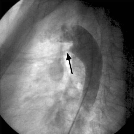
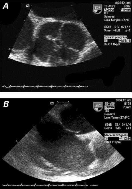
A 30-year-old woman was referred to our hospital for evaluation of increasing dyspnea on exertion. On physical examination, a continuous Gibson murmur was heard at the upper left sternal border. Results of electrocardiography and chest radiography were normal. Transthoracic echocardiography revealed a quadricuspid aortic valve with mild aortic regurgitation; color-flow Doppler imaging showed a narrow mosaic jet; and continuous-wave Doppler imaging showed high-velocity left-to-right flow. There was no atrial or ventricular dilation, and systolic function was normal. Aortography showed shunting from the aorta into the pulmonary artery (Fig. 1). Transesophageal echocardiography showed the X-shaped commissural pattern of a quadricuspid aortic valve (Fig. 2A) and a patent foramen ovale (Fig. 2B). A patent ductus arteriosus (PDA) was closed percutaneously with use of an AMPLATZER® duct occluder (AGA Medical Corporation; Plymouth, Minn).
Fig. 1 Aortogram shows shunting from the aorta into the pulmonary artery (arrow). Real-time motion image is available at www.texasheart.org/journal.
Fig. 2 Transesophageal echocardiography shows A) a quadricuspid aortic valve and B) a patent foramen ovale. Real-time motion image of Figure 2A is available at www.texasheart.org/journal.
Comment
Quadricuspid aortic valve is a very rare congenital malformation, far less common than unicuspid or bicuspid aortic valve.1 Most cases have been discovered incidentally during surgery or at autopsy. The incidence at autopsy is reportedly between 0.008% and 0.043%.2,3 In accordance with the classification system of Hurwitz and Roberts, our patient had a “type C” quadricuspid aortic valve (2 equal larger cusps and 2 equal smaller cusps).1
Quadricuspid aortic valve in association with other congenital anomalies is extremely rare. Aortic regurgitation appears to be the most common hemodynamic abnormality associated with quadricuspid aortic valve.4 Aortic regurgitation develops chiefly as a result of fibrosis and incomplete coaptation, and surgery is frequently required in a patient's 5th or 6th decade of life.1 Coronary artery anomalies have been seen in 10% of cases of quadricuspid aortic valve.3 Quadricuspid aortic valves have been reported in association with nonobstructive cardiomyopathy, pulmonary valve stenosis, ventricular septal defect, and fibromuscular subaortic stenosis.1,4 Infective endocarditis is a potential complication.5
Echocardiography is increasingly useful in detecting quadricuspid aortic valve. If this anomaly is found incidentally, continual follow-up is recommended, in view of the eventual requirement of valve replacement. Patients with quadricuspid aortic valve should also be carefully evaluated for other congenital abnormalities.
In 1923, Simonds described a postmortem finding of quadricuspid aortic valve with PDA.6 To the best of our knowledge, ours is the 1st report of quadricuspid aortic valve in association with PDA in a living patient.
Supplementary Material
Footnotes
Address for reprints: Dong-Soo Kim, MD, Department of Medicine, Inje University College of Medicine, Busan Paik Hospital, 633-165 Gaegeum-dong, Busanjin-gu, 614-735 Busan, ROK
E-mail: dongskim@inje.ac.kr
References
- 1.Hurwitz LE, Roberts WC. Quadricuspid semilunar valve. Am J Cardiol 1973;31(5):623–6. [DOI] [PubMed]
- 2.Feldman BJ, Khandheria BK, Warnes CA, Seward JB, Taylor CL, Tajik AJ. Incidence, description and functional assessment of isolated quadricuspid aortic valves. Am J Cardiol 1990;65(13):937–8. [DOI] [PubMed]
- 3.Tutarel O. The quadricuspid aortic valve: a comprehensive review. J Heart Valve Dis 2004;13(4):534–7. [PubMed]
- 4.Timperley J, Milner R, Marshall AJ, Gilbert TJ. Quadricuspid aortic valves. Clin Cardiol 2002;25(12):548–52. [DOI] [PMC free article] [PubMed]
- 5.Bauer F, Litzler PY, Tabley A, Cribier A, Bessou JP. Endocarditis complicating a congenital quadricuspid aortic valve. Eur J Echocardiogr 2008;9(3):386–7. [DOI] [PubMed]
- 6.Simonds JP. Congenital malformations of the aortic and pulmonary valves. Am J Med Sci 1923;166(1):584–95.
Associated Data
This section collects any data citations, data availability statements, or supplementary materials included in this article.


