Abstract
Susceptibility to Alzheimer’s disease (AD) is governed by multiple genetic factors. Remarkably, the LDL receptor–related protein (LRP) and its ligands, apoE and α2M, are all genetically associated with AD. In this study, we provide evidence for the involvement of the LRP pathway in amyloid deposition through sequestration and removal of soluble amyloid β-protein (Aβ). We demonstrate in vitro that LRP mediates the clearance of both Aβ40 and Aβ42 through a bona fide receptor-mediated uptake mechanism. In vivo, reduced LRP expression is associated with LRP genotypes and is correlated with enhanced soluble Aβ levels and amyloid deposition. Although LRP has been proposed to be a clearance pathway for Aβ, this work provides the first in vivo evidence that the LRP pathway may modulate Aβ deposition and AD susceptibility by regulating the removal of soluble Aβ.
Introduction
The LDL receptor–related protein (LRP) is a multifunctional receptor that mediates the internalization and degradation of ligands involved in metabolic pathways of lipoproteins and protease/protease-inhibitor complexes (1), including α2-macroglobulin (α2M) (2), apoE (3), and Kunitz protease inhibitor (KPI) containing forms of amyloid precursor protein (APP) (4). Remarkably, the aforementioned ligands are all genetically associated with Alzheimer’s disease (AD) (5, 6) and are found in senile plaques in brains of AD patients (7). To date, the strongest evidence directly implicating a role of LRP in AD is from genetic studies first reported by us (8) and subsequently confirmed in four independent case-control cohorts (9–12). In addition, another genetic polymorphism in LRP was found to be associated with AD (13), further evidence corroborating the LRP gene as an important AD-susceptibility locus. Our study reported a genetic polymorphism (C766T) in exon 3 of LRP that is under-represented in AD and associated with later age of disease onset. However, the underlying biologic relevance of the silent LRP C766T polymorphism is unclear. Moreover, the precise mechanisms by which LRP and its ligands may contribute to AD pathogenesis are unknown.
The generation of amyloid β-protein (Aβ) from APP and its subsequent deposition in the brain are believed to be key events in the pathogenesis of AD (14). Not surprisingly, the sequestration and clearance of Aβ recently has been hypothesized to be another potential key regulatory step in amyloid deposition (7, 8, 15). It has been shown that α2M can complex with Aβ and subsequently be degraded through the LRP-mediated pathway in cultured cells (16, 17). In addition, because LRP is highly expressed in the central nervous system (CNS) (18), internalization of apoE-enriched lipoprotein particles by way of LRP may impact neuronal membrane remodeling (19). Thus, alterations in brain LRP expression or activity may impact both neuronal homeostasis and amyloid deposition and thereby modify the pathogenesis of AD. In this study, we demonstrate that the LRP pathway is critical for clearance of soluble Aβ in vitro and provide genetic and biochemical in vivo evidence that LRP modulates soluble Aβ levels, amyloid burden, and susceptibility to AD.
Methods
Cell lines and cDNA constructs.
Control mouse fibroblasts (+/+; MEF-1) and fibroblasts heterozygous (+/–; PEA 10) or homozygous (–/–; PEA 13) for LRP deficiency were obtained from American Type Culture Collection (Rockville, Maryland, USA) and cultured as described previously (20). Human APP695 was inserted into the pBabepuro retroviral expression vector (21) and transfected into GP+E86 packaging cell line. Stable transformants were selected with puromycin (5 μg/ml). After infection with recombinant viruses, mouse fibroblasts (LRP+/+, LRP+/–, and LRP–/–) were selected with puromycin (2.5 μg/ml) and analyzed without clonal selection. Chinese hamster ovary (CHO) cells overexpressing APP751 with V717F FAD mutation were generated as described previously (22). A full-length RAP cDNA (GenBank accession no. M63959) was amplified by PCR from a human liver cDNA library (CLONTECH Laboratories, Palo Alto, California, USA) and subcloned into pGEX-4T (Amersham Pharmacia Biotech, Piscataway, New Jersey, USA). GST-RAP fusion protein was expressed in Escherichia coli and purified using a glutathione column, according to manufacturer’s directions.
In vitro Aβ clearance assays.
Purified α2M was obtained from Athens Research Laboratory (Athens, Georgia, USA) and activated by methylamine as described (4). Conditioned medium from CHO cells (IS-CHO; Irvine Scientific, Santa Ana, California, USA) overexpressing APP751 with V717F FAD mutation was added to confluent nontransfected LRP+/– and LRP–/– fibroblasts for 48 hours in the presence of 20 nM α2M, and Aβ40 and Aβ42 levels were measured by sandwich ELISA. For clearance of iodinated synthetic Aβ, 125I-Aβ40 (Amersham Pharmacia Biotech) and α2M were incubated overnight at 37°C, and the incubation mix was added to confluent cultured cells for 24 hours. The medium was then collected and subjected to scintillation counting for γ radiation.
Human subjects and neuropathological evaluation.
All subjects were unrelated white Americans of European descent. All pathology-confirmed AD subjects (National Institute of Neurological Disorders and Stroke [NINDS]/Alzheimer’s Disease and Related Disorders Association [ADRDA] criteria) were obtained from the Alzheimer’s Disease Research Center (ADRC) at the University of California, San Diego (UCSD). Senile plaques were identified in thioflavin S–stained sections of the midfrontal cortex and counted under a ×10 objective and a ×10 ocular lens (field size, 1.6 mm2), as described previously (23). For quantitation of LRP levels, all LRP T allele–positive AD cases (n = 17) with pathological data and available frozen tissue from the appropriate brain region were entered into the study. Age-matched AD cases with C/C genotype were randomly selected for measurement of LRP levels (n = 20). For assessing amyloid burden, all available pathology-confirmed cases with known LRP genotypes from UCSD-ADRC were included (n = 103). All AD subjects were late-onset AD (≥60 years at onset of disease). All autopsied control subjects were obtained from the Johns Hopkins University ADRC and the Baltimore Longitudinal Study of Aging (24). APOE and LRP genotyping were performed as described previously (8).
Quantitation of LRP levels.
Frozen brain tissues derived from the midfrontal cortex of pathologically confirmed control and AD subjects were homogenized in 1% NP40 lysis buffer (50 mM Tris, pH 8.0, 150 mM NaCl, 0.02% sodium azide, 1% NP40, 100 μg/ml AEBSF, and 10 μg/ml leupeptin). For quantitation of LRP in brain, 50 μg of detergent-soluble protein was separated by SDS-PAGE and transferred onto nitrocellulose membranes. From the same blots, LRP was detected by a polyclonal Ab against the 85-kDa light chain of LRP (25), while actin (AC-40; Sigma Chemical Co., St. Louis, Missouri, USA) and synaptophysin (SY38; Roche, Indianapolis, Indiana, USA) were detected by specific mAb’s. The primary Ab’s were detected by incubation with biotinylated secondary Ab’s, followed by 125I-streptavidin. The signals were quantitated by phosphorimaging (Bio-Rad, Hercules, California, USA). Signals from quantitations were in a linear range, as determined from standards included with each experiment. The LRP, actin, and synaptophysin signal on each blot was first standardized to the internal control. Then the LRP signal was normalized to actin or synaptophysin.
Aβ measurements.
Human plasma samples were collected from living subjects clinically diagnosed as probable AD at the UCSD-ADRC using NINDS/ADRDA criteria. Plasma samples were collected in EDTA tubes and kept at 4°C until centrifugation. Aβ quantitation was performed using a standard sandwich ELISA. Briefly, microtiter wells were coated with a monospecific Ab that selectively recognizes the carboxy terminal of Aβ1-40 or Aβ1-42; the wells were then blocked with 1% BSA/PBS. Human plasma samples were diluted 1:1 in 0.1% CHAPS/PBS and then captured in Ab-coated wells for 18 hours at 4°C. After binding, wells were washed with PBS, and Aβ was detected with an anti-human Aβ1-12 mAb (26D6) conjugated to horseradish peroxidase (HRP). Each sample was assayed in triplicate and quantitated to a standard Aβ curve within the linear range. For immunoprecipitation of Aβ from medium of cultured cells, a polyclonal Ab to Aβ (3134) was used as described (26).
Statistics.
For two-group comparisons between genotypes or disease status, a two-sided t test was used. Where SD was significantly different between groups, Welch’s correction was applied to two-sided t tests. ANOVA, coupled with Tukey post hoc test, was used to assess the combinatorial effects of LRP and APOE genotypes on plasma Aβ levels. A χ2 test for linear trend was used to assess LRP genotype distributions across ordered plaque-number categories among AD subjects. Linear regression analysis was used to assess the magnitude and significance of the correlation between LRP levels and age of subjects.
Results
LRP mediates the clearance of secreted Aβ without altering Aβ production from APP695.
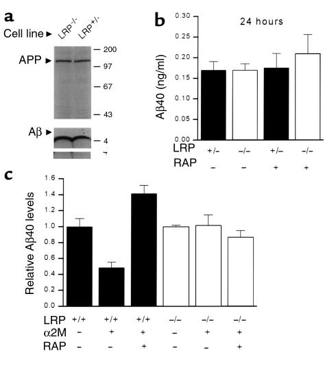
It has been demonstrated previously that LRP can serve as a cell surface–internalization receptor for the KPI containing isoforms of secreted APP, including APP751 and APP770 (4). Moreover, LRP can associate with cellular APP751 at the cell surface and mediate its internalization (27). Recently, it was shown by Ulery and colleagues that LRP alters Aβ production from the KPI containing APP751, presumably through the KPI-LRP interaction (28). Thus, it is important to examine the possibility that LRP might be involved in the formation of Aβ from APP695, the major neuronal isoform lacking the KPI domain. To study the effects of LRP expression on Aβ production and clearance, we first established an in vitro cell-culture model using mouse fibroblasts genetically deficient in LRP (LRP–/–) and corresponding LRP-expressing control cells (LRP+/+ and LRP+/–). In these fibroblasts transfected with equivalent levels of human APP695, the levels of Aβ secreted into the medium in the absence of α2M within 24 hours was equivalent between LRP+/– and LRP–/– cells as measured by immunoblotting and sandwich ELISA (Figure 1, a and b). Addition of RAP, a competitive antagonist of all ligands binding to LRP, had no effect on levels of secreted Aβ during this time period (Figure 1b). Thus, this indicated that different levels of LRP did not alter the secretion of Aβ from APP695, although the possible role of LRP in other aspects of APP processing cannot be excluded. Having established that LRP does not alter Aβ production within a 24-hour time period, we examined the clearance of endogenously secreted Aβ through the α2M-LRP pathway. Consistent with recent observations in primary neurons (29), addition of activated α2M directly to serum-free medium of LRP+/+ fibroblasts reduced Aβ levels by approximately 60% (Figure 1c) within 48 hours. In contrast, α2M had no effect on Aβ levels in LRP–/– cells, indicating that LRP is required for α2M-mediated reduction in Aβ. Coincubation of α2M with RAP completely blocked α2M-mediated reduction of Aβ in LRP-expressing cells, confirming the specificity of the LRP pathway in removal of Aβ complexes (Figure 1c). Interestingly, RAP treatment resulted in higher Aβ levels compared with untreated controls in LRP-expressing cells, suggesting a basal level of RAP-sensitive Aβ clearance activity in untreated controls. In LRP-deficient cells, RAP did not affect the level of Aβ in the presence or absence of α2M (Figure 1c), indicating that the effects of RAP are specific for LRP and not directly on secretion or removal of Aβ. These data clearly demonstrate that secreted Aβ is removed through the α2M-LRP pathway and that LRP is absolutely required for α2M-mediated uptake of Aβ. Moreover, Aβ production from APP695 is not affected by LRP.
Figure 1.
LRP does not alter the secretion of Aβ in cultured cells. LRP+/– and LRP–/– fibroblasts transfected with human APP695 were generated as described. (a) To determine whether LRP levels contribute to changes in production and secretion of Aβ, APP-transfected LRP+/– and LRP–/– cells were metabolically labeled with 35S-methionine for 20 minutes and immunoprecipitated with an Ab specific for APP (upper panel). In parallel experiments, medium was conditioned for 24 hours, and the amount of secreted Aβ was analyzed by immunoprecipitation (3134), followed by Western blotting (26D6) (lower panel). (b) To distinguish between Aβ secretion and LRP-dependent Aβ clearance within a 24-hour period, APP overexpressing LRP+/– and LRP–/– cells were treated with or without RAP (1 μM), and the medium was quantitated for the amount of Aβ by ELISA. Graph shows a representative experiment (n = 3) with means and SEM. (c) Confluent cultures of APP overexpressing LRP+/+ and LRP–/– fibroblasts were incubated with or without activated α2M (100 nM) in serum-free medium to induce uptake of Aβ through LRP. RAP (1 μM), an antagonist for all known LRP ligands, was added to block α2M-LRP–mediated effects. After 48 hours, medium was collected and analyzed for levels of Aβ by sandwich ELISA assay. Aβ levels are normalized to the no-treatment group of each cell line. Experiments were performed three times in triplicate. Error bars represent SEM.
LRP mediates the clearance of secreted Aβ40 and Aβ42 through a bona fide receptor-mediated mechanism.
It has been suggested previously that LRP can mediate the internalization and degradation of Aβ complexes in vitro (16). However, it has not been directly demonstrated that uptake of Aβ complexes by cultured cells is through a bona fide receptor-mediated mechanism. This issue is highlighted by previous observations that Aβ can be internalized by fluid-phase pinocytosis and by the scavenger-receptor pathway in macrophages (30, 31). Moreover, it is not known whether the LRP pathway is capable of mediating the clearance of Aβ42, a minor but putatively pathogenic species of Aβ that are initially deposited in senile plaques (32). To test whether Aβ42 is removed through the LRP pathway, conditioned medium from CHO cells overexpressing V717F FAD mutant APP was added to native (i.e., untransfected) LRP+/– and LRP–/– cells in the presence of α2M. As shown in Figure 2a, α2M significantly reduced the levels of soluble Aβ40 and Aβ42 in the medium of LRP+/– cells to similar degrees. As expected, neither Aβ40 nor Aβ42 levels were affected by α2M in LRP–/– cells, indicating the requirement of LRP in α2M-mediated clearance of Aβ. Approximately 65% of Aβ40 and 60% of Aβ42 was cleared from the medium within 48 hours in LRP+/– cells under conditions where cell-free degradation of Aβ was undetectable (Figure 2a). These data show that the LRP pathway efficiently mediates the uptake and degradation of the highly pathogenic Aβ42 species as well as Aβ40.
Figure 2.
LRP mediates the clearance of both Aβ40 and Aβ42 through a receptor-mediated uptake mechanism. (a) CHO cells overexpressing APP751 with V717F FAD mutation were grown to confluency, and IS-CHO was collected for 48 hours. Conditioned medium was then added to confluent nontransfected LRP+/– and LRP–/– fibroblasts for 48 hours in the presence of 20 nM α2M, and Aβ40 and Aβ42 levels were measured by sandwich ELISA. Experiments were performed three times in triplicate. Error bars represent SEM. (b) To determine optimal Aβ uptake, mixtures of 0.1 nM 125I-Aβ with 0, 1, 2, and 3 nM α2M were incubated overnight at 37°C, and the incubation mix was added to confluent LRP+/– (filled squares) and LRP–/– (open circles) fibroblasts for 24 hours. The medium was then collected and subjected to scintillation counting for γ radiation. Percentage of Aβ uptake reflects the proportion of counts lost from the input amount. (c) To determine whether Aβ uptake is subject to self-competition, a mixture of 0.1 nM 125I-Aβ and 2 nM α2M was incubated overnight at 37°C, and the incubation mix was added to confluent LRP+/– fibroblasts in the presence of increasing amounts of excess unlabeled α2M/Aβ complex for 24 hours (filled squares). In parallel experiments, 1 μM RAP was coincubated with the 125I-Aβ/α2M mix (open squares). The medium was then collected and subjected to scintillation counting for γ radiation. Percentage of Aβ uptake is normalized to maximal Aβ uptake in the absence of unlabeled Aβ/α2M complex. (d) To determine whether Aβ uptake is subject to saturation, increasing amounts of 125I-Aβ/α2M complex (mixed as before) were added to confluent LRP+/– cells for 24 hours, and the medium was collected and subjected to scintillation counting for γ-radiation. Aβ uptake is calculated in femtomoles and represented in a log scale. All experiments were performed three times in triplicate, and a representative experiment is shown. Error bars represent SEM.
To demonstrate that Aβ uptake by the α2M-LRP pathway is through a bona fide receptor-dependent mechanism, we next assessed the uptake of synthetic 125I-labeled Aβ in cultured fibroblasts. We first determined the optimal ratio of 125I-α2M/Aβ for in vitro uptake of Aβ through LRP using a fixed physiological concentration of 0.1 nM Aβ. As shown in Figure 2b, an increasing amount of α2M facilitated the uptake of 125I-Aβ in a concentration-dependent manner in LRP+/– cells. Maximal uptake of Aβ was approximately 60% (Figure 2b), consistent with soluble Aβ produced from cultured cells. As anticipated, there was little to no uptake of 125I-Aβ in LRP–/– cells at any tested concentration of α2M (Figure 2b). Increasing amounts of unlabeled Aβ/α2M complex effectively blocked the removal of 125I-Aβ in a concentration-dependent fashion, such that excess cold Aβ/α2M inhibited 125I-Aβ uptake as effectively as the addition of RAP (Figure 2c). Finally, uptake of 125I-Aβ/α2M complex was completely saturable with half-maximal uptake at 50 pM 125I-Aβ/1 nM α2M complex, an approximate Kd range (0.2–10 nM) that has been reported for binding of α2M to a variety of cell types (33) (Figure 2d).
Reduced LRP levels correlate with increased AD susceptibility.
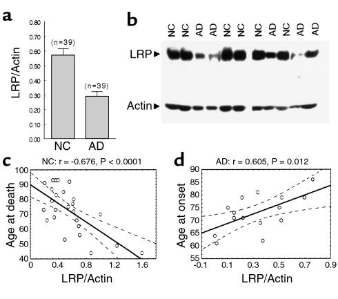
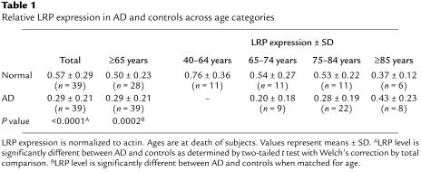
Having established that the level of LRP expression is critical for Aβ clearance in vitro, we next assessed whether LRP levels in the human brain might be altered during normal aging or disease. Measurement of LRP levels in the brain were performed by quantitative immunoblotting of the LRP 85-kDa light chain from the midfrontal cortex of AD and normal controls (NC). From pathology-confirmed AD and control age-matched subjects, LRP levels relative to actin were approximately twofold lower in AD brains compared with that of controls (Figure 3a: t = 4.884, df = 76, P < 0.0001; Table 1). Surprisingly, among control subjects there was a strong inverse correlation between age and LRP levels (n = 39, r = –0.4905, P = 0.0015), indicating that LRP expression normally declines with age. As shown in Table 1, the average reduction in brain LRP level was approximately twofold between an age group of 40–64 (0.76 ± 0.11 SEM, n = 11) and those equal to or greater than 85 (0.37 ± 0.05 SEM, n = 6), with intermediate LRP expression among the age group of 65–84 (0.53 ± 0.06 SEM, n = 22). Interestingly, this inverse correlation was markedly stronger among noncarriers of the APOE ε4 allele (Figure 3c: n = 28, r = –0.6758, P = 0.00008). When LRP levels were normalized to synaptophysin instead of actin, a similar inverse correlation with age and LRP expression was observed among all control subjects (n = 39, r = –0.3948, P = 0.0128), indicating that the observed effect is not attributable to neuronal/synaptic loss.
Figure 3.
Association of brain LRP levels with AD susceptibility. LRP levels were quantitated by immunoblotting for the 85-kDa light chain of LRP and normalized to actin. (a) Comparison of AD and age-matched normal controls (NC) showed a significant difference in LRP levels (t = 4.884, df = 76, P < 0.0001). Error bars represent SEM. (b) Representative immunoblots containing LRP and actin signals from AD and NC samples are shown. (c) Levels of LRP in the brain are inversely correlated with age of control subjects (control subjects lacking APOE ε4 allele shown: r = –0.6758, P < 0.0001; all control subjects: r = 0.4905, P = 0.0015). (d) AD patients show a positive correlation between LRP levels and ages at onset of disease (AD subjects lacking APOE ε4 allele shown: r = 0.6048, P = 0.0116; all AD subjects: r = 0.33465, P = 0.0429). The regression slope (center line) and 95% confidence interval (two curved lines) are shown. The correlation coefficient (r) and P values are shown above the graph.
Table 1.
Relative LRP expression in AD and controls across age categories
In contrast to that observed in control subjects, higher LRP levels significantly correlated with later ages at onset of AD (n = 37, r = 0.33465, P = 0.0429) and death (n = 37, r = 0.41032, P = 0.0169). Importantly, LRP levels were still lower in AD patients after the age of 80 (0.34 ± 0.05 SEM, n = 17) compared with age-matched control subjects (0.48 ± 0.05 SEM, n = 13), although the largest differences were observed under the age of 85 (Table 1). Interestingly, the positive correlation between LRP levels and age of onset (Figure 3d: n = 15, r = 0.60483, P = 0.0116) and death (n = 15, r = 0.63599, P = 0.0108) resulted primarily from individuals lacking the APOE ε4 allele. Thus, the LRP effect is predominant among approximately 50% of AD cases where currently no genetic susceptibility is attributed because pathogenic effects associated with APOE ε4 may be sufficient by itself to confer AD risk. Duration of disease from onset to death had no effect on LRP levels (graph not shown: n = 37, r = 0.081, P = 0.6357), indicating that the decline in LRP expression is unrelated to AD-related brain aberrations.
Genetic association of LRP with LRP levels, amyloid burden, and plasma Aβ.
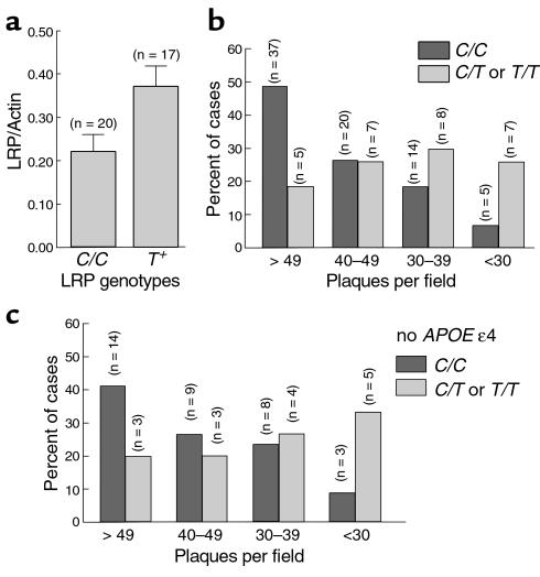
In view of the preceding observations, we next asked whether the correlation between LRP levels and AD susceptibility can be confirmed by a different analysis. The recently reported under-representation of the LRP exon 3 (C766T) polymorphism in late-onset AD has been demonstrated in five different populations (8–12), although the putative causative mutation/polymorphism in linkage disequilibrium with the C766T polymorphism remains to be identified. Because our current results demonstrated that the level of LRP in the brain is correlated with disease onset, we examined whether a manifestation of the C766T polymorphism is also reflected in LRP expression. Analysis of LRP genotype status revealed significantly higher LRP levels among AD cases harboring the C/T or T/T genotypes compared with carriers of the C/C genotype (Figure 4a: t = 2.335, df = 35, P = 0.0254), although still lower than that of control subjects. Because LRP T allele carriers have higher age at onset of AD than LRP C/C carriers as reported previously (8), we confined the analysis to comparable ages. In an age group (70–82 years) where there was no difference in ages at death between C/C (n = 16, 76.4 ± 0.18 SD) and T-positive genotypes (n = 11, 77.3 ± 0.15 SD), the same significant difference was observed (t = 2.424, df = 25, P = 0.0229). Higher LRP levels in AD are therefore associated with the protective effects of the T allele, consistent with under-representation of the T allele in AD (8). Different APOE genotypes, however, did not alter the level of LRP in AD brains (data not shown).
Figure 4.
Association of LRP genotypes with LRP levels and amyloid burden in the AD brain. LRP levels were quantitated by immunoblotting for the 85-kDa light chain of LRP and normalized to actin. (a) AD patients harboring LRP T allele showed significantly higher levels of LRP in the brain (t = 2.335, df = 35, P = 0.0254). (b) AD brains were segregated into ordered categories of increasing amyloid burden, ranging from less than 30, 30–39, 40–49, and greater than 49 plaques per field and examined for association with LRP genotypes. The percentage and number of individuals within each plaque-per-field category are shown as a function of LRP genotypes. Statistical analysis shows an excessive overrepresentation of C/C genotypes across increasing plaques-per-field categories compared with T-positive genotypes (χ2 for linear trend = 11.762, df = 1, P = 0.0006). (c) The LRP effect on amyloid burden is still observed among subjects that do not carry the APOE ε4 allele (χ2 for linear trend = 6.135, df = 1, P = 0.0133).
Next, we assessed whether the LRP T-allele associates with total amyloid burden in AD. Extending our previous analysis of neuritic plaques in brains of AD individuals (8), we assessed the total amount of thioflavin S–positive plaques, regardless of type, as an indicator of amyloid burden. LRP T allele carriers showed significantly fewer numbers of total senile plaques compared with C/C carriers, using t-test analysis (t = 2.860, df = 101, P = 0.0051). To examine the plaque-density profile of LRP T-positive versus C/C carriers, AD cases were segregated into four increasing levels of plaque-density categories. Statistical analysis revealed that the LRP T allele was excessively over-represented in the lower plaque-density categories as compared with the C/C genotype (Figure 4b: χ2 for linear trend = 11.762, df = 1, P = 0.0006). The LRP-associated effects on amyloid burden were not dependent on the underlying APOE genetic status, because the same trend was observed among subjects that did not carry the APOE ε4 allele (Figure 4c: χ2 for linear trend = 6.135, df = 1, P = 0.0133).
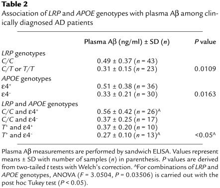
Amyloid burden in postmortem AD brain tissues represents a terminal state that may not accurately reflect the dynamic in vivo relationship between LRP expression and Aβ levels. Moreover, the measurements of Aβ in the brain is confounded by the presence of multiple pools with heterogeneous solubilities. Thus, we analyzed Aβ levels in human plasma to determine whether LRP genotypes associate with levels of soluble Aβ in vivo. Consistent with higher LRP levels among LRP T-allele carriers, Aβ in plasma was significantly decreased by 58% in carriers of the LRP T allele compared with carriers of the C/C genotype (Table 2: Welch’s t = 2.627, df = 60, P = 0.0109). Surprisingly, plasma Aβ level was also significantly lower (by 54%) in non-APOE ε4 carriers compared with carriers of the APOE ε4 allele (Table 2: Welch’s t = 2.478, df = 55, P = 0.0163), indicating that apoE4 not only increases amyloid burden in the brain (34), but also the amount of soluble Aβ systemically. This finding led us to ask whether there are any additive effects between LRP and APOE genotypes. The highest Aβ levels were found among carriers of both LRP C/C genotype and APOE ε4 allele, which was greater than twofold more than carriers of both LRP T-allele and non-APOE ε4 genotypes (Table 2: ANOVA, F = 3.0504, P = 0.03506; post hoc Tukey, P < 0.05).
Table 2.
Association of LRP and APOE genotypes with plasma Aβ among clinically diagnosed AD patients
Discussion
The genetic associations of APOE, α2M, and LRP to late-onset AD are particularly intriguing in light of the fact that both apoE and α2M are two key ligands for LRP (5, 6, 8). Both apoE and α2M avidly bind Aβ in vitro and in vivo (5, 35). These observations, together with the finding that LRP and all of its ligands are present in senile plaques (7), strongly implicate the pathogenic importance of the LRP pathway in AD. We found that LRP levels are significantly reduced in AD, compared with healthy controls. Linear-regression analysis revealed that LRP levels progressively decline with the increasing age of control subjects (an inverse correlation) and are further reduced in AD subjects. Among AD patients, however, increased LRP levels were correlated with later age of disease onset, indicating that higher LRP levels might be protective against AD. This apparent protective effect was accentuated among noncarriers of the APOE ε4 allele. As increasing age is the primary risk factor for AD, these data indicate that reduced LRP expression may be one factor contributing to AD susceptibility. This notion is highly consistent with the negative association of the LRP T allele to AD (8, 9, 11, 12) and increased LRP levels among T-allele carriers demonstrated in this study. Although the biological mode of the LRP T allele requires further characterization, we hypothesize that the C766T polymorphism is in linkage disequilibrium with a causative mutation or polymorphism that regulates LRP expression (for example, promoter/enhancer) in the aging brain.
In the current study, we provide compelling evidence that Aβ uptake via the α2M-LRP pathway is through a bona fide receptor-mediated mechanism and not through nonspecific Aβ degradation or fluid-phase pinocytosis. This was shown by the competition of 125I-Aβ uptake with excess unlabeled Aβ complexes and the complete saturation of 125I-Aβ uptake at physiological concentrations. So far, no other Aβ uptake pathway meets the criteria for a bona fide receptor-mediated mechanism. Although the scavenger receptor has been postulated to mediate the uptake of amyloid fibrils, such process is not subject to competition and saturation of the receptor (30, 31). At another level of Aβ catabolism, recent observations have indicated that neutral endopeptidase and insulin-degrading enzyme are both capable of degrading extracellular Aβ in a cell-autonomous manner (36, 37). Thus, it is likely that there are multiple ways of mediating Aβ degradation in vivo. Our study demonstrated that LRP does not alter the secretion of Aβ from APP695-expressing cells but is required for α2M-mediated clearance of soluble Aβ. Because both LRP and APP695 are predominantly expressed in neurons, reduced LRP levels in the AD brain is predicted to negatively impact the clearance of soluble Aβ but not its production in neurons. However, it is important to note that LRP overexpression in LRP-deficient CHO cells results in altered trafficking of KPI containing APP751 (28), possibly through the APP-LRP physical interaction (4). Thus, it is possible that LRP also alters APP trafficking (i.e., internalization/recycling) and Aβ generation through other mechanisms. As APP isoforms are differentially expressed in neurons and glia, LRP-clearance activity versus altered APP trafficking might be differentially modulated across cell types.
The genetic association of LRP T polymorphism with both increased LRP expression and reduced amyloid deposition is intriguing in light of the in vitro evidence of Aβ clearance through the LRP pathway (16, 17). These observations are now further strengthened by genetic association of LRP with soluble Aβ levels in plasma. It is noteworthy that the pathogenic Aβ42 species is as effectively cleared through the LRP pathway as Aβ40 (Figure 1a), an activity that may dramatically impact amyloid deposition in vivo. Accordingly, we interpret these data to indicate that reduced LRP expression, at least in part, contributes to increased Aβ levels and amyloid deposition by negatively impacting Aβ clearance. This interpretation is consistent with our observation that reduced LRP expression is also correlated with increased AD susceptibility and earlier age of disease onset. In our cell-culture system, we demonstrated the requirement of LRP in the α2M-mediated clearance of Aβ. However, it has been reported that apoE and lactoferrin, two other LRP ligands, also sequester Aβ and mediate its clearance (16, 38). Thus, reduced LRP levels may impede the clearance of various Aβ complexes. Interestingly, many LRP ligands, including apoE, α2M, and lactoferrin, are produced from astrocytes, whereas LRP is largely expressed in neurons. Thus, it is likely that receptor-mediated uptake and clearance of soluble Aβ complexes occur in neurons, whereas uptake of fibrillar amyloid is mediated by microglia (30, 31). In this regard, downregulation of LRP expression has been linked to proinflammatory stimuli such as LPS and IFN-γ in cultured cells (39, 40). We speculate that proinflammatory processes present in the AD brain may induce downregulation of LRP expression, further reducing Aβ clearance and enhancing amyloid deposition. Since LRP mediates the normal function of neuronal remodeling through internalization of apoE (19), reduced LRP expression in aging and disease may also compromise neuronal viability independent of the effects on Aβ clearance.
It has been demonstrated previously that the APOE ε4 allele promotes amyloid deposition (34). Our unexpected finding that the APOE ε4 allele is also associated with higher Aβ levels in plasma of AD subjects raises the possibility that different isoforms of apoE may also impact the removal of soluble Aβ. This is in agreement with the binding affinity of native apoE isoforms for Aβ (apoE2 > apoE3 > apoE4) (15, 41) and reduced LRP-dependent uptake of apoE4/Aβ complexes in CHO cells (38). However, the possibility that apoE4 interferes with Aβ clearance by accelerating Aβ aggregation cannot be excluded, consistent with the delayed amyloid deposition in apoE-deficient mice (42). Two recent studies surprisingly have shown that human apoE actually delays Aβ deposition in apoE-null mice, with apoE3 being more effective than apoE4. On the other hand, apoE is required for fibrillar Aβ deposits, and apoE4 converts Aβ to fibrillar deposits faster than apoE3 in a mouse model of human apoE expression (43, 44). These observations are consistent with the notion that apoE4 may both impede LRP-mediated Aβ clearance and promote Aβ fibrillogenesis.
The results of the current study provided the first in vivo evidence of the LRP-clearance pathway in AD pathogenesis. Our observations lead us to postulate that reduced LRP expression is a contributing risk factor for AD, possibly by impeding clearance of soluble Aβ complexes. Functional characterization of α2M polymorphisms associated with AD (6) and future transgenic animal models of LRP and/or α2M expression should further elucidate the mechanism of Aβ clearance and AD pathogenesis. The observation that AD risk or protection associated with LRP levels is strongest among noncarriers of the APOE ε4 allele is particularly interesting in light of the ligand/receptor relationship between apoE and LRP. Because the receptor function of LRP obviously depends on intact activity of its ligands, we hypothesize that high levels of LRP cannot effectively rescue the pathogenic effects of apoE4, the latter operating at a step that negates the clearance mechanism. However, in the presence of apoE2 or apoE3, where the ligand complexes are not perturbed, alterations in LRP level and, in turn, clearance activity become highly consequential for AD pathogenesis. Since increased LRP expression may promote both neuronal survival mediated by apoE2 and apoE3 isoforms and also enhance the clearance of soluble Aβ complexes, the current data provide an alternative direction for AD therapeutic intervention by targeting the Aβ/LRP clearance pathway in non-APOE ε4 carriers.
Acknowledgments
We thank Leon Thal and Doug Galasko for critical review of the manuscript, Marilyn Farquhar for providing the LRP Ab, H. Land for the retroviral vector, Joseph Buxbaum for Aβ Ab, Lawrence Hasen and Eliezer Masliah for providing pathology data, and Chui Pui Pang and Ho-Keung Ng for providing support (L. Baum). We also thank Mary Turner, Mary Sundsmo, Xiaohua Chen, Elizabeth Mosmiller, and Gay Rudow for technical assistance, and Joachim Herz and Daniel Steinberg for helpful discussions. This work is dedicated to the memory of Tsunao Saitoh, who had the foresight to initiate the LRP investigations. This work was supported in part by NIH grants AG12376 and NS-01812 and the Allied Signal Award for Aging Research.
Footnotes
David E. Kang and Claus U. Pietrzik contributed equally to this work.
References
- 1.Strickland DK, Kounnas MZ, Argraves SW. LDL receptor-related protein: a multiligand receptor for lipoprotein and proteinase catabolism. FASEB J. 1995;9:890–898. doi: 10.1096/fasebj.9.10.7615159. [DOI] [PubMed] [Google Scholar]
- 2.Borth W. Alpha-2-macroglobulin, a multi-functional binding protein with targeting characteristics. FASEB J. 1992;6:3345–3353. doi: 10.1096/fasebj.6.15.1281457. [DOI] [PubMed] [Google Scholar]
- 3.Herz J, et al. Surface location and high affinity for calcium of a 500-kd liver membrane protein closely related to the LDL-receptor suggest a physiological role as lipoprotein receptor. EMBO J. 1993;7:4119–4127. doi: 10.1002/j.1460-2075.1988.tb03306.x. [DOI] [PMC free article] [PubMed] [Google Scholar]
- 4.Kounnas MZ, et al. LDL-receptor-related protein, a multifunctional ApoE receptor, binds secreted β-amyloid precursor protein and mediates its degradation. Cell. 1995;82:331–340. doi: 10.1016/0092-8674(95)90320-8. [DOI] [PubMed] [Google Scholar]
- 5.Strittmatter WJ, et al. Apolipoprotein E: high avidity binding to β-amyloid and increased frequency of type 4 allele in late onset familial Alzheimer’s disease. Proc Natl Acad Sci USA. 1993;90:1977–1981. doi: 10.1073/pnas.90.5.1977. [DOI] [PMC free article] [PubMed] [Google Scholar]
- 6.Blacker D, et al. Alpha-2 macroglobulin is genetically associated with Alzheimer disease. Nat Genet. 1998;19:357–360. doi: 10.1038/1243. [DOI] [PubMed] [Google Scholar]
- 7.Rebeck WG, Harr SD, Strickland DK, Hyman BT. Multiple, diverse senile plaque-associated proteins are ligands of an apolipoprotein E receptor, the α2-macroglobulin receptor/low-density-lipoprotein receptor-related protein. Ann Neurol. 1995;37:211–217. doi: 10.1002/ana.410370212. [DOI] [PubMed] [Google Scholar]
- 8.Kang DE, et al. Genetic association of the low-density lipoprotein receptor-related protein gene (LRP), an apolipoprotein E receptor, with late-onset Alzheimer’s disease. Neurology. 1997;49:56–61. doi: 10.1212/wnl.49.1.56. [DOI] [PubMed] [Google Scholar]
- 9.Kamboh MI, Ferrell RE, DeKosky ST. Genetic association studies between Alzheimer’s disease and two polymorphisms in the low density lipoprotein receptor-related protein gene. Neurosci Lett. 1998;244:65–68. doi: 10.1016/s0304-3940(98)00141-4. [DOI] [PubMed] [Google Scholar]
- 10.Baum L, et al. Low density lipoprotein receptor related protein gene exon 3 polymorphism association with Alzheimer’s disease in Chinese. Neurosci Lett. 1998;247:33–36. doi: 10.1016/s0304-3940(98)00294-8. [DOI] [PubMed] [Google Scholar]
- 11.Hollenbach E, Ackermann S, Hyman BT, Rebeck GW. Confirmation of an association between a polymorphism in exon 3 of the low-density lipoprotein receptor-related protein gene and Alzheimer’s disease. Neurology. 1998;50:1905–1907. doi: 10.1212/wnl.50.6.1905. [DOI] [PubMed] [Google Scholar]
- 12.Lambert JC, Wavrant-De Vrieze F, Amouyel P, Chartier-Harlin MC. Association at LRP gene locus with sporadic late-onset Alzheimer’s disease. Lancet. 1998;351:1787–1788. doi: 10.1016/s0140-6736(05)78749-3. [DOI] [PubMed] [Google Scholar]
- 13.Wavrant-DeVrieze F, et al. Association between coding variability in the LRP gene and the risk of late-onset Alzheimer’s disease. Hum Genet. 1999;104:432–434. doi: 10.1007/s004390050980. [DOI] [PubMed] [Google Scholar]
- 14.Selkoe DJ. Cell biology of the amyloid β-protein precursor and the mechanism of Alzheimer disease. Annu Rev Cell Biol. 1994;10:373–403. doi: 10.1146/annurev.cb.10.110194.002105. [DOI] [PubMed] [Google Scholar]
- 15.LaDu MJ, et al. Isoform-specific binding of apolipoprotein E to β-amyloid. J Biol Chem. 1994;269:23403–23406. [PubMed] [Google Scholar]
- 16.Narita M, Holtzman DM, Schwartz AL, Bu G. Alpha2-macroglobulin complexes with and mediates the endocytosis of β-amyloid peptide via cell surface low-density lipoprotein receptor-related protein. J Neurochem. 1997;69:1904–1911. doi: 10.1046/j.1471-4159.1997.69051904.x. [DOI] [PubMed] [Google Scholar]
- 17.Du Y, et al. Alpha2-macroglobulin as a β-amyloid peptide-binding plasma protein. J Neurochem. 1997;69:299–305. [PubMed] [Google Scholar]
- 18.Wolf BB, Lopes MBS, VandenBerg SR, Gonias SL. Characterization and immunohistochemical localization of α2-macroglobulin receptor (low-density lipoprotein receptor-related protein) in human brain. Am J Pathol. 1992;141:37–42. [PMC free article] [PubMed] [Google Scholar]
- 19.Holtzman D, et al. Low density lipoprotein receptor-related protein mediates apolipoprotein E–dependent neurite outgrowth in a central nervous system-derived neuronal cell line. Proc Natl Acad Sci USA. 1995;92:9480–9484. doi: 10.1073/pnas.92.21.9480. [DOI] [PMC free article] [PubMed] [Google Scholar]
- 20.Willnow TE, Herz J. Genetic deficiency in low density lipoprotein receptor-related protein confers cellular resistance to Pseudomonas exotoxin A. Evidence that this protein is required for uptake and degradation of multiple ligands. J Cell Sci. 1994;107:719–726. [PubMed] [Google Scholar]
- 21.Morgenstern JP, Land H. Advanced mammalian gene transfer: high titre retroviral vectors with multiple drug selection markers and a complementary helper-free packaging cell line. Nucleic Acids Res. 1990;18:3587–3596. doi: 10.1093/nar/18.12.3587. [DOI] [PMC free article] [PubMed] [Google Scholar]
- 22.Podlisny MB, et al. Aggregation of secreted amyloid beta-protein into sodium dodecyl sulfate-stable oligomers in cell culture. J Biol Chem. 1995;270:9564–9570. doi: 10.1074/jbc.270.16.9564. [DOI] [PubMed] [Google Scholar]
- 23.Olichney JM, et al. The apolipoprotein E epsilon 4 allele is associated with increased neuritic plaques and cerebral amyloid angiopathy in Alzheimer’s disease and Lewy body variant. Neurology. 1996;47:190–196. doi: 10.1212/wnl.47.1.190. [DOI] [PubMed] [Google Scholar]
- 24.Troncoso JC, Martin LJ, Dal Forno G, Kawas CH. Neuropathology in controls and demented subjects from the Baltimore Longitudinal Study of Aging. Neurobiol Aging. 1996;17:365–371. doi: 10.1016/0197-4580(96)00028-0. [DOI] [PubMed] [Google Scholar]
- 25.Czekay RP, Orland RA, Woodward L, Adamson ED, Farquhar MG. The expression of megalin (gp330) and LRP diverges during F9 cell differentiation. J Cell Sci. 1995;108:1433–1441. doi: 10.1242/jcs.108.4.1433. [DOI] [PubMed] [Google Scholar]
- 26.Desdouits F, Buxbaum JD, Desdouits-Magnen J, Nairn AC, Greengard P. Amyloid alpha peptide formation in cell-free preparations. Regulation by protein kinase C, calmodulin, and calcineurin. J Biol Chem. 1996;271:24670–24674. doi: 10.1074/jbc.271.40.24670. [DOI] [PubMed] [Google Scholar]
- 27.Knauer MF, Orlando RA, Glabe CG. Cell surface APP751 isoform complexes with protease nexin 2 ligands and is internalized via the low density lipoprotein receptor-related protein (LRP) Brain Res. 1996;740:6–14. doi: 10.1016/s0006-8993(96)00711-1. [DOI] [PubMed] [Google Scholar]
- 28.Ulery PG, et al. Modulation of β-amyloid precursor protein processing by the low density lipoprotein receptor-related protein (LRP). Evidence that LRP contributes to the pathogenesis of Alzheimer’s disease. J Biol Chem. 2000;275:7410–7415. doi: 10.1074/jbc.275.10.7410. [DOI] [PubMed] [Google Scholar]
- 29.Qiu Z, Strickland DK, Hyman BT, Rebeck GW. Alpha2-macroglobulin enhances the clearance of endogenous soluble β-amyloid peptide via low-density lipoprotein receptor-related protein in cortical neurons. J Neurochem. 1999;73:1393–1398. doi: 10.1046/j.1471-4159.1999.0731393.x. [DOI] [PubMed] [Google Scholar]
- 30.Khoury J, et al. Scavenger receptor-mediated adhesion of microglia to beta-amyloid fibrils. Nature. 1996;382:716–719. doi: 10.1038/382716a0. [DOI] [PubMed] [Google Scholar]
- 31.Paresce DM, Ghosh RN, Maxfield FR. Microglial cells internalize aggregates of the Alzheimer’s disease amyloid beta-protein via a scavenger receptor. Neuron. 1996;17:553–565. doi: 10.1016/s0896-6273(00)80187-7. [DOI] [PubMed] [Google Scholar]
- 32.Gravina SA, et al. Amyloid β protein (Aβ) in Alzheimer’s disease brain. Biochemical and immunocytochemical analysis with Ab’s specific for forms ending at Aβ 40 or Aβ 42(43) J Biol Chem. 1995;270:7013–7016. doi: 10.1074/jbc.270.13.7013. [DOI] [PubMed] [Google Scholar]
- 33.Hanover JA, Rudick JE, Willingham MC, Pastan I. Alpha 2-macroglobulin binding to cultured fibroblasts: identification by affinity chromatography of high-affinity binding sites. Arch Biochem Biophys. 1983;227:570–579. doi: 10.1016/0003-9861(83)90486-1. [DOI] [PubMed] [Google Scholar]
- 34.Schmechel DE, et al. Increased amyloid β-peptide deposition in cerebral cortices as a consequence of apolipoprotein E genotype in late-onset Alzheimer disease. Proc Natl Acad Sci USA. 1993;90:9649–9653. doi: 10.1073/pnas.90.20.9649. [DOI] [PMC free article] [PubMed] [Google Scholar]
- 35.Du Y, et al. Alpha2-macroglobulin as a β-amyloid peptide-binding plasma protein. J Neurochem. 1997;69:299–305. [PubMed] [Google Scholar]
- 36.Iwata N, et al. Identification of the major Aβ1-42-degrading catabolic pathway in brain parenchyma: suppression leads to biochemical and pathological deposition. Nat Med. 2000;6:143–150. doi: 10.1038/72237. [DOI] [PubMed] [Google Scholar]
- 37.Qiu WQ, et al. Insulin-degrading enzyme regulates extracellular levels of amyloid β-protein by degradation. J Biol Chem. 1998;273:32730–32738. doi: 10.1074/jbc.273.49.32730. [DOI] [PubMed] [Google Scholar]
- 38.Yang DS, et al. Apolipoprotein E promotes the binding and uptake of β-amyloid into Chinese hamster ovary cells in an isoform-specific manner. Neuroscience. 1999;90:1217–1226. doi: 10.1016/s0306-4522(98)00561-2. [DOI] [PubMed] [Google Scholar]
- 39.LaMarre J, Wof BB, Kittler EL, Quesenberry PJ, Gonias SL. Regulation of macrophage α2-macroglobulin receptor/low density lipoprotein receptor-related protein by lipopolysaccharide and interferon-γ. J Clin Invest. 1993;91:1219–1224. doi: 10.1172/JCI116283. [DOI] [PMC free article] [PubMed] [Google Scholar]
- 40.Marzolo MP, Bernhardi RV, Bu G, Inestrosa NC. Expression of α2-macroglobulin receptor/low-density lipoprotein receptor-related protein (LRP) in rat microglial cells. J Neurosci Res. 2000;60:401–411. doi: 10.1002/(SICI)1097-4547(20000501)60:3<401::AID-JNR15>3.0.CO;2-L. [DOI] [PubMed] [Google Scholar]
- 41.Aleshkov S, Abraham CR, Zannis VI. Interaction of nascent apoE2, apoE3, and apoE4 isoforms expressed in mammalian cells with amyloid peptide β (1–40). Relevance to Alzheimer’s disease. Biochemistry. 1997;36:10571–10580. doi: 10.1021/bi9626362. [DOI] [PubMed] [Google Scholar]
- 42.Bales KR, et al. Lack of apolipoprotein E dramatically reduces amyloid β-peptide deposition. Nat Genet. 1997;17:263–264. doi: 10.1038/ng1197-263. [DOI] [PubMed] [Google Scholar]
- 43.Holtzman DM, et al. Expression of human apolipoprotein E reduces amyloid-beta deposition in a mouse model of Alzheimer’s disease. J Clin Invest. 1999;103:R15–R21. doi: 10.1172/JCI6179. [DOI] [PMC free article] [PubMed] [Google Scholar]
- 44.Holtzman DM, et al. Apolipoprotein E isoform-dependent amyloid deposition and neuritic degeneration in a mouse model of Alzheimer’s disease. Proc Natl Acad Sci USA. 2000;97:2892–2897. doi: 10.1073/pnas.050004797. [DOI] [PMC free article] [PubMed] [Google Scholar]


