Abstract
The Xenopus tropicalis genome shows a single gene in each of the four cytochrome P450 1 (CYP1) subfamilies that occur in vertebrates, designated as CYP1A, CYP1B1, CYP1C1, and CYP1D1. We cloned the cDNAs of these genes and examined their expression in untreated tadpoles and in tadpoles exposed to waterborne aryl hydrocarbon receptor agonists, 3,3',4,4',5-pentachlorobiphenyl (PCB126), β-naphthoflavone (βNF), or indigo. We also examined the effects of PCB126 on expression of genes involved in stress response, cell proliferation, thyroid homeostasis, and prostaglandin synthesis. PCB126 induced CYP1A, CYP1B1, and CYP1C1 but had little effect on CYP1D1 (77-, 1.7-, 4.6- and 1.4-fold induction versus the control, respectively). βNF induced CYP1A and CYP1C1 (26- and 2.5-fold), while, under conditions used, indigo tended to induce only CYP1A (1.9-fold). The extent of CYP1 induction by PCB126 and βNF was positively correlated to the number of putative dioxin response elements 0–20 kb upstream of the start codons. No morphological effect was observed in tadpoles exposed to 1 nM-10 µM PCB126 at two days post-fertilization (dpf) and screened 20 days later. However, in 14-dpf tadpoles a slight up-regulation of the genes for PCNA, transthyretin, HSC70, Cu-Zn SOD, and Cox-2 was observed two days after exposure to 1 µM PCB126. This study of the full suite of CYP1 genes in an amphibian species reveals gene- and AHR agonist-specific differences in response, as well as a much lower sensitivity to CYP1 induction and short-term toxicity by PCB126 compared with in fish larvae. The single genes in each CYP1 subfamily may make X. tropicalis a useful model for mechanistic studies of CYP1 functions.
Keywords: Cytochrome P450 1, CYP1; Xenopus (Silurana) tropicalis, 3,3',4,4',5-pentachlorobiphenyl, PCB126; β-naphthoflavone, βNF; indigo; transthyretin
Introduction
Early life-stages of animals are often particularly sensitive to environmental toxicants. In light of the dramatic world-wide decline in amphibian populations during the last few decades (Stuart et al., 2004) it is important to investigate the effect of various possible causes, including effects of pollutants in developing amphibians. Amphibians have a biphasic lifecycle with an aquatic embryonic-larval phase and a more or less terrestrial adult phase. During the early life-stages amphibians may be exposed to pollutants via the diet, the skin, or the gills, or via deposition in the eggs by the female. Wild frogs and toads residing in polluted areas have been shown to accumulate persistent chemicals including polychlorinated biphenyls (PCBs) (Fontenot et al., 2000; Ter Schure et al., 2002). Laboratory studies show that environmentally relevant concentrations of contaminants can cause serious effects in developing frogs, e.g., by disrupting gonad development (Goleman et al., 2002; Hayes et al., 2002; Pettersson and Berg, 2007; Gyllenhammar et al., 2009) and interfering with methamorphosis (Lehigh Shirey et al., 2006).
Laterally substituted dioxins (2,3,7,8-tetrachlorodibenzo-p-dioxin, TCDD), planar PCBs, and other persistent chemicals are widespread pollutants in aquatic ecosystems. Early life-stages of fish exposed to low concentrations of dioxins and dioxin-like PCBs develop cranio-facial and cardiovascular malformations, edemas, and an array of other defects (Guiney et al., 2000; Carney et al., 2006). In contrast, frog species that have been examined have considerably less sensitivity than fish to developmental toxicity by halogenated AHR agonists (Jung and Walker, 1997; Lavine et al., 2005). Possibly, the AHR in frogs has a lower binding affinity for dioxin (Lavine et al., 2005). Dioxin also appears to be relatively rapidly eliminated via the gut in feeding tadpoles (Jung and Walker, 1997; Philips et al., 2006).
The AHR-regulated genes responsible for the effects of dioxin and dioxin-like compounds remain largely unidentified. However, genes in the cytochrome P450 1 (CYP1) family are among the most responsive to AHR agonist exposure. A large number of studies in various vertebrates demonstrate that CYP1A mRNA and protein levels are strongly induced in a range of tissues and cell types after exposure to AHR agonists (Smolowitz et al., 1991; Whitlock, 1999; Huang et al., 2001; Jönsson et al., 2010). CYP1B1 expression is induced also by AHR agonists, in mammals, birds, and fish (Sutter et al., 1994; Chambers et al., 2007; Jönsson et al., 2007b). The CYP1C subfamily was discovered relatively recently by Godard et al (2005). CYP1C genes occur in fish, frog, and avian genomes, but are absent from mammalian genomes available (Goldstone et al., 2007). In fish, the CYP1Cs are inducible by PCB126 or TCDD in both embryonic and adult stages as are the CYP1As and CYP1Bs (Jönsson et al., 2007a; Jönsson et al., 2007b; Zanette et al., 2009; Jönsson et al., 2010). CYP1D1 genes are expressed in fish but are not induced by PCB126, TCDD, or 6-formylindolo[3,2-b]carbazole (FICZ) (Goldstone and Stegeman, 2008; Goldstone et al., 2009; Jönsson et al., 2009; Zanette et al., 2009). CYP1 gene induction is correlated to a variety of endpoints for AHR-mediated toxicity, but the role of the CYP1 genes in the toxicity is unclear. Antioxidants protect against some effects of dioxin and PCB126 in fish (Dong et al., 2002; Na et al., 2009) suggesting oxidative stress mediates the effects of dioxin-like compounds. This could involve uncoupling of the catalytic cycle of CYP1A by planar PCBs, leading to release of reactive oxygen species (ROS) and an oxidative stress response (Schlezinger et al., 2006).
In some frogs 3,3’,4,4’,5-pentachlorobiphenyl (PCB126) elicits induction of CYP1A in dermis and internal organs, including vascular endothelium (Huang et al., 2001). Information on CYP1 genes other than the CYP1As in amphibians is scant. Thus, the first objective of this study was to identify and clone the full complement of CYP1 genes of the Western clawed frog Xenopus (Silurana) tropicalis, and to determine the expression of these genes in untreated tadpoles and tadpoles treated with AHR agonists (PCB126, indigo, or β-naphthoflavone, βNF). The subsequent objective was to determine whether expression of the AHR and a battery of other genes potentially involved in effects of dioxin-like compounds might be affected in tadpoles exposed to PCB126. PCBs have proven to disrupt thyroid function in a range of species, including the African clawed frog (X. laevis) (Lehigh Shirey et al., 2006). Numerous targets for PCBs and PCB metabolites have been identified within the thyroid system, including thyroid gland, thyroid hormone metabolism and transport (affecting thyroid hormone plasma levels), and thyroid hormone receptor-mediated transcription (Brouwer et al., 1998; Miyazaki et al., 2008). Furthermore, persistent AHR agonists, such as PCB126, have shown to suppress cell proliferation (Gierthy and Crane, 1984; Huang and Elferink, 2005; Jönsson et al., 2007a). In addition, cyclooxygenase 2 (Cox-2) could be associated with AHR-mediated mechanisms as it has been reported to be induced by dioxins in mammalian cells and in zebrafish (Puga et al., 1997; Bugiak and Weber, 2009). Thus, genes involved in thyroid function, cell proliferation, oxidative stress, and prostaglandin synthesis were examined.
Materials and Methods
Animals
Clawed frogs (X. tropicalis) were obtained from Xenopus 1 Inc. (Dexter, MI, USA). The frogs were kept in the aquarium facility at the Evolutionary Biology Centre, Uppsala University. Adult frogs were held in “frog water” (composed of 30% Uppsala tap water in distilled water) at a 12:12 hours light:dark cycle and 26.0 ± 0.5 °C and fed tropical fish food, Excel (Aquatic Nature, Sweden) three times a week. To obtain tadpoles, mating was induced by injecting 20 IU (international units) of human chorionic gonadotropin (hCG) in 100 µl 0.9% NaCl solution into the dorsal lymph sac of adult males and females. After 24 hours a second dose of 100 IU hCG (100 µl in 0.9% NaCl) was injected. Immediately thereafter, pairs of female and male frogs were placed in aquaria to mate, and eggs were collected after a few hours. The eggs hatched at 1 day post-fertilization (dpf), and at 3 dpf feeding (2–3 times per day) with Sera micron® (Sera, Heinsberg, Germany) was introduced. The experiments of the current study were approved by the Local Ethics Committee for Research on Animals in Uppsala. Cloning of the CYP1 genes was performed at Woods Hole Oceanographic Institution and the exposures and analyses were made at Evolutionary Biology Centre, Uppsala University.
Cloning
CYP1 genes were cloned from whole body homogenates of tadpoles (10 dpf) by RNA extraction, cDNA synthesis, and amplification by PCR using methods previously described (Jönsson et al., 2007a). The PCR products were sequenced by Eurofins MWG Operon, and assembled, resulting in the full coding sequences of CYP1A, CYP1B1 (95% of full length), CYP1C1, and CYP1D1. The deduced amino acid sequences of the four cDNAs were aligned with ClustalW and sequence identity was examined by pair-wise comparisons with homologous sequences in zebrafish using BioEdit (Hall, 1999). The cloned sequences were submitted to GenBank and accession numbers are given in Supplemental Table 1.
Phylogenetic analysis
Phylogenetic relationships were investigated using maximum likelihood (RAxML-7.0.4) (Stamatakis, 2006a). The WAG substitution model using the categories approximation (PROTMIXWAG) (Stamatakis, 2006b) was used for RAxML analyses, and 100 randomly seeded bootstrap replicates were performed. Cloned sequences were mapped to the X. tropicalis genome assembly (version 4.1) using BioEdit. The core dioxin response element (DRE) sequence KNGCGTG was used to search on both strands of the genomic assembly retrieved from ENSEMBL (Release 58).
Basal expression in developing X. tropicalis
Temporal changes in basal expression of the four cloned CYP1 genes, as well as the genes encoding AHR, ARNT2, and β actin were examined using unexposed tadpoles. The reason for examining ARNT2 and β actin expression was to seek a reference gene which is stable over development. Tadpoles were kept in a 40-L aquarium (at 26 °C) with daily feeding. The first samples were collected a few hours after hatching, at about 30 hpf (denoted 1 dpf), and subsequently tadpoles were sampled at 2, 3, 4, 8, and 16 dpf. At 1–8 dpf four replicate samples were collected, each sample being composed of five tadpoles. At 16 dpf four replicates of single tadpoles were collected. It is common that tadpoles of the same age differ in developmental stage, and therefore we collected thirteen tadpoles of varying size at 28 dpf in order to study developmental stage-related variations in gene expression. These tadpoles were anesthetized with benzocaine (500 mg L−1 water) and stages were determined according to Nieuwkoop and Faber (1994). All samples were frozen in liquid nitrogen and stored at −80 °C.
AHR agonist exposure and experimental design
Three experiments were designed to examine the effect of AHR agonists (PCB126, βNF, and indigo) on various aspects of CYP1 gene expression. We further investigated effects of PCB126 on AHR gene expression, EROD activity, and development and growth in X. tropicalis tadpoles. Exposure was performed via ambient water using glass vials placed in an incubator (26 °C). No mortality was observed during the exposure. All concentrations given are nominal (log Pow values for PCB126, βNF, and indigo being 7.0, 4.6, and 3.6, respectively).
CYP1 response to PCB126 in two different age groups of tadpoles
In the first experiment CYP1 gene expression was examined in tadpoles exposed to PCB126 at two different stages of development. Thus tadpoles were exposed to 200 ppm of acetone (the carrier), or to 10 or 100 nM PCB126 (plus the carrier) for 24 hours starting at 2 or 12 dpf. The 2- dpf tadpoles were exposed in 14-cm glass petri-dishes containing 120 ml of frog water (60 tadpoles/dish, 2 dishes per exposure), whereas the older tadpoles were exposed in flasks containing 500 ml of frog water (10 tadpoles/flask, 1 flask per exposure) at a water temperature of 26 °C. After 24 hours the exposure solutions were replaced with fresh frog water, and the tadpoles were fed with Sera micron®, After another 24 hours replicates of six pooled 4-dpf tadpoles or of single 14-dpf tadpoles were collected from each exposure group. The samples were frozen in liquid nitrogen, and stored at −80 °C until real time PCR analysis. In the 4-dpf group, three replicates from each of the two dishes per concentration were analyzed, and comparison of CYP1 gene expression data for the two dishes showed no statistical difference by student’s t test. Therefore samples of similar exposure were used as replicates regardless of which vial they were collected from (n=6 for each exposure in both age groups).
PCB126 concentration-response
This experiment was designed to establish concentration-response relationships for PCB126 versus CYP1 gene expression (at 15 dpf) and EROD activity (at 4 dpf and at 15 dpf). In addition, the effects of PCB126 on development (stage) and growth (size) were examined. For these studies groups of 2- or 13-dpf tadpoles were exposed for 24 hours to various concentrations of PCB126 (1 nM-1 µM) plus the carrier (0.1% acetone) or to the carrier only (one vial per exposure). Younger tadpoles (2 dpf) were also exposed to a higher concentration of PCB126 (10 µM) including the carrier (1% acetone) or to the carrier only. Exposure was carried out in dishes containing 75 tadpoles in 100 ml of frog water (2 dpf) or in flasks containing 20 tadpoles in 250 ml of frog water (13 dpf). After 24 hours of exposure the solutions were replaced with fresh frog water (150 and 250 ml for the 2- and 13-dpf groups, respectively). Following feeding (Sera micron®) the tadpoles were returned to the incubator and held for another 24 hours. At 4 and 15 dpf, respectively, five or six tadpoles per exposure were used in an in vivo EROD assay (described further down). Older tadpoles (15 dpf) were also sampled for use in real time PCR; for each exposure six replicates of single tadpoles were collected, frozen in liquid nitrogen, and stored at −80 °C until analysis. In addition, 35–40 tadpoles from each exposure group at 4 dpf were set aside for use in the growth and development (stage and size) study. These tadpoles were kept in flasks in the incubator (with daily feeding and water change) until 22 dpf at which they were anesthetized and stage-determined according to Nieuwkoop and Faber (1994). Finally the tadpoles were photographed from the top (i.e., the dorsal side), and the distance between the eyes was measured to be used as an indicator of size. No tadpole had reached metamorphosis (during which the head becomes smaller).
CYP1 expression in tadpoles exposed to indigo, βNF, or PCB126
In order to examine the response of the CYP1 and AHR genes to different types of CYP1 inducers we exposed tadpoles to indigo, βNF, or PCB126. Four flasks were prepared with 500 ml frog water and six 28-dpf tadpoles. Indigo, βNF, or PCB126 plus the carrier (DMSO) or the carrier only was added to the flasks yielding final concentrations of 1 µM of inducer and 50 ppm of DMSO. A relatively short exposure time was chosen (12 hours) based on that EROD induction by indigo was previously found to be transient in rainbow trout (Jönsson et al., 2006). After exposure the tadpoles were anesthetized for determination of developmental stage (Nieuwkoop and Faber, 1994),and subsequently frozen in liquid nitrogen (one tadpole per replicate), and stored at −80 °C until analysis.
Effect of PCB126 on marker gene expression
Early and subtle effects of pollutants might be reflected in changes in gene expression. Therefore we analyzed expression of a battery of selected marker genes by real time PCR using the 15-dpf samples from the highest concentration (1 µM) of the PCB126 concentration-response study of CYP1 gene expression described above. The genes examined were proliferating cell nuclear antigen (PCNA), thyroid hormone receptors α and β (THRα and THRβ), transthyretin (TTR), type II and III deiodinase (DOI2 and DOI3), glutathiontransferase π (GSTπ), heat-chock protein 70 cognate (HSC70), Cu-Zn superoxide dismutase 1 (SOD1), and Cox-2.
In vivo EROD assay
EROD activity was determined in vivo at 4 dpf or 15 dpf. Single tadpoles (5 or 6 per exposure group) were placed in the wells of a 12-well plate containing frog water. The water was replaced with a “reaction solution” consisting of water supplemented with 7-ethoxyresorufin (dissolved in DMSO) to final concentrations of 1 µM 7-ethoxyresorufin and 0.1% DMSO. After 1 hour of pre-incubation in reaction solution (1 ml to 4-dpf tadpoles and 2.5 ml to 15-dpf tadpoles) the reaction was initiated by replacing the buffer with fresh reaction solution (1 or 2.5 ml). After 10 and 40 min of incubation (at 26 °C), 0.2-ml aliquots were transferred from each well to a white Fluoronunc 96-well plate (VWR International, Arlington Heights, IL, USA). Each plate included resorufin standards (Sigma-Aldrich) in reaction buffer (0–250 nM). The fluorescence was determined in a multi-well plate reader (Victor 3; PerkinElmer, Boston, MA, USA) at 544 nm (excitation) and 590 nm (emission). EROD activity was calculated and expressed as fmol resorufin/min/tadpole.
Real-time PCR
Total RNA was isolated, DNAse-treated, and reverse-transcribed using methods previously described by Jönsson et al. (Jönsson et al., 2010). The gene-specific primers used for real-time PCR were synthesized by Sigma-Aldrich and the primer sequences are shown in Supplemental Table 1. All selected PCR primer pairs yielded a single and specific PCR product which was confirmed by gel electrophoresis and sequencing of the gel products by Uppsala Genome Center (Rudbeck Laboratory, Uppsala). Real-time PCR was carried out using the Rotor-Gene 6000 real-time DNA amplification system (Qiagen, Hilden, Germany). The 20-µl PCR reaction mixtures consisted of iQ SYBR Green Supermix (Bio-Rad Laboratories), forward and reverse primers (5 pmol of each) and cDNA (derived from 0.05 µg RNA). In each sample, the genes were analyzed in duplicate with the following protocol: 95 °C (10 min) followed by 40 cycles of 95 °C (15 s) and 62 °C (60 s). To ensure that a single product was amplified, melt curve analysis was performed on the PCR products at the end of each run.
Temporal changes in gene expression in developing unexposed tadpoles were determined using data calculated by E−Ct (Schmittgen and Livak, 2008). This method avoids inaccuracies related to temporal variations of a reference gene. Relative changes due to exposure (indigo, βNF, or PCB126) were determined by E−ΔΔCt (Schmittgen and Livak, 2008). As a reference gene β-actin was used. PCR efficiency values (E) were determined for each primer pair using the LinRegPCR program (Ruijter et al., 2009). Outlier data were excluded based on the Grubbs test (Grubbs, 1969). Data were log-transformed when the variance differed between groups. In the figures data representing five or six biological replicates are shown as mean + SD. The statistical analyses were performed using Prism 5 by GraphPad Software Inc. (San Diego, CA, USA).
Results
CYP1 sequence analysis
Clawed frog CYP1A, CYP1C1, and CYP1D1 full-length transcripts and approximately 95% of the full CYP1B1 transcript were cloned, using primers based on sequences in the Xenopus tropicalis genome (http://www.ensembl.org/Xenopus_tropicalis/Info/Index; GenBank accession no: HQ018040-43; Supplemental Table 1). Compared with the cloned CYP1B1, the predicted coding sequence in ENSEMBL (ENSXETT00000054079) contained some mismatches including a piece which was predicted to be an intron. The ENSEMBL sequence also covered only part of the CYP1D1, and the remaining part (a few hundred nucleotides at the 5’ end) was obtained using ESTs from GenBank.
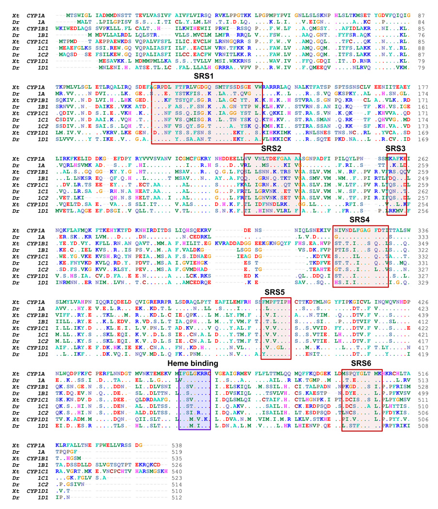
Figure 1 shows the deduced amino acid sequences of the cloned CYP1 transcripts in the clawed frog after alignment with orthologous zebrafish sequences. Pair-wise comparisons of the deduced amino acid sequences of the clawed frog CYP1s with the zebrafish CYP1 sequences shows the highest identity scores between putative orthologs (Table 1). Full length identity scores varied between 48 and 58%, with the CYP1D sequences showing the lowest score and the CYP1C sequences (X. tropicalis CYP1C1 and zebrafish CYP1C1) the highest. For all sequences, higher identity scores were found when substrate recognition site (SRS) sequences were compared than when the whole sequences were compared. There was between 52 and 69% identity in the SRS (Table 1).
Figure 1.
Deduced amino acid sequences of the cloned Xenopus tropicalis CYP1A, CYP1B1, CYP1C1, and CYP1D1 transcripts aligned with orthologous sequences in zebrafish (Danio rerio). The heme binding site and substrate recognition regions (SRS1–6) (Lewis et al., 2003) of the proposed enzymes are indicated by shading.
Table 1.
Percentage of sequence identity after pair-wise alignment (ClustalW) of deduced amino acid sequences, or substrate recognition site (SRS) regions as determined with BioEdit (Hall, 1999). Comparisons were made between pairs of the cloned X. tropicalis CYP1 sequences and orthologous sequences in zebrafish (Danio rerio). Bold underlined numbers indicate the highest identities observed.
| X.tropicalis | CYP1A | CYP1B1 | CYP1C1 | CYP1D1 | |||||
|---|---|---|---|---|---|---|---|---|---|
| Full | SRS | Full | SRS | Full | SRS | Full | SRS | ||
| D. rerio | CYP1A | 56 | 63 | 33 | 41 | 37 | 49 | 43 | 47 |
| CYP1B1 | 37 | 47 | 54 | 61 | 49 | 59 | 33 | 44 | |
| CYP1C1 | 38 | 43 | 49 | 52 | 58 | 69 | 36 | 46 | |
| CYP1C2 | 35 | 44 | 48 | 45 | 57 | 64 | 37 | 47 | |
| CYP1D1 | 45 | 57 | 30 | 38 | 34 | 33 | 48 | 52 | |
Approximately 95% of full sequence
Gene structure and phylogenic analysis
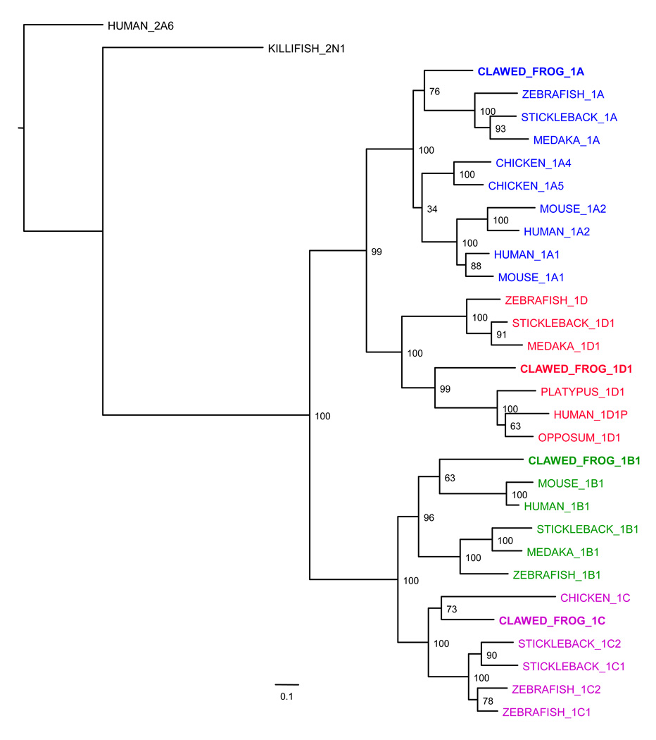
The X. tropicalis CYP1 deduced amino acid sequences were analyzed for molecular phylogenetic relationships with selected other vertebrate CYP1s (Figure 3). As expected the frog CYP1s cluster with the tetrapod CYP1s, with the exception of CYP1A. The phylogeny of amniota CYP1A1/CYP1A2 is complicated by the issue of reciprocal gene conversion between the two paralogs, which duplicated subsequent to the amphibian divergence (Goldstone and Stegeman, 2006). Thus, the single X. tropicalis CYP1A appears to be more similar to the fish CYP1As than to the amniota CYP1As included in the tree. Frog CYP1C clusters with the chicken CYP1C sequence, as previously suggested (Goldstone and Stegeman, 2006; Verslycke et al., 2006; Goldstone et al., 2009), leading us to conclude that the frog gene is a co-ortholog of the fish CYP1C genes. The frog CYP1D1 clusters with the deduced platypus and opossum CYP1D1 and with the human pseudogene sequence CYP1D1P (Goldstone et al., 2009).
Figure 3.
Maximum likelihood phylogenetic tree of frog CYP1 sequences with representative vertebrate CYP1 sequences. Numbers at nodal points are support values derived from maximum likelihood bootstrap analyses (200 replicates). To facilitate phylogenetic analyses of the Xenopus tropicalis sequences, the human pseudogene CYP1D1P was included in the alignment with stop codons removed. Zebrafish, mouse, and human CYP1 sequences have all been published (See Supplemental Table 2). Chicken, medaka, and stickleback CYP1B1 and CYP1C genes, and stickleback medaka CYP1D1, as well as platypus and opossum CYP1D1 sequences were predicted from the respective genomes. See also (Goldstone et al., 2007; Goldstone and Stegeman, 2008; Goldstone et al., 2009). The tree was rooted with human CYP2A6 and killifish CYP2N1.
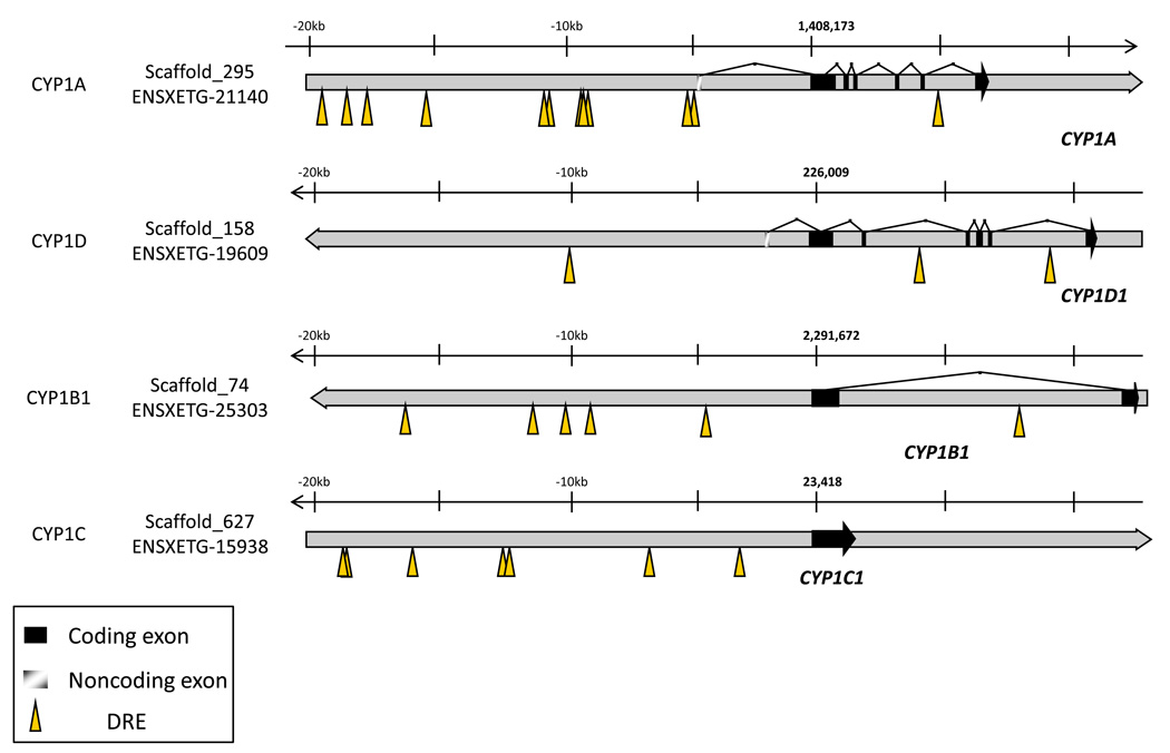
The gene structures for the frog CYP1 genes are very similar to that described previously for the zebrafish CYP1s. CYP1B1 is a two-exon gene, while CYP1A and CYP1D1 are seven-exon genes with a non-coding first exon (Figure 2). As in fish, clawed frog CYP1C1 is a single-exon gene, likely produced either via retrotransposition of an ancestral CYP1B1, or duplication of an ancestral CYP1B1 followed by loss of the single intron.
Figure 2.
Gene structure and dioxin response elements (DRE) for the four cloned X. tropicalis CYP1 genes. Expressed exons are black boxes, while striped boxes indicate untranslated regions. Shown also are the location of calculated XRE sequences (KNGCGTG). Note the large number of DREs upstream of CYP1A. CYP1B1 has two exons, while CYP1C1 has a single exon.
Dioxin response elements (DREs) are DNA regions where the AHR/ARNT transcription factor complex binds to induce gene expression. Consensus DRE core sequences, defined as (T/G)NGCGTG (Zeruth and Pollenz, 2007), were searched for in the 20-kb regions upstream from the clawed frog CYP1 gene start codons (using the X. tropicalis genome assembly 4.1, ENSEMBL version 58), and the locations of such sequences are depicted in Fig. 2. The gene having the greatest abundance of putative DRE sequences was CYP1A in which 13 such sequences were found, whereas CYP1B1 and CYP1C had 6 and 7, and CYP1D1 had only 1 putative DREs, respectively in the regions examined (Fig. 2). The CYP1A gene had a cluster of 5 DREs located about 5 kb upstream of the transcription start site (~9.5 kb upstream of the translation start site). There also were 2 DREs at about 1 kb upstream of the transcription start site.
Basal expression in developing X. tropicalis
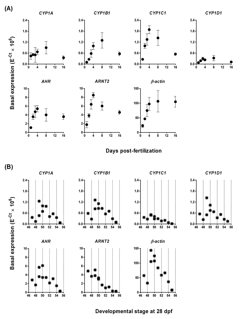
Temporal trends for basal expression of CYP1A, CYP1B1, CYP1C1, and CYP1D1, as well as AHR, ARNT2, and β-actin, were examined over the first 16 days of development in unexposed tadpoles. The seven genes displayed various trends for increasing levels of expression with development (Fig. 4A). Expression of CYP1C1 peaked at around 4 dpf whereas CYP1A, CYP1B1, and CYP1D1 expression peaked at around 8 dpf (Fig. 4A). Constitutive expression of the AHR and ARNT2 genes showed increasing trends up to day four, at which time the levels started to decrease. For AHR the level of expression stabilized between day 8 and day 16 post-fertilization whereas the CYP1s and ARNT2 decreased after peaking (Fig. 4A). The expression of β-actin increased up to day 4 after fertilization and then remained at the same level until day 16 (Fig. 4A).
Figure 4.
Time-dependent (A) and stage-dependent (B) variations in basal expression of CYP1A, CYP1B1, CYP1C1, CYP1D, AHR, ARNT2, and β-actin in developing Xenopus tropicalis tadpoles. Time-dependent variations in basal expression were studied in unexposed tadpoles collected from 1 day to 16 days post-fertilization (dpf). Data are shown as mean + SD. N=4, where the number of tadpoles per replicate was five at 1–8 dpf and one at 16 dpf. Variations with developmental stage were studied in a group of 13 unexposed tadpoles sampled at 28 dpf, where each star represents data from one tadpole. Stages were determined according to Nieuwkoop and Faber (1994). Expression was calculated by E−Ct.
After 28 days of development tadpole siblings showed a large variation in developmental stage, i.e., in a group of 13 siblings the stages varied from 47 to 55 when stage was determined according to the method by Nieuwkoop and Faber (1994). In order to assess how this variation might affect the constitutive levels of the CYP1s and the other genes we examined the expression of these genes in samples prepared from each individual of the 13 tadpoles. The results revealed a considerable variation within the stage interval examined. At NF 47–48 most of the genes analyzed (excluding CYP1D1 and β-actin) showed roughly similar expression levels as those observed in 16-dpf tadpoles. At later developmental stages expression of CYP1A, CYP1B1, CYP1D1, AHR and β-actin displayed a trend where expression first was increasing, peaking at NF 49–50, and then dramatically decreasing (Fig. 4B). The levels of CYP1C1 and ARNT2 showed a decreasing trend basically within the whole stage interval studied (Fig. 4B).
CYP1 response to PCB126 in two different age groups of tadpoles
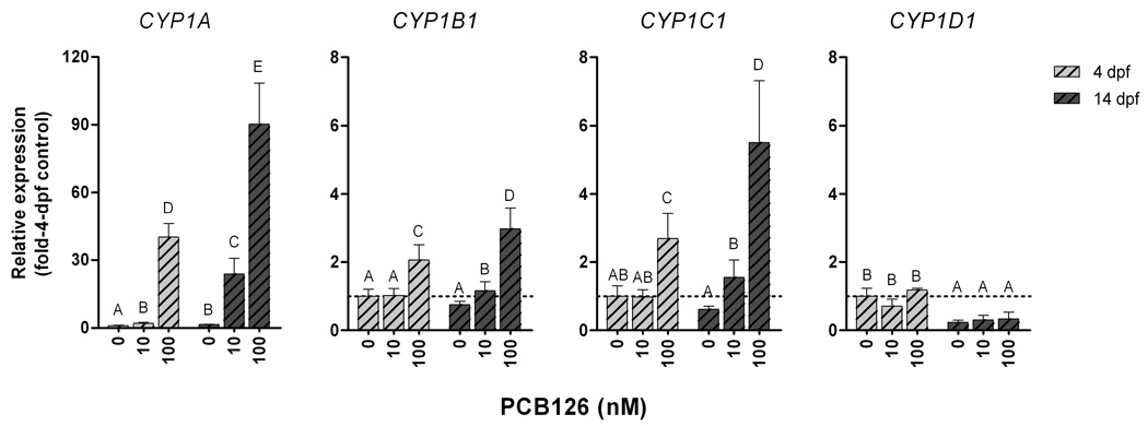
The purposes of this experiment were to compare the inducibility of the four CYP1 genes using a potent AHR agonist (PCB126), and to determine whether the induction response varied over development. CYP1 gene expression was examined in two age groups of tadpoles (4 and 14 dpf) and at two PCB126 concentrations (10 or 100 nM). As a reference gene β-actin was chosen because it displayed a stable basal expression level from 4 to 16 dpf as opposed to ARNT2 which showed a considerably lower expression at 16 dpf than at 4 dpf (Fig. 4A). Furthermore, β-actin was not affected by PCB126 exposure, at either concentration. Both age groups responded to PCB126 with CYP1 induction, but the strongest response was observed in the older tadpoles. After exposure to 10 nM PCB126 the 4-dpf tadpoles showed induction only of CYP1A, whereas at 14 dpf CYP1B1 and CYP1C1 were induced also (Fig. 5). When comparing the CYP1A levels to the levels in their own control group the 4- and 14-dpf tadpoles showed a 2-fold and 15-fold induction, respectively (not shown). The CYP1 expression levels in the controls differed somewhat between the 4- and 14-dpf groups. To allow comparison between age groups all data shown in Fig. 5 have been calibrated to the 4-dpf control levels. The higher PCB126 concentration (100 nM) induced CYP1A, CYP1B1, and CYP1C1 in both age groups although the older tadpoles showed the strongest induction. Compared to the 4-dpf controls, CYP1A, CYP1B1, and CYP1C1 were induced 40-, 2-, and 3-fold at 4 dpf, and 90-, 3-, and 6-fold at 14 dpf (Fig. 5). Compared to the 14-dpf controls, the fold-induction of CYP1A, CYP1B1, and CYP1C1 in the 14-dpf tadpoles was 60, and 4, and 9 (not shown). Thus, by both ways of calculation CYP1 gene induction was higher at 14 dpf than at 4 dpf. Regarding CYP1D1 expression, the control levels were lower at 14 dpf than at 4 dpf, but neither of the two PCB126 concentrations had any effect on CYP1D1 expression at either 4 nor 14 dpf (Fig. 5).
Figure 5.
Relative expression of CYP1A, CYP1B1, CYP1C1, and CYP1D in PCB126-exposed Xenopus tropicalis tadpoles at 4 days post-fertilization (dpf; light bars) and 14 dpf (dark bars). Starting 2 or 12 dpf groups of tadpoles were exposed for 24 hour to the carrier (0.1% acetone) or to 10 or 100 nM PCB126 plus carrier and subsequently held in clean water for another 24 hour before sampling. Calculations were made using β-actin as reference gene and the mean values of the different CYP1s in the 4-dpf carrier controls as calibrators (E−ΔΔCt). Statistically significant differences between groups were examined by one-way ANOVA followed by Tukey’s post-hoc test and are shown by different letters (p<0.05). Data are shown as mean ± SD (n = 5–6).
PCB126 concentration-response
EROD activity
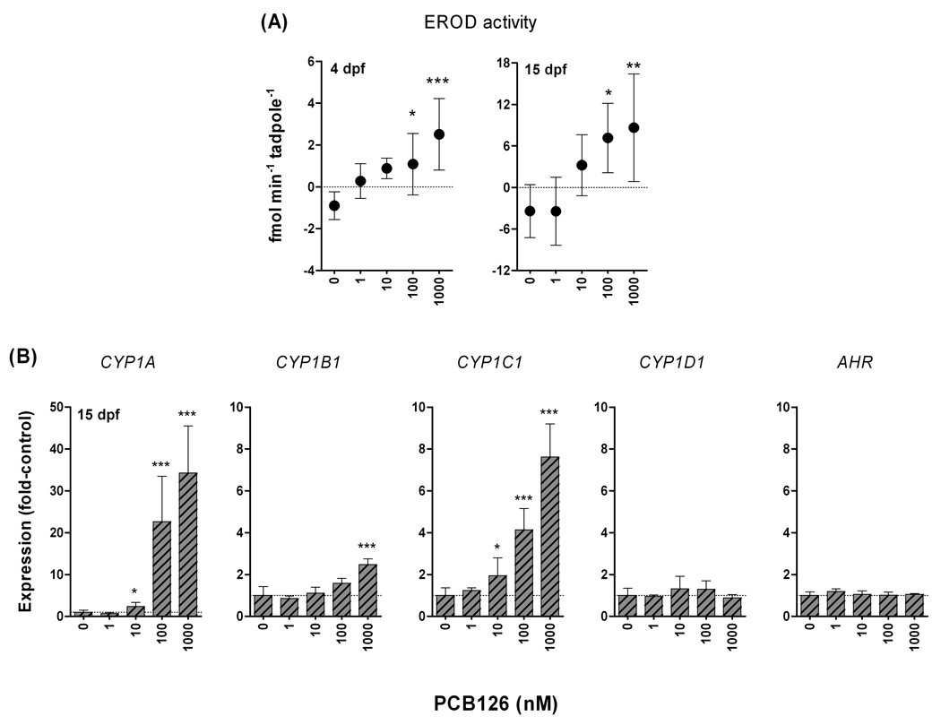
7-Ethoxyresorufin-O-deethylase (EROD) activity frequently is used as a marker of CYP1 induction, usually interpreted as CYP1A activity, although other CYP1s also catalyze this activity. The capacity for EROD induction was examined in 4- and 15-dpf tadpoles exposed to a range of PCB126 concentrations, or in carrier controls (0.1% acetone). We used an in vivo assay, in which tadpoles are placed in water containing 7-ethoxyresorufin. 7-Ethoxyresorufin is taken up by the animal (probably via the gills and skin) a proportion of the resorufin formed diffuses to the water, and the change in resorufin fluorescence of the water between two sampling time points is determined. For tadpoles of both age groups, the controls showed a decrease in fluorescence over time, suggesting absorption of 7-exthoxyresorufin (which has a background fluorescence) from the water. Presumably, the rate of resorufin formation / release to the water was not high enough to balance the removal of ethoxyresorufin (by absorption) in these tadpoles. Thus detection was limited by the kinetics for absorption of substrate and release of product by the tadpoles rather than by the sensitivity of the fluorometer. PCB126 exposure increased EROD activity (amount of resorufin released per tadpole per minute) at both 4 and 15 dpf. However, the reaction rate was extremely slow, the highest EROD activity level being in the femtomole/min/tadpole range. In both age groups there was significant induction compared with the controls at the highest doses (100 nM and 1 µM; Fig 6A). A positive trend for EROD activity over log PCB126 concentration was observed in both age groups (linear regression: r=0.6, p<0.01 and p<0.001, respectively). The older tadpoles (15 dpf) showed greater EROD activity than the younger ones (4 dpf) in the groups where EROD was detectable (10 nM -1 µM; Fig 6A). One group of 4-dpf tadpoles was also exposed to 10 µM PCB126, which resulted in a statistically significant induction compared with the corresponding carrier control (1% acetone; not shown), although the activity tended to be lower at 10 µM than at 1 µM (0.7±0.4 versus 2.5±1.7 fmol/min/tadpole).
Figure 6.
Concentration-response relationships for PCB126 versus A) EROD activity (at 4 or 15 dpf) and B) relative expression of CYP1A, rbCYP1B1, rbCYP1C1, CYP1D1, and AHR (at 15 dpf) in Xenopus tropicalis. Tadpoles were exposed to the carrier (0.1% acetone) or to 1 nM - 1 µM PCB126 plus the carrier for 24 hours and subsequently held for 24 hours in clean water (at 26 °C). Calculation of gene expression was made using β-actin as reference gene and the mean values of the different CYP1s in carrier controls were used as calibrators (E−ΔΔCt). EROD activity was analyzed in 5–6 replicates. Gene expression was analyzed in six replicates of controls and three replicates of PCB126-exposed tadpoles. Statistically significant differences compared with the controls were examined by one-way ANOVA followed by Dunnett’s post hoc test and are shown by stars (***p<0.001, **p<0.01, and *p<0.05). Data are shown as mean ± SD.
CYP1 and AHR gene expression
Concentration-response relationships for PCB126 exposure and CYP1 and AHR gene expression were examined in 15-dpf tadpoles. Both CYP1A and CYP1C1 showed a PCB126 concentration-dependent induction (Fig. 6B), with a LOEC of 10 nM PCB126 (nominal concentration in the system used). CYP1B1 was significantly induced only at the highest concentration (1 µM; Fig. 6B), although there was a positive trend for CYP1B1 expression over the log PCB126 concentration (linear regression: r = 0.9; p<0.0001). Expression of the CYP1D1 and AHR genes was not significantly induced by 1 nM - 1 µM PCB126 (Fig. 6B). Precise EC50 values could not be determined for induction of any of the CYP1 genes since the maximal level appeared to be outside the concentration range examined (i.e., the concentration-dependent increase in expression did not level out in the concentration range studied). However, setting the expression level at the highest PCB126 concentration as maximal induction level indicated that the EC50 values for CYP1A, CYP1B1, and CYP1C1 were at least 70, 100, and 100 nM.
CYP1 expression in tadpoles exposed to indigo, βNF, or PCB126
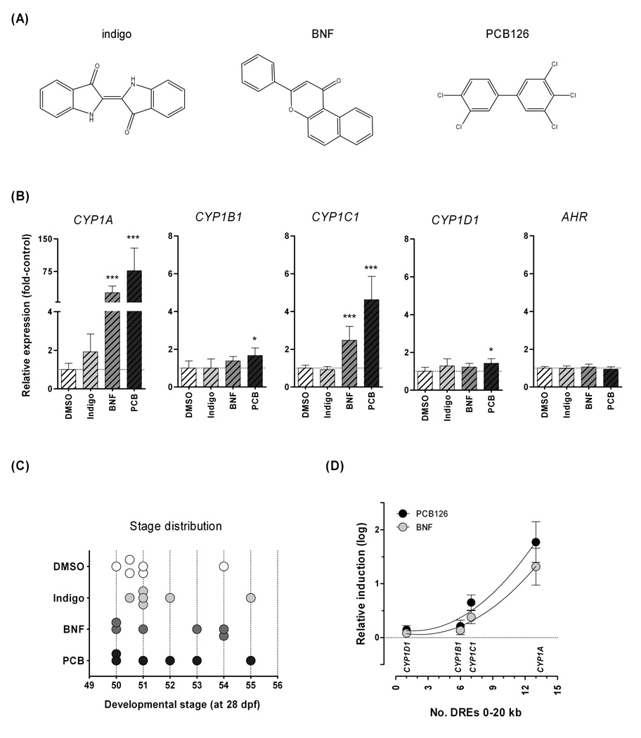
In this experiment we compared the gene expression response to two non-halogenated AHR agonists, indigo and βNF with the response to PCB126. Groups of 28-dpf tadpoles of the same clutch as those of the basal expression experiment were exposed to indigo, βNF, or PCB126 (all 1 µM), or to the carrier. We chose to use a relatively short exposure time (12 hours) since indigo seems to be rapidly degraded in fish and mammals (Adachi et al., 2001; Jönsson et al., 2006; Sugihara et al., 2008). Indigo had no statistically significant effect on any of the four CYP1 genes, although there was a tendency for induction of CYP1A (1.9-fold; Fig 7B). βNF induced CYP1A and CYP1C1 26- and 5-fold respectively, but had no significant effect on CYP1B1 or CYP1D1 (Fig 7A). PCB126 elicited a strong and moderate induction of CYP1A and CYP1C1 (77- and 5-fold versus the DMSO control, respectively), and a slight induction of CYP1B1 (1.7-fold). CYP1D1 expression also was slightly elevated (1.4-fold) in this experiment (Fig 7B). None of the three compounds had any effect on AHR gene expression (Fig 7B). The stage distribution varied somewhat between exposure groups (Fig. 7C). However no correlation was observed when plotting induction versus stage, suggesting the variation had minor effect on the results.
Figure 7.
Effect of indigo, β-naphthoflavone (βNF), and 3,3',4,4',5-pentachlorobiphenyl (PCB126) on CYP1A, CYP1B1, CYP1C1, CYP1D1, and AHR gene expression in Xenopus tropicalis tadpoles. A) Chemical structures for indigo, βNF, and PCB126, B) relative CYP1 gene expression, C) distribution of developmental stages within the exposure groups, and D) relationship between log transformed gene expression and DRE number in the regions 0–20 kb upstream of the start codon in the four CYP1 genes. At 28 days post-fertilization tadpoles were exposed for 12 hours to 50 ppm of DMSO (carrier; light bars), or to 1 µM of indigo (grey bars), βNF (dark grey bars), or PCB126 (dark bars) including the carrier (at 26 °C). Calculations of relative expression were made using β-actin as reference gene and the mean values in the DMSO group were used as calibrators (E−ΔΔCt). Statistically significant differences in gene expression compared with the control were determined by one-way ANOVA followed by Dunnett’s post hoc test and are shown by stars (***p<0.001 and *p<0.05). Data representing 5–6 replicates per exposure group are shown as mean + SD.
βNF- and PCB126-induced CYP1 expression was positively correlated with the number of putative DREs in the CYP1 genes when data for expression of each gene (i.e., log-transformed data from Fig. 7B) was plotted against the DRE number in the same gene (Fig. 7D).
Effects of PCB126 on development
The potential effect of PCB126 on development and growth was examined by determination of developmental stage and distance between the eyes (as an indicator of size) in groups of PCB126-exposed tadpoles. PCB126 at 1 nM – 10 µM had no statistically significant effect, relative to carrier, on these parameters in tadpoles exposed at 2 dpf and examined at 22 dpf. The median stage among the groups varied between 50 and 52 (minimum: 47–49; maximum: 54–55). The median distance between eyes was 6.0 mm in all groups except the 10 µM group where it was 6.3 mm (minimum: 2.5–4.0 mm and maximum: 7.5–8.5 mm; n=24–34)
Effects of PCB126 on the expression of other genes
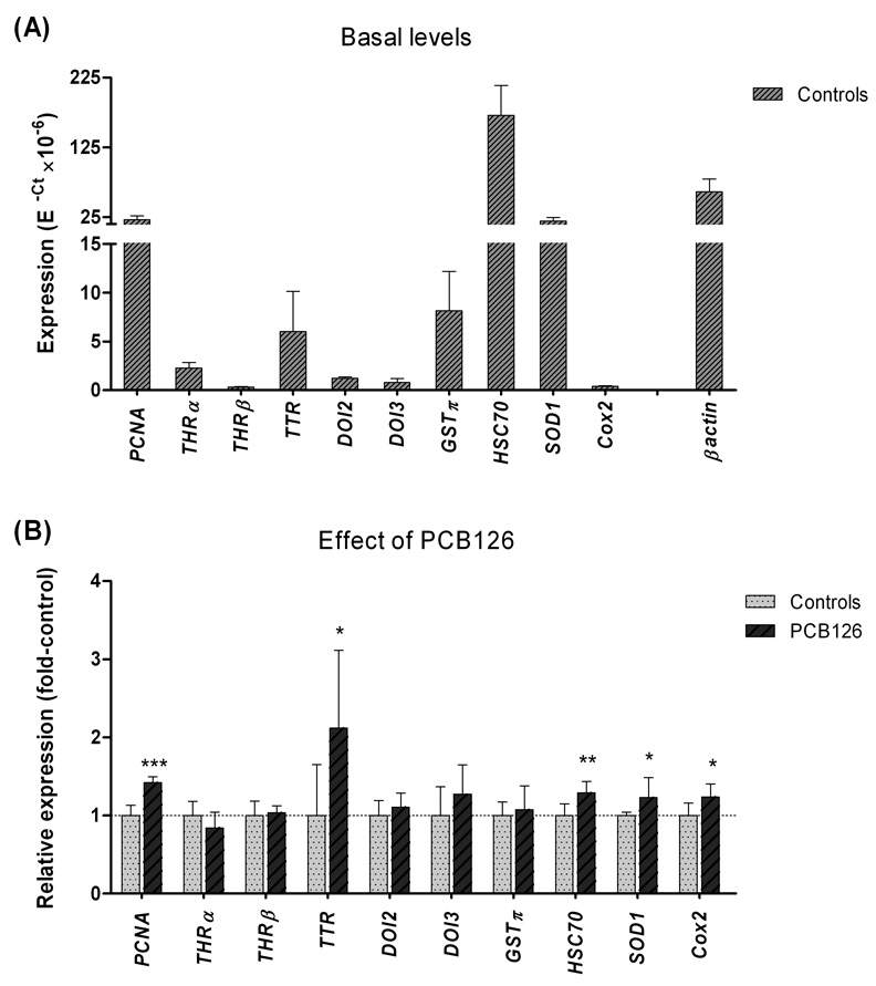
Although we did not find any external morphological defects in PCB126-exposed tadpoles, more subtle changes could occur, which might be reflected in gene expression. Thus we examined the expression of a battery of selected genes (PCNA, THRα, THRβ, TTR, DOI2, DOI3, GSTπ, HSC70, SOD1, and Cox-2) in 15-dpf tadpoles exposed to 1 µM PCB126 and in the controls (0.1% acetone). Fig. 8A shows expression levels of these genes in the controls. HSC70 (a constitutively expressed HSP70 form) showed the highest basal level of expression among the genes studied, followed by β actin, PCNA, and SOD1 (Fig. 8A). The lowest levels were observed for THRβ and Cox-2 (Fig. 8A). The results of PCB126 exposure are shown in Fig. 8B. The strongest effect by PCB126 exposure was observed for the thyroid hormone transporter, transthyretin (TTR), which showed a 2.1-fold up-regulation versus the control. Slight up-regulation was also observed for PCNA, HSC70, SOD1, and Cox-2 (1.4-, 1.3-, 1.2-, and 1.2-fold versus the control; Fig. 8B). None of the other thyroid system genes examined (THRα, THRβ, TTR, DOI2, and DOI3) nor GSTπ showed any statistically significant change by this exposure.
Figure 8.
Gene expression of selected marker genes in A) controls (basal levels) and B) PCB126-exposed Xenopus tropicalis tadpoles 15 days post-fertilization. Tadpoles were exposed to the carrier (0.1% acetone), or to 1 µM PCB126 plus the carrier for 24 hours and subsequently held for 24 hours in clean water (at 26 °C) before sampling. The marker genes studied were proliferating cell nuclear antigen (PCNA), thyroid hormone receptors α and β (THRα and THRβ), transthyretin (TTR), type II and III deiodinases (DOI2 and DOI3), glutathione S-transferase π (GSTπ), heat-chock protein 70 cognate (HSC70), CuZn superoxide dismutase (SOD1), and cyclooxygenase 2 (Cox-2). Carrier control levels were calculated by E−Ct, and relative expression after exposure to PCB126 was calculated using the mean values in carrier controls as calibrators (E−ΔΔCt; n=5–6). Statistically significant effects of PCB126 exposure were examined by one-way ANOVA followed by Dunnett’s post hoc test and are shown by stars (***p<0.001, **p<0.01 and, *p<0.05).
Discussion
Amphibian populations around the globe are experiencing declines, which may involve molecular effects of contaminants. In this study we determined gene expression effects of AHR agonists in an amphibian model, X. tropicalis. We identified and cloned the full suite of CYP1 genes in this diploid species and examined basal and AHR agonist-induced expression of the CYP1 genes, as well as of the AHR transcription factor. We further investigated effects of the environmental pollutant and AHR agonist PCB126 on the expression of a suite of marker genes involved in stress response, cell proliferation, thyroid homeostasis, and prostaglandin synthesis in X. tropicalis tadpoles.
Phylogeny
The initial emphasis was on CYP1 genes. X. tropicalis has one member in each of the CYP1A, CYP1B, CYP1C, and CYP1D subfamilies that are expressed in this amphibian. Given the molecular phylogeny and gene structure, and the fact that there are single genes in CYP1A, CYP1B and CYP1D subfamilies in fish, we conclude that the X. tropicalis genes in these subfamilies are orthologous with the genes in other vertebrates, and the frog genes are named accordingly. While this paper was being revised, we became aware of a publication that reports the presence of the same four CYP1 genes in the X. tropicalis genome (Suzuki and Iwata, 2010). However, that paper did not report any cloning to confirm expression of the genes and no experimental determination of the responsiveness of the CYP1s to AHR agonists. The occurrence of single genes in the four CYP1 subfamilies in X. tropicalis is consistent with divergence of CYP1A and CYP1D, and of CYP1B and CYP1C, ostensibly as a result of the posited second round of whole genome duplication, prior to the divergence of fish and tetrapods. The CYP1C line was duplicated in fish after that, and strictly speaking the frog CYP1C is orthologous to both CYP1C1 and CYP1C2. We have termed the frog CYP1C as CYP1C1, given the slightly greater identity of the frog CYP1C with CYP1C1s than with CYP1C2s, and with the advice of the CYP Nomenclature Committee. (As a note, it is strongly recommended that anyone dealing with new CYPs contact the committee to avoid misnaming CYP genes.) X. laevis, a tetraploid relative of X. tropicalis and a common experimental model, has two CYP1As (CYP1A6 and CYP1A7) (Fujita et al., 1999). Other CYP1 subfamilies have not been studied in X. laevis, but we might reasonably anticipate paralogous pairs in these other subfamilies too. X. tropicalis has one AHR gene, while X. laevis has two AHR1s (AHR1α and AHR1β) (Ohi et al., 2003; Lavine et al., 2005). At least two ARNT genes occur in X tropicalis (ARNT1, ENSXETG00000024204, and ARNT2) and X. laevis (ARNT1 and ARNT2) (Rowatt et al., 2003).
A single gene in each CYP1 subfamily and a single AHR gene in X. tropicalis suggests that comparative studies of the diploid X. tropicalis and the tetraploid X. laevis could facilitate understanding of the physiological and toxicological roles of CYP1s and AHR with endogenous and exogenous ligands.
Basal expression patterns
Transcript levels of the four CYP1 genes, AHR and ARNT2 in X. tropicalis were quite low in newly hatched tadpoles but increased dramatically over the first three days post-hatching (i.e., from day 1 to day 4 post-fertilization). These initial increases correlate with the rapid growth and development of the X. tropicalis tadpoles, and with the onset of feeding, soon after hatching. At approximately 4 dpf CYP1C1, AHR, and ARNT2 expression peaked and started to decrease, while CYP1A, CYP1B1, and CYP1D1 expression peaked at around 8 dpf (i.e., 7 days after hatching, NF 47–48). Similar results were reported for AHR1α and AHR1β in X. laevis, which displayed increasing expression levels from stages 12 to 54, with a temporal drop in the period around hatching, and a peak at stage 48 (Lavine et al., 2005). We did not examine gene expression before hatching but this has been studied in X. laevis, where the earliest expression of CYP1A7, AHR1β, and ARNT2 was detected at stages 12, 14, and 22, respectively (Ohi et al., 2003; Rowatt et al., 2003; Lavine et al., 2005).
In developing zebrafish, the levels of CYP1B1, CYP1C1, and CYP1C2 expression peaked around hatching, while CYP1A continues to increase for several weeks after hatching (Jönsson et al., 2007b). Similarly, aryl hydrocarbon hydroxylase (AHH) activity (largely due to CYP1A in fish) increased after hatching in killifish (Fundulus heteroclitus)(Binder and Stegeman, 1984). It is unclear whether these patterns observed in killifish, zebrafish, and X. tropicalis involve endogenous functions of the CYP1s or changing susceptibility to / accumulation of low levels of AHR agonists in the growing animals (e.g., contaminants derived from the females by deposition to the eggs or from feed). However, CYP1B1 is expressed in the developing eye, neural tissues, budding limbs, and other structures in embryonic mice, zebrafish, and chicken, and has been suggested to be involved in retinoid metabolism (Chambers et al., 2007; Choudhary et al., 2007; Yin et al., 2008). The functions of the various frog CYP1s are not known. Substrates could be identified by heterologous expression and assay of the CYPs, or could be suggested by homology modeling and docking of potential substrates. Such studies are underway in zebrafish (Scornaienchi et al., 2010) although activities have not been established with a broad range of possible substrates. Identifying physiological functions of the CYP1s is an important issue for further study.
Induced expression patterns
The relative induction of CYP1 subfamily genes by PCB126 in X. tropicalis was similar to that generally seen in fish, i.e., a strong induction of CYP1A, a weaker induction of CYP1B1 and CYP1C1 and no significant induction of CYP1D1. The CYP1D1 gene appears not to be AHR-regulated in zebrafish (Goldstone et al., 2009), and consistent with that, clawed frog CYP1D1 expression generally was unaffected by AHR agonists at any dose. A small but statistically significant increase was observed after exposure to PCB126 at 28 dpf, but whether this increase represents a real transcriptional induction or simply random variation is unclear.
Older tadpoles were more responsive to PCB126 than were younger tadpoles (14 versus 4 dpf). This was evident as a larger number of CYP1 transcripts being induced and a higher induction magnitude in older versus younger tadpoles (Fig. 4). Similar findings were reported for CYP1A6 in X. laevis, where both constitutive and induced expression levels increased with age and stage (studied at stages 36–37, 52–55, and 62–64) (Zimmermann et al., 2008). In an attempt to explain this stage-dependent difference in responsiveness Zimmermann et al. (2008) described the developmental expression of an AHR repressor gene in X. laevis. However, basal and induced levels of the AHR repressor transcript increased with time of development, indicating that the AHR repressor might not determine the differing responsiveness of CYP1A6 at late versus early developmental stages of X. laevis. In green frog and leopard frog, some PCBs appear to be more efficiently metabolized at later stages than at earlier stages of development (Leney et al., 2006). The planar (non-ortho-) PCB congeners were not analyzed by Leney et al. (2006). If the stage difference in metabolism includes the planar PCBs and TCDD as well, this metabolic difference could contribute to the observed stage differences in CYP1 gene response. The stage differences in CYP1 gene response in X. tropicalis and X. laevis could also involve toxicokinetic differences. Conceivably, bioavailability and distribution of inducer to target cells could differ at earlier and later stages as well. It is also possible that cross-talk with other transcription factors (e.g. steroid receptors) or epigenetic modifications could alter CYP1 induction. Furthermore, the differences in timing of maximal expression of AHR and ARNT expression, and the timing of maximal induction response, give rise to questions whether the amounts of AHR strictly determine the degree of induction response, and how much AHR is required for maximal response. It is clear that CYP1 regulation in frogs and other animals requires further study.
The responses of the CYP1 genes in X. tropicalis to three well- known AHR agonists, PCB126, βNF, and indigo, showed both differences and similarities to CYP1 induction in other vertebrates. PCB126 was the most potent of these three compounds in X. tropicalis tadpoles, but it still had relatively low potency compared with the response in larval fish. The PCB126 concentration needed to induce CYP1A, CYP1B1 and CYP1C1 in X. tropicalis tadpoles was considerably greater than that required to induce CYP1 expression in developing zebrafish; the LOEC value of PCB126 for CYP1A and CYP1C1 induction was 10 nM in clawed frog tadpoles (this study) and 0.3 nM in zebrafish larvae (Jönsson et al., 2007a). The EC50 for induction of CYP1A, CYP1B1, and CYP1C1 in X. tropicalis was 70–100 nM (or higher; 14 dpf) as compared to 1.4–2.7 nM for CYP1A, CYP1B1, CYP1C1, and CYP1C2 in zebrafish larvae (3 dpf; similar exposure design) (Jönsson et al., 2007a). The potency of PCB126 as a CYP1A inducer is relatively low in other frogs as well (Huang et al., 2001). Lesser sensitivity to TCDD in resistant as compared to susceptible strains or species is associated with a lower AHR binding affinity of TCDD, e.g., in birds (Brunström and Lund, 1988). Accordingly, altering amino acid residues in the ligand binding domain of avian AHRs was shown to convert AHR from resistant to susceptible forms (Karchner et al., 2006). Lavine et al. (2005) found that affinity of the X. laevis AHR for TCDD is more than 20 times less than that of the AHR in AH-responsive mice (AHRb-1 in C57BL/6J). If the affinity of AHR for halogenated agonists in other frogs is similar to that in X. laevis, it could partially explain the lower CYP1 induction potency and toxicity of dioxin and dioxin-like compounds in frogs versus mammals and fish.
Some studies have noted a difference in potency between halogenated and non-halogenated AHR agonists in a species (Flaveny et al., 2009). If the weak effects of TCDD and PCB126 in Xenopus tadpoles is due to a low AHR affinity only for halogenated AHR agonists, then possibly CYP1 genes might respond more strongly to non-halogenated agonists. However, indigo and βNF, both of which are potent AHR agonists in fish and mammals (Adachi et al., 2001; Jönsson et al., 2006), were less effective as CYP1 inducers than PCB126 in X. tropicalis tadpoles. Hence, three AHR agonists with different chemical structures all were weak or relatively weak CYP1 inducers in X. tropicalis. We note that 6-formylindolo[3,2-b]carbazole (FICZ), related to indigo, was more potent as a CYP1A inducer than TCDD in a X. laevis cell line (Laub et al., 2010) FICZ has an affinity similar to TCDD for AHR in fish and mammals (Rannug et al., 1987; Jönsson et al., 2009). It will be important to determine directly the affinity of X. laevis and X. tropicalis AHRs for these ligands, to address whether a low affinity for AHR can contribute to the weak responsiveness.
The abundance of DREs in the promoter region could be important for how strongly CYP1 genes respond to AHR agonists. The differences in inducibility of the four CYP1 genes we observed (Fig. 7B) may be related to the number of putative DREs in the promoter regions of these genes. Furthermore, Suzuki and Iwata (2010) suggested that resistance to TCDD in amphibians may be explained by differences in the number of DREs and their localization in the CYP1 gene promoters. In the mammalian CYP1A1 and CYP1A2 genes a cluster of 6–7 DREs located a few kb upstream of the transcriptional start site have been found important for the induction responsiveness (Nukaya et al., 2009). A similar cluster is also present in X. tropicalis. However, which DREs are functional in frog CYP1 genes is not known. Functional studies of putative DREs in CYP1A genes in mammals and fish have shown that not all DREs can initiate gene expression, and induction also depends on the occurrence of binding sites for other transcription factors and co-activators in the vicinity of the DREs (Zeruth and Pollenz, 2007). Conceivably, there could be fewer functional DREs in the clawed frog CYP1A than in CYP1A in many fish species, e.g., zebrafish (Jönsson et al., 2007b). Whether this possibility or, alternatively, some epigenetic regulation underlies the low induction response in frogs deserves exploration.
EROD activity
PCB126 induced EROD activity in a concentration-dependent manner in X. tropicalis, tadpoles, although the EROD reaction rate was extremely slow (in the fmol/min/tadpole range). Using a similar EROD assay in rainbow trout fry we recorded reaction rates in the picomole/min/fry range (unpublished results). This suggests differences in substrate specificities, in amounts of EROD catalyzing enzyme, or in phase 2 enzyme activities (i.e., affecting the resorufin fluorescence through conjugation) between X. tropicalis and rainbow trout. EROD rates induced in tissues, usually liver, of adult frogs are lower than typically seen in fish or mammals (Huang et al., 1998; Huang et al., 1999; Rankouhi et al., 2005). Hence, our observation might reflect a taxon specific difference in EROD rates.
Other effects of PCB126
Even at an extremely high nominal PCB126 concentration (10 µM), we found no visible morphological defects in tadpoles exposed at 2 dpf and screened at 22 dpf. In contrast, developing zebrafish embryos exposed to 10 nM PCB126 had edema, malformations, circulatory failure, and other defects, eventually leading to death (Jönsson et al., 2007a). The present findings in X. tropicalis support earlier studies in other frog species (including X. laevis) showing a relatively high resistance to toxicity of AHR agonists (Beatty et al., 1976; Jung and Walker, 1997; Lavine et al., 2005). The fact that dioxin-like contaminants have a low acute toxicity in tadpoles does not, however, rule out the possibility of that more subtle alterations occur which might cause disturbances becoming evident first at later life stages, e.g., in the adult frog. Thus, we analyzed the expression of selected marker genes in PCB126-exposed 14-dpf tadpoles. A slight up-regulation was observed for TTR, PCNA, HSC70, SOD1, and Cox-2 showing that PCB126 could have an effect on other genes than the CYP1s in X. tropicalis. Although the extent of induction of these genes was low and the PCB126 concentration high, our findings suggest that PCB126 could cause cell stress/oxidative stress, and interfere with thyroid function, prostaglandin metabolism, and cell proliferation in X. tropicalis tadpoles.
PCBs have several molecular targets in the thyroid system, affecting the thyroid gland, and metabolism and transport of the thyroid hormones. In order to evaluate effects of PCB126 on the thyroid system in X. tropicalis we studied expression of the genes for THRα, THRβ, DOI2, DOI3, and TTR in 14-dpf tadpoles (approximately stage 48) after two days of exposure to 1 µM PCB126. We found that only the expression of the thyroid hormone transporter TTR was affected, showing 2.1-fold induction versus the control. Similarly, TTR expression was induced 2.5-fold in X. laevis tadpoles exposed to Aroclor 1254, 50 ppb (Lehigh Shirey et al., 2006). The same X. laevis tadpoles showed a suppressed expression of DOI2 and DOI3 (Lehigh Shirey et al., 2006). This was suggested to be secondary to the up-regulation of TTR, i.e., increased levels of TTR protein would result in decreased levels of free circulating thyroid hormone, leading to down-regulation of THR-regulated genes, including DOI2 and DOI3. They also found that Aroclor delayed metamorphic timing (Lehigh Shirey et al., 2006). We did not see any effect of PCB126 on DOI2 and DOI3 in the current study, which may have been due to the shorter exposure time. Lehigh Shirley et al. (2006) exposed the tadpoles for 80 days (approximately between stage 48 and 59) with a frequent renewal of exposure solutions, an experimental setup which likely allowed more severe effects to develop. In addition, PCB126 has been shown to have a lower thyroid hormone disrupting potency than certain other PCB congeners. In mammalian studies hydroxylated metabolites of PCB106 and PCB159 were found to be more potent than PCB126 in suppressing THR-mediated transcription (Miyazaki et al., 2008).
The underlying mechanism for the increased TTR expression caused by PCB is not known. Notably, however we found an abundance of putative binding sites for E2F in the TTR promoter. E2F factors are transcription factors which regulate genes active in the S phase of the cell cycle, including proliferating cell nuclear antigen, PCNA (Dimova and Dyson, 2005).
The fact that the PCNA transcript levels were increased by exposure to PCB126 in X. tropicalis tadpoles is interesting in light of findings that PCNA expression is suppressed by PCB126 in zebrafish embryos (Jönsson et al., 2007a). Several studies in mammals have shown that persistent AHR agonists can inhibit cell proliferation by inducing cell cycle arrest (Gierthy and Crane, 1984; Huang and Elferink, 2005). On the other hand, in human cells the carcinogenic AHR agonist 3-methylcholanthrene has been shown to induce the expression of S phase-related genes, including PCNA, via an AHR-dependent mechanism, thus promoting cell proliferation (Watabe et al., 2010). It appears that the AHR can have either a suppressing or a stimulating effect on cell cycle progression, possibly depending on the cell type, ligand, and/or circumstances. However, since the functions of the thyroid system involve formation of new tissues, it is also possible that the induced expression of PCNA in PCB126-exposed tadpoles is related to effects on the thyroid system.
Regarding SOD1 and Cox-2 these genes have been reported to be induced by AHR agonists and their expression can possibly be regulated via AHR binding to DREs (Puga et al., 1997; Cho et al., 2001; Bugiak and Weber, 2009).
Conclusion
Identification of CYP1A, CYP1B1, CYP1C1, and CYP1D1 genes in X. tropicalis establishes that the four CYP1 subfamilies present in fish occur and are expressed in this amphibian model as well. The induction of these CYP1 genes relative to one another is similar to the pattern seen in zebrafish, with CYP1A being more strongly induced than CYP1B1 and CYP1C1, and CYP1D1 effectively not being induced in tadpoles exposed to the AHR agonists PCB126, βNF, and indigo. PCB126 was the most potent and indigo the least potent CYP1 inducer of these three compounds. PCB126 was strikingly less potent at inducing CYP1 induction and had little acute toxicity in X. tropicalis tadpoles compared to fish larvae, supporting earlier reports suggesting that frog tadpoles have relatively low sensitivity to dioxin-like contaminants. Whether lesser sensitivity is based on functional properties of the AHR or other features of the AHR signaling is not clear. Altered expression of selected marker genes in 14-dpf tadpoles exposed to 1 µM PCB126 for 2 days suggests that PCB126 may cause cell stress/oxidative stress, and interfere with thyroid function, prostaglandin metabolism, and cell proliferation in X. tropicalis tadpoles, although the magnitude of induction of the marker genes was low and the PCB126 concentration high. These results also indicate that gene expression analysis can be used to determine early effects of toxicants in frogs. Longer term exposure experiments, for instance life cycle studies, are needed to reveal whether the changes in gene expression observed could have a negative consequence for the animals. Additional studies of the AHR and the AHR gene battery and related genes are warranted, to search for possible mechanisms underlying the lower sensitivity of frogs to AHR agonists.
Supplementary Material
Acknowledgements
Financial support was provided by grants from the Carl Trygger’s Foundation and by the Swedish Research Council Formas to MEJ and CB, and by NIH Grant P42ES007381 (Superfund Research Program at Boston University) to JJS.
Footnotes
Publisher's Disclaimer: This is a PDF file of an unedited manuscript that has been accepted for publication. As a service to our customers we are providing this early version of the manuscript. The manuscript will undergo copyediting, typesetting, and review of the resulting proof before it is published in its final citable form. Please note that during the production process errors may be discovered which could affect the content, and all legal disclaimers that apply to the journal pertain.
Conflict of interest statements for authors
None of the authors have any conflict of interest regarding the research described in this article.
References
- Adachi J, Mori Y, Matsui S, Takigami H, Fujino J, Kitagawa H, Miller CA, III, Kato T, Saeki K, Matsudas T. Indirubin and indigo are potent aryl hydrocarbon receptor ligands present in human urine. J Biol Chem. 2001;276:31475–31478. doi: 10.1074/jbc.C100238200. [DOI] [PubMed] [Google Scholar]
- Beatty PW, Holscher MA, Neal RA. Toxicity of 2, 3, 7, 8-tetrachlorodibenzo-p-dioxin in larval and adult forms of Rana catesbeiana. Bull Environ Contam Toxicol. 1976;16:578–581. doi: 10.1007/BF01685367. [DOI] [PubMed] [Google Scholar]
- Binder RL, Stegeman JJ. Microsomal electron transport and xenobiotic monooxygenase activities during the embryonic period of development in the killifish, Fundulus heteroclitus. Toxicol Appl Pharmacol. 1984;73:432–443. doi: 10.1016/0041-008x(84)90096-6. [DOI] [PubMed] [Google Scholar]
- Brouwer A, Morse DC, Lans MC, Schuur AG, Murk AJ, Klasson-Wehler E, Bergman A, Visser TJ. Interactions of persistent environmental organohalogens with the thyroid hormone system: mechanisms and possible consequences for animal and human health. Toxicol Ind Health. 1998;14:59–84. doi: 10.1177/074823379801400107. [DOI] [PubMed] [Google Scholar]
- Brunström B, Lund J. Differences between chick and turkey embryos in sensitivity to 3,3',4,4'-tetrachloro-biphenyl and in concentration/affinity of the hepatic receptor for 2,3,7,8-tetrachlorodibenzo-p-dioxin. Comp Biochem Physiol C. 1988;91:507–512. doi: 10.1016/0742-8413(88)90069-2. [DOI] [PubMed] [Google Scholar]
- Bugiak B, Weber LP. Hepatic and vascular mRNA expression in adult zebrafish (Danio rerio) following exposure to benzo-a-pyrene and 2,3,7,8-tetrachlorodibenzo-p-dioxin. Aquat Toxicol. 2009;95:299–306. doi: 10.1016/j.aquatox.2009.03.009. [DOI] [PubMed] [Google Scholar]
- Carney SA, Chen J, Burns CG, Xiong KM, Peterson RE, Heideman W. Aryl hydrocarbon receptor activation produces heart-specific transcriptional and toxic responses in developing zebrafish. Mol Pharmacol. 2006;70:549–561. doi: 10.1124/mol.106.025304. [DOI] [PubMed] [Google Scholar]
- Chambers D, Wilson L, Maden M, Lumsden A. RALDH-independent generation of retinoic acid during vertebrate embryogenesis by CYP1B1. Development. 2007;134:1369–1383. doi: 10.1242/dev.02815. [DOI] [PubMed] [Google Scholar]
- Cho JS, Chang MS, Rho HM. Transcriptional activation of the human Cu/Zn superoxide dismutase gene by 2,3,7,8-tetrachlorodibenzo-p-dioxin through the xenobiotic-responsive element. Mol Genet Genomics. 2001;266:133–141. doi: 10.1007/s004380100536. [DOI] [PubMed] [Google Scholar]
- Choudhary D, Jansson I, Rezaul K, Han DK, Sarfarazi M, Schenkman JB. Cyp1b1 protein in the mouse eye during development: An immunohistochemical study. Drug Metab Dispos. 2007 doi: 10.1124/dmd.106.014282. [DOI] [PubMed] [Google Scholar]
- Dimova DK, Dyson NJ. The E2F transcriptional network: old acquaintances with new faces. Oncogene. 2005;24:2810–2826. doi: 10.1038/sj.onc.1208612. [DOI] [PubMed] [Google Scholar]
- Dong W, Teraoka H, Yamazaki K, Tsukiyama S, Imani S, Imagawa T, Stegeman JJ, Peterson RE, Hiraga T. 2,3,7,8-tetrachlorodibenzo-p-dioxin toxicity in the zebrafish embryo: local circulation failure in the dorsal midbrain is associated with increased apoptosis. Toxicol Sci. 2002;69:191–201. doi: 10.1093/toxsci/69.1.191. [DOI] [PubMed] [Google Scholar]
- Flaveny CA, Murray IA, Chiaro CR, Perdew GH. Ligand selectivity and gene regulation by the human aryl hydrocarbon receptor in transgenic mice. Mol Pharmacol. 2009;75:1412–1420. doi: 10.1124/mol.109.054825. [DOI] [PMC free article] [PubMed] [Google Scholar]
- Fontenot LW, Noble GP, Akins JM, Stephens MD, Cobb GP. Bioaccumulation of polychlorinated biphenyls in ranid frogs and northern water snakes from a hazardous waste site and a contaminated watershed. Chemosphere. 2000;40:803–809. doi: 10.1016/s0045-6535(99)00329-x. [DOI] [PubMed] [Google Scholar]
- Fujita Y, Ohi H, Murayama N, Saguchi K, Higuchi S. Molecular cloning and sequence analysis of cDNAs coding for 3-methylcholanthrene-inducible cytochromes P450 in Xenopus laevis liver. Arch Biochem Biophys. 1999;371:24–28. doi: 10.1006/abbi.1999.1425. [DOI] [PubMed] [Google Scholar]
- Gierthy JF, Crane D. Reversible inhibition of in vitro epithelial cell proliferation by 2,3,7,8-tetrachlorodibenzo-p-dioxin. Toxicol Appl Pharmacol. 1984;74:91–98. doi: 10.1016/0041-008x(84)90274-6. [DOI] [PubMed] [Google Scholar]
- Goldstone HM, Stegeman JJ. A Revised Evolutionary History of the CYP1A Subfamily: Gene Duplication, Gene Conversion, and Positive Selection. J Mol Evol. 2006 doi: 10.1007/s00239-005-0134-z. [DOI] [PubMed] [Google Scholar]
- Goldstone JV, Goldstone HM, Morrison AM, Tarrant A, Kern SE, Woodin BR, Stegeman JJ. Cytochrome P450 1 genes in early deuterostomes (tunicates and sea urchins) and vertebrates (chicken and frog): origin and diversification of the CYP1 gene family. Mol Biol Evol. 2007;24:2619–2631. doi: 10.1093/molbev/msm200. [DOI] [PubMed] [Google Scholar]
- Goldstone JV, Jönsson ME, Behrendt L, Woodin BR, Jenny MJ, Nelson DR, Stegeman JJ. Cytochrome P450 1D1: a novel CYP1A-related gene that is not transcriptionally activated by PCB126 or TCDD. Arch Biochem Biophys. 2009;482:7–16. doi: 10.1016/j.abb.2008.12.002. [DOI] [PMC free article] [PubMed] [Google Scholar]
- Goldstone JV, Stegeman JJ. Gene structure of the novel cytochrome P4501D1 genes in stickleback (Gasterosteus aculeatus) and medaka (Oryzias latipes) Mar Environ Res. 2008;66:19–20. doi: 10.1016/j.marenvres.2008.02.011. [DOI] [PMC free article] [PubMed] [Google Scholar]
- Goleman WL, Carr JA, Anderson TA. Environmentally relevant concentrations of ammonium perchlorate inhibit thyroid function and alter sex ratios in developing Xenopus laevis. Environ Toxicol Chem. 2002;21:590–597. [PubMed] [Google Scholar]
- Grubbs FE. Procedures for detecting outlying observations in samples. Technometrics. 1969;11:1–21. [Google Scholar]
- Guiney PD, Walker MK, Spitsbergen JM, Peterson RE. Hemodynamic dysfunction and cytochrome P4501A mRNA expression induced by 2,3,7,8-tetrachlorodibenzo-p-dioxin during embryonic stages of lake trout development. Toxicol Appl Pharmacol. 2000;168:1–14. doi: 10.1006/taap.2000.8999. [DOI] [PubMed] [Google Scholar]
- Gyllenhammar I, Holm L, Eklund R, Berg C. Reproductive toxicity in Xenopus tropicalis after developmental exposure to environmental concentrations of ethynylestradiol. Aquat Toxicol. 2009;91:171–178. doi: 10.1016/j.aquatox.2008.06.019. [DOI] [PubMed] [Google Scholar]
- Hall TA. BioEdit: a user-friendly biological sequence alignment editor and analysis program for Windows 95/98/NT. Nucl Acids Symp Ser. 1999;41:95–98. [Google Scholar]
- Hayes TB, Collins A, Lee M, Mendoza M, Noriega N, Stuart AA, Vonk A. Hermaphroditic, demasculinized frogs after exposure to the herbicide atrazine at low ecologically relevant doses. Proc Natl Acad Sci U S A. 2002;99:5476–5480. doi: 10.1073/pnas.082121499. [DOI] [PMC free article] [PubMed] [Google Scholar]
- Huang G, Elferink CJ. Multiple mechanisms are involved in Ah receptor-mediated cell cycle arrest. Mol Pharmacol. 2005;67:88–96. doi: 10.1124/mol.104.002410. [DOI] [PubMed] [Google Scholar]
- Huang YW, Karasov WH, Patnode KA, Jefcoate CR. Exposure of northern leopard frogs in the green bay ecosystem to polychlorinated biphenyls, polychlorinated dibenzo—p-dioxins, and polychlorinated dibenzofurans is measured by direct chemistry but not hepatic ethoxyresorufin—o-deethylase activity. Environ Toxicol Chem. 1999;18:2123–2130. doi: 10.1002/etc.5620181002. [DOI] [PubMed] [Google Scholar]
- Huang YW, Melancon MJ, Jung RE, Karasov WH. Induction of cytochrome P450-associated monooxygenases in northern leopard frogs, Rana pipiens, by 3,3′,4,4′,5-Pentachlorobiphenyl. Environ Toxicol Chem. 1998;17:1564–1569. [Google Scholar]
- Huang YW, Stegeman JJ, Woodin BR, Karasov WH. Immunohistochemical localization of cytochrome P4501A induced by 3,3',4,4',5-pentachlorobiphenyl (PCB 126) in multiple organs of northern leopard frogs, Rana pipiens. Environ Toxicol Chem. 2001;20:191–197. [PubMed] [Google Scholar]
- Jung RE, Walker MK. Effects of 2,3,7,8-tetrachlorodibenzo-p-dioxin (TCDD) on development of anuran amphibians. Environ Tox Chem. 1997;16:230–240. [Google Scholar]
- Jönsson EM, Abrahamson A, Brunström B, Brandt I. Cytochrome P4501A induction in rainbow trout gills and liver following exposure to waterborne indigo, benzo[a]pyrene and 3,3',4,4',5-pentachlorobiphenyl. Aquat Toxicol. 2006;79:226–232. doi: 10.1016/j.aquatox.2006.06.006. [DOI] [PubMed] [Google Scholar]
- Jönsson ME, Franks DG, Woodin BR, Jenny MJ, Garrick RA, Behrendt L, Hahn ME, Stegeman JJ. The tryptophan photoproduct 6-formylindolo[3,2-b]carbazole (FICZ) binds multiple AHRs and induces multiple CYP1 genes via AHR2 in zebrafish. Chem Biol Interact. 2009;181:447–454. doi: 10.1016/j.cbi.2009.07.003. [DOI] [PMC free article] [PubMed] [Google Scholar]
- Jönsson ME, Gao K, Olsson JA, Goldstone JV, Brandt I. Induction patterns of new CYP1 genes in environmentally exposed rainbow trout. Aquat Toxicol. 2010;98:311–321. doi: 10.1016/j.aquatox.2010.03.003. [DOI] [PMC free article] [PubMed] [Google Scholar]
- Jönsson ME, Jenny MJ, Woodin BR, Hahn ME, Stegeman JJ. Role of AHR2 in the expression of novel cytochrome P450 1 family genes, cell cycle genes, and morphological defects in developing zebrafish exposed to 3,3',4,4',5-pentachlorobiphenyl or 2,3,7,8-tetrachlorodibenzo-p-dioxin. Toxicol Sci. 2007a;100:180–193. doi: 10.1093/toxsci/kfm207. [DOI] [PubMed] [Google Scholar]
- Jönsson ME, Orrego R, Woodin BR, Goldstone JV, Stegeman JJ. Basal and 3,3,'4,4',5-Pentachlorobiphenyl-induced expression of Cytochrome P450 1A, 1B and 1C Genes in Zebrafish. Tox Appl Pharmacol. 2007b;221:29–41. doi: 10.1016/j.taap.2007.02.017. [DOI] [PMC free article] [PubMed] [Google Scholar]
- Karchner SI, Franks DG, Kennedy SW, Hahn ME. The molecular basis for differential dioxin sensitivity in birds: role of the aryl hydrocarbon receptor. Proc Natl Acad Sci U S A. 2006;103:6252–6257. doi: 10.1073/pnas.0509950103. [DOI] [PMC free article] [PubMed] [Google Scholar]
- Laub LB, Jones BD, Powell WH. Responsiveness of a Xenopus laevis cell line to the aryl hydrocarbon receptor ligands 6-formylindolo[3,2-b]carbazole (FICZ) and 2,3,7,8-tetrachlorodibenzo-p-dioxin (TCDD) Chem Biol Interact. 2010;183:202–211. doi: 10.1016/j.cbi.2009.09.017. [DOI] [PMC free article] [PubMed] [Google Scholar]
- Lavine JA, Rowatt AJ, Klimova T, Whitington AJ, Dengler E, Beck C, Powell WH. Aryl hydrocarbon receptors in the frog Xenopus laevis: two AhR1 paralogs exhibit low affinity for 2,3,7,8-tetrachlorodibenzo-p-dioxin (TCDD) Toxicol Sci. 2005;88:60–72. doi: 10.1093/toxsci/kfi228. [DOI] [PMC free article] [PubMed] [Google Scholar]
- Lehigh Shirey EA, Jelaso Langerveld A, Mihalko D, Ide CF. Polychlorinated biphenyl exposure delays metamorphosis and alters thyroid hormone system gene expression in developing Xenopus laevis. Environ Res. 2006;102:205–214. doi: 10.1016/j.envres.2006.04.001. [DOI] [PubMed] [Google Scholar]
- Leney JL, Drouillard KG, Haffner GD. Metamorphosis increases biotransformation of polychlorinated biphenyls: a comparative study of polychlorinated biphenyl metabolism in green frogs (Rana clamitans) and leopard frogs (Rana pipiens) at various life stages. Environ Toxicol Chem. 2006;25:2971–2980. doi: 10.1897/05-561r1.1. [DOI] [PubMed] [Google Scholar]
- Lewis DF, Gillam EM, Everett SA, Shimada T. Molecular modelling of human CYP1B1 substrate interactions and investigation of allelic variant effects on metabolism. Chem Biol Interact. 2003;145:281–295. doi: 10.1016/s0009-2797(03)00021-8. [DOI] [PubMed] [Google Scholar]
- Miyazaki W, Iwasaki T, Takeshita A, Tohyama C, Koibuchi N. Identification of the Functional Domain of Thyroid Hormone Receptor Responsible for Polychlorinated Biphenyl–Mediated Suppression of Its Action in Vitro. Environ Health Perspect. 2008;116:1231–1236. doi: 10.1289/ehp.11176. [DOI] [PMC free article] [PubMed] [Google Scholar]
- Na YR, Seok SH, Baek MW, Lee HY, Kim DJ, Park SH, Lee HK, Park JH. Protective effects of vitamin E against 3,3',4,4',5-pentachlorobiphenyl (PCB126) induced toxicity in zebrafish embryos. Ecotoxicol Environ Saf. 2009;72:714–719. doi: 10.1016/j.ecoenv.2008.09.015. [DOI] [PubMed] [Google Scholar]
- Nieuwkoop PD, Faber J, editors. Normal table of Xenopus laevis (Daudin) : a systematical and chronological survey of the development from the fertilized egg till the end of metamorphosis. New York: Garland Publishing, Inc.; 1994. [Google Scholar]
- Nukaya M, Moran S, Bradfield CA. The role of the dioxin-responsive element cluster between the Cyp1a1 and Cyp1a2 loci in aryl hydrocarbon receptor biology. Proc Natl Acad Sci U S A. 2009;106:4923–4928. doi: 10.1073/pnas.0809613106. [DOI] [PMC free article] [PubMed] [Google Scholar]
- Ohi H, Fujita Y, Miyao M, Saguchi K-i, Murayama N, Higuchi S. Molecular cloning and expression analysis of the aryl hydrocarbon receptor of Xenopus laevis. Biochem Biophys Resh Com. 2003;307:595–599. doi: 10.1016/s0006-291x(03)01244-0. [DOI] [PubMed] [Google Scholar]
- Pettersson I, Berg C. Environmentally relevant concentrations of ethynylestradiol cause female-biased sex ratios in Xenopus tropicalis and Rana temporaria. Environ Toxicol Chem. 2007;26:1005–1009. doi: 10.1897/06-464r.1. [DOI] [PubMed] [Google Scholar]
- Philips BH, Susman TC, Powell WH. Developmental differences in elimination of 2,3,7,8-tetrachlorodibenzo-p-dioxin (TCDD) during Xenopus laevis development. Mar Environ Res. 2006;62 Suppl:S34–S37. doi: 10.1016/j.marenvres.2006.04.027. [DOI] [PubMed] [Google Scholar]
- Puga A, Hoffer A, Zhou S, Bohm JM, Leikauf GD, Shertzer HG. Sustained increase in intracellular free calcium and activation of cyclooxygenase-2 expression in mouse hepatoma cells treated with dioxin. Biochem Pharmacol. 1997;54:1287–1296. doi: 10.1016/s0006-2952(97)00417-6. [DOI] [PubMed] [Google Scholar]
- Rankouhi TR, Koomen B, Sanderson JT, Bosveld AT, Seinen W, van den Berg M. Induction of ethoxy-resorufin-O-deethylase activity by halogenated aromatic hydrocarbons and polycyclic aromatic hydrocarbons in primary hepatocytes of the green frog (Rana esculenta) Environ Toxicol Chem. 2005;24:1428–1435. doi: 10.1897/04-367r.1. [DOI] [PubMed] [Google Scholar]
- Rannug A, Rannug U, Rosenkranz HS, Winqvist L, Westerholm R, Agurell E, Grafstrom A-K. Certain Photooxidized Derivatives of Tryptophan Bind with Very High Affinity to the Ah Receptor and Are Likely to be Endogenous Signal Substances. J Biol Chem. 1987;262:15422–15427. [PubMed] [Google Scholar]
- Rowatt AJ, DePowell JJ, Powell WH. ARNT gene multiplicity in amphibians: characterization of ARNT2 from the frog Xenopus laevis. J Exp Zool B Mol Dev Evol. 2003;300:48–57. doi: 10.1002/jez.b.45. [DOI] [PubMed] [Google Scholar]
- Ruijter JM, Ramakers C, Hoogaars WMH, Karlen Y, Bakker O, van den Hoff MJB, Moorman AFM. Amplification efficiency: linking baseline and bias in the analysis of quantitative PCR data. Nucl Acids Res. 2009;37:e45. doi: 10.1093/nar/gkp045. [DOI] [PMC free article] [PubMed] [Google Scholar]
- Schlezinger JJ, Struntz WD, Goldstone JV, Stegeman JJ. Uncoupling of cytochrome P450 1A and stimulation of reactive oxygen species production by co-planar polychlorinated biphenyl congeners. Aquatic Toxicology. 2006;77:422–432. doi: 10.1016/j.aquatox.2006.01.012. [DOI] [PubMed] [Google Scholar]
- Schmittgen TD, Livak KJ. Analyzing real-time PCR data by the comparative C(T) method. Nat Protoc. 2008;3:1101–1108. doi: 10.1038/nprot.2008.73. [DOI] [PubMed] [Google Scholar]
- Scornaienchi M, Thornton C, Willett K, Wilson J. Functional differences in the cytochrome P450 1 family enzymes from zebrafish (Danio rerio) using heterologously expressed proteins. Arch Biochem Biophys. 2010;502:17–22. doi: 10.1016/j.abb.2010.06.018. [DOI] [PMC free article] [PubMed] [Google Scholar]
- Smolowitz RM, Hahn ME, Stegeman JJ. Immunohistochemical localization of cytochrome P-450IA1 induced by 3,3',4,4'-tetrachlorobiphenyl and by 2,3,7,8-tetrachlorodibenzofuran in liver and extrahepatic tissues of the teleost Stenotomus chrysops (scup) Drug Metabol Disposit. 1991;19:113–123. [PubMed] [Google Scholar]
- Stamatakis A. RAxML-VI-HPC: maximum likelihood-based phylogenetic analyses with thousands of taxa and mixed models. Bioinformatics. 2006a;22:2688–2690. doi: 10.1093/bioinformatics/btl446. [DOI] [PubMed] [Google Scholar]
- Stamatakis A. Phylogenetic models of rate heterogeneity: A high performance computing perspective. Proc. of IPDPS2006. 2006b [Google Scholar]
- Stuart SN, Chanson JS, Cox NA, Young BE, Rodrigues AS, Fischman DL, Waller RW. Status and trends of amphibian declines and extinctions worldwide. Science. 2004;306:1783–1786. doi: 10.1126/science.1103538. [DOI] [PubMed] [Google Scholar]
- Sugihara K, Okayama T, Kitamura S, Yamashita K, Yasuda M, Miyairi S, Minobe Y, Ohta S. Comparative study of aryl hydrocarbon receptor ligand activities of six chemicals in vitro and in vivo. Archives of Toxicology. 2008;82:5–11. doi: 10.1007/s00204-007-0232-3. [DOI] [PubMed] [Google Scholar]
- Sutter TR, Tang YM, Hayes CL, Wo YY, Jabs EW, Li X, Yin H, Cody CW, Greenlee WF. Complete cDNA sequence of a human dioxin-inducible mRNA identifies a new gene subfamily of cytochrome P450 that maps to chromosome 2. J Biol Chem. 1994;269:13092–13099. [PubMed] [Google Scholar]
- Suzuki KT, Iwata H. Cytochrome P450 Family 1 Genes in Xenopus tropicalis. In: Isobe T, Nomiyama K, Subramanian A, Tanabe S, editors. Interdisciplinary Studies on Environmental Chemistry — Environmental Specimen Bank. 2010. pp. 155–160. [Google Scholar]
- Ter Schure AF, Larsson P, Merila J, Jonsson KI. Latitudinal fractionation of polybrominated diphenyl ethers and polychlorinated biphenyls in frogs (Rana temporaria) Environ Sci Technol. 2002;36:5057–5061. doi: 10.1021/es0258632. [DOI] [PubMed] [Google Scholar]
- Watabe Y, Nazuka N, Tezuka M, Shimba S. Aryl hydrocarbon receptor functions as a potent coactivator of E2F1-dependent trascription activity. Biol Pharm Bull. 2010;33:389–397. doi: 10.1248/bpb.33.389. [DOI] [PubMed] [Google Scholar]
- Verslycke T, Goldstone JV, Stegeman JJ. Isolation and phylogeny of novel cytochrome P450 genes from tunicates (Ciona spp.): a CYP3 line in early deuterostomes? Mol Phylogenet Evol. 2006;40:760–771. doi: 10.1016/j.ympev.2006.04.017. [DOI] [PubMed] [Google Scholar]
- Whitlock JP., Jr Induction of cytochrome P4501A1. Annu Rev Pharmacol Toxicol. 1999;39:103–125. doi: 10.1146/annurev.pharmtox.39.1.103. [DOI] [PubMed] [Google Scholar]
- Yin HC, Tseng HP, Chung HY, Ko CY, Tzou WS, Buhler DR, Hu CH. Influence of TCDD on zebrafish CYP1B1 transcription during development. Toxicol Sci. 2008;103:158–168. doi: 10.1093/toxsci/kfn035. [DOI] [PubMed] [Google Scholar]
- Zanette J, Jenny MJ, Goldstone JV, Woodin BR, Watka LA, Bainy AC, Stegeman JJ. New cytochrome P450 1B1, 1C2 and 1D1 genes in the killifish Fundulus heteroclitus: Basal expression and response of five killifish CYP1s to the AHR agonist PCB126. Aquat Toxicol. 2009;93:234–243. doi: 10.1016/j.aquatox.2009.05.008. [DOI] [PMC free article] [PubMed] [Google Scholar]
- Zeruth G, Pollenz RS. Functional analysis of cis-regulatory regions within the dioxin-inducible CYP1A promoter/enhancer region from zebrafish (Danio rerio) Chem Biol Interact. 2007;170:100–113. doi: 10.1016/j.cbi.2007.07.003. [DOI] [PubMed] [Google Scholar]
- Zimmermann AL, King EA, Dengler E, Scogin SR, Powell WH. An Aryl Hydrocarbon Receptor Repressor from Xenopus laevis: Function, Expression and Role in Dioxin Responsiveness during Frog Development. Toxicol Sci. 2008;104:124–134. doi: 10.1093/toxsci/kfn066. [DOI] [PMC free article] [PubMed] [Google Scholar]
Associated Data
This section collects any data citations, data availability statements, or supplementary materials included in this article.


