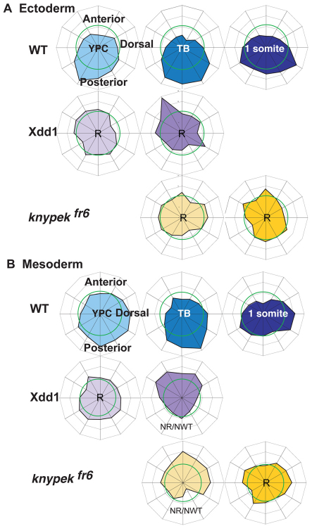
Fig. 4.
Biased MTOC position depends on some components of Wnt/PCP signaling. (A) MTOC distributions in ectoderm in wild type and in embryos with reduced Wnt/PCP signaling, Xdd1-injected or knypek mutants, and (B) MTOC distributions in mesoderm in wild type and embryos lacking Wnt planar cell polarity signaling. Wnt/PCP signaling, Xdd1-injected or knypek mutants. A uniform distribution of centrosomes is indicated by the green ring (at 8.3%). The outermost ring of the graph is set at 16% of cells. NR/NWT denotes distributions that are statistically neither like random nor like wild type. R denotes random distributions.

