Abstract
Background:
Double gallbladder is a rare anomaly of the biliary tract. Double gallbladder arising from the left hepatic duct was previously reported only once in the literature.
Case Report:
A case of symptomatic cholelithiasis in a double gallbladder, diagnosed on preoperative ultrasound, computed tomography (CT) and endoscopic retrograde cholangiopancreatogram (ERCP) is reported. At laparoscopic cholangiography via the accessory gallbladder no accessory cystic duct was visualized. After conversion to open cholecystectomy, the duplicated gallbladder was found to arise directly from the left hepatic duct; it was resected and the duct repaired.
Conclusions:
We emphasize that a careful intraoperative cholangiographic evaluation of the accessory gallbladder is mandatory in order to prevent inadvertent injury to bile ducts, since a large variety of ductal abnormality may exist.
Keywords: Double gallbladder, Biliary anomalies, Laparoscopic cholecystectomy
INTRODUCTION
Double gallbladder is a rare anomaly of the biliary tract, occurring in about 1 per 3800 cases at autopsy.1 Two cases of double gallbladders managed laparoscopically have been reported previously.2,3 We report herein another patient in whom laparoscopic cholecystectomy was attempted. The case represents a very rare variety of a double gallbladder, only once previously reported in the literature.4 It highlights possible anomaly of the accessory biliary system, emphasizing the need for an intraoperative cholangiography in order to prevent iatrogenic injuries to the bile ducts.
CASE REPORT
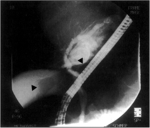
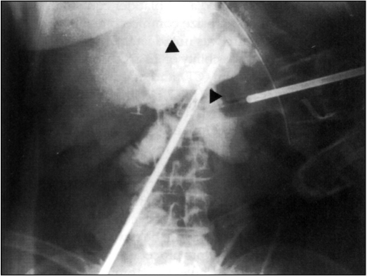
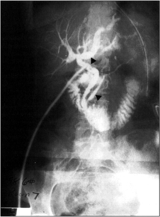
A 69-year-old female presented with several months history of right upper abdominal and epigastric pain. Ultrasonography revealed a gallbladder containing multiple stones and a normal-size common bile duct. In addition, a cystic structure was noted lateral to the left hepatic duct, raising the possibility of an accessory gallbladder. Computed tomography (CT) and endoscopic retrograde cholangiopancreatogram (ERCP) confirmed the presence of an accessory, partially intrahepatic gallbladder, which also contained stones (Figure 1). No ductal stones were visualized, and liver function tests were normal. Since the accessory gallbladder did not have an identified cystic duct on ERCP, the laparoscopic procedure started with a double cholangiogram through the cystic duct of the normal gallbladder and the accessory gallbladder ((Figure 2). No accessory cystic duct was, however, visualized, and the laparoscopic procedure was converted to an open procedure. Cholecystectomy of the primary gallbladder was completed, and a cholecystectomy of the accessory gallbladder was performed in a retrograde fashion. The accessory gallbladder was found to have no cystic duct and originated directly from the distal left hepatic duct. It was dissected off the lateral wall of the left hepatic duct, and the resulting 3 mm defect was closed with 5-0 polydioxanone. Completion cholangiogram revealed several small stones in the distal common bile duct, which was then explored (Figure 3). Recovery was uneventful, and the T-tube cholangiogram was normal. Pathology report described a 3 × 4 cm accessory gallbladder containing three stones. Histology revealed chronic cholecystitis with a mild dysplasia of the mucosa.
Figure 1.
Preoperative ERCP demonstrating double gallbladder with stones. (See arrows.)
Figure 2.
Intraoperative cholangiogram performed via the accessory gallbladder. (Upper arrow shows the left hepatic duct. Lower arrow points at the accessory gallbladder.)
Figure 3.
Completion T-tube cholangiogram showing continuity of the biliary tree. (Upper arrow shows the left hepatic duct. Lower arrow points at the pancreatic duct.)
DISCUSSION
Double gallbladder is a biliary anomaly usually not diagnosed preoperatively. Instead, it often represents an intraoperative surprise2 or is missed during the operation, only to be diagnosed at postoperative ERCP, performed for persistent biliary symptoms.5,6 In our case, both gallstone-containing gallbladders were probably symptomatic. The presence of the accessory gallbladder was suggested by the sonogram and confirmed on pre-operative ERCP and CT scan. The operation was started laparoscopically, but was converted to an open procedure due to the absence of the accessory cystic duct.
This case represents a type VI in the large spectrum of accessory gallbladders as proposed by Mochizuki7 (Table 1), or type C as proposed by Harlafits et al. (Table 2). This rare variety of the accessory gallbladder has been reported only once.4 Among 207 cases reviewed by Harlafits et al.,4 the majority of the anomalies consisted of duplicated gallbladders sharing the same cystic duct (Type A), or accessory gallbladders, with two cystic ducts entering common bile duct separately (Type B). The large spectrum of ductal anomalies associated with a double gallbladder mandates an intra-operative cholangiogram prior to the resection of the accessory gallbladder. Such an approach would minimize the risks of inadvertent injury to the biliary ductal system.
Table 1.
Classification of double gallbladder. Adopted from Mochizuki S, Makita T7
| Type | Description of the anatomy |
|---|---|
| Type I | Diverticulum of the cystic duct |
| Type II | Diverticulum of the neck of the main gallbladder |
| Type III | A sac attached to the neck of the main gallbladder via a small cystic duct |
| Type IV | Connection of the accessory sac to the middle of the hepatic duct via a small cystic duct |
| Type V | Duplicated fundus of the gallbladder |
| Type VI | Accessory sac attached to a hepatic duct of lateral left lobe of the liver |
Table 2.
Classification of double gallbladder. Adopted from Herlaftis N, Gray SW, Skandalakis JE4
| Types | Anatomic description |
|---|---|
| A (The split primordium group) | Single cystic duct entering the common bile duct |
| B (The accessory gallbladder group) | Two or more cystic ducts opening separately into the CBD |
| C (Miscellaneous anomalies) | Other rare anomalies not included in A or B group |
References:
- 1. Boyden EA. The accessory gallbladder, an embriological and comparative study of aberrant biliary vesicles occurring in man and the domestic mammals. Am J Anat. 1926;38:117 [Google Scholar]
- 2. Garcia CJ, Weber A, Serrano BF, Tanur TB. Double gallbladder treated successfully by laparoscopy. J Laparoendosc Surg. 1993;3:153–155 [DOI] [PubMed] [Google Scholar]
- 3. Miyajima N, Yamakawa T, Varma A, Uno K, Othaki S, Kano N. Experience with laparoscopic double gallbladder removal. Surg Endosc. 1995;9:63–66 [DOI] [PubMed] [Google Scholar]
- 4. Herlaftis N, Gray SW, Skandalakis JE. Multiple gallbladders. Surg Gynecol Obstet. 1977;145:928–934 [PubMed] [Google Scholar]
- 5. Heinerman M, Lexer G, Sungler P, Mayer F, Boeckl O. ERCP demonstration of a double gallbladder following laparoscopic cholecystectomy. Surg Endosc. 1995;9:61–62 [DOI] [PubMed] [Google Scholar]
- 6. Silvis R, van Wieringen AJ, van der Werken CH. Reoperation for a symptomatic double gallbladder. Surg Endosc. 1996;10:336–337 [DOI] [PubMed] [Google Scholar]
- 7. Mochizuki S, Makita T. Double gallbladder of swine. Kaibogaku Zassbi J Anat. 1996;71:650–655 [PubMed] [Google Scholar]


