Abstract
A 30-year-old woman presented with halitosis, sour taste, bloating, and right-sided abdominal pain of 3-months' duration. An upper gastrointestinal series revealed a diverticulum in the posterior cardia of the stomach. The patient underwent a laparoscopic resection of the diverticulum. Postoperatively, the patient did well; at a 28-month follow-up, no further symptoms were reported. Laparoscopic removal of a diverticulum produced an excellent outcome.
Keywords: Gastric diverticulum, Laparoscopic resection
INTRODUCTION
Gastric diverticula are found in the gastric cardia (true diverticulum) or distal stomach (false diverticulum). Patients often present with vague symptoms that may be attributable to other gastrointestinal pathology. Once all other pathologies have been addressed, diverticula in the gastric cardia can be successfully treated laparoscopically. Five cases of laparoscopic resection of gastric diverticula have been reported.1–4 Laparoscopic removal of cardia gastric diverticula produced good to excellent outcomes.
CASE REPORT
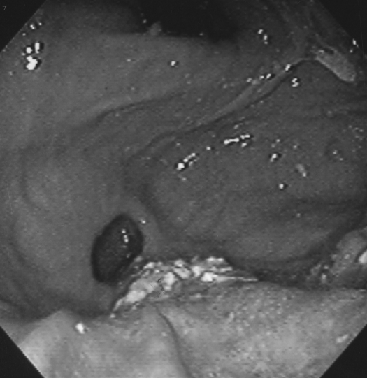
In August 2000, a 30-year-old woman presented with fatty food intolerance, gas belch symptoms, abdominal discomfort, and a sour taste in her mouth. She underwent an uncomplicated laparoscopic cholecystectomy; pathology confirmed cholelithiasis and chronic cholecystitis. Her symptoms resolved postoperatively. Four months later, she returned complaining of halitosis, sour taste, bloating, and right-sided abdominal pain of 3-months' duration. She denied having any heartburn. Her family physician had treated her with omeprazole, which initially alleviated the symptoms. Her repeat abdominal examination was normal. An esophogastroduodenoscopy disclosed small fundic polyps and a gastric diverticulum near the fundus that contained old food particles (Figure 1). Minor changes were noted at the gastroesophageal junction that were consistent with mild reflux disease.
Figure 1.
A retroflexed view using the gastroscope reveals the gastric diverticulum in the fundus.
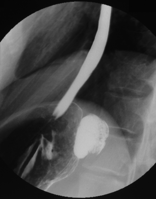
Pathology revealed benign fundic polyps; biopsies were negative for evidence of Helicobacter pylori. An upper gastrointestinal series revealed a diverticulum in the posterior cardia of the stomach (Figure 2). Seven months after her initial presentation, the patient underwent a laparoscopic resection of the diverticulum.
Figure 2.
An upper gastrointestinal series illustrates the diverticulum in the posterior cardia of the stomach.
A pneumoperitonieum was obtained with an open Hassan technique. A right-sided port was used to elevate the liver and 2 left-sided ports were used to take down the short gastric vessels with the AutoSonix ULTRASHEARS (US Surgical, Tyco Health Care, Norwalk CT). The posterior cardia of the stomach was then mobilized and inspected.
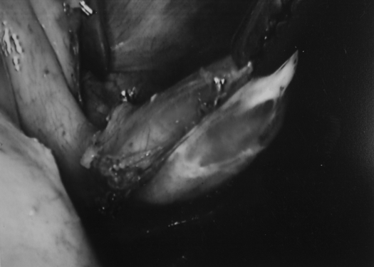
The gastric diverticulum was isolated, dissected free of retroperitoneal attachments to the base of the diverticulum, and transected with an ENDO GIA Universal (US Surgical, Tyco Health Care, Norwalk, CT) (Figure 3). Pathology revealed a 3.0 x 2.5 x 2.0-cm pouch with layers of mucosa, muscularis mucosa, submucosa, and serosa, but not the muscular propria. Postoperatively, the patient did well; at 28-month follow-up, no further symptoms were reported.
Figure 3.
The gastric diverticulum is dissected free of the retroperitoneal attachments.
DISCUSSION
In 1951, Palmer collectively reviewed 31 reports of true gastric diverticula representing 412 cases.5 Diverticula were found in 0.02% (6/29 900) of autopsy studies and in 0.04% (165/380 000) of upper gastrointestional studies. Other diverticuli are found on gastroscopy or at the time of surgery. Seventy-five percent of true gastric diverticula were located in the posterior wall of the cardia of the stomach, 2 cm below the esophagastric junction and 3 cm from the lesser curve. False diverticula were either traction or pulsion and associated with inflammation, other diseases, or both. Diverticula were usually less than 4 cm in size (range, 3 cm to 11 cm).
Gastric diverticulum are generally found in adults, but have also been reported in children as young as 12 years old.5,6 The constant location of true gastric diverticula, the size at detection, and rare changes in size support a congenital etiology.5,7
Most gastric diverticula are usually asymptomatic and are managed by treatment of associated disease processes and followed conservatively. When symptoms are present, they are usually nonspecific and often associated with coexisting disease processes.8,9 Symptoms include epigastric pain, dysphagia, belching, bloating, and early satiety.7,8 Complications, though rare, include bleeding, diverticulitis, perforation, and cancer in the diverticulum.6,10–12
Endoscopy is a valuable adjunct to delineate a diverticulum. Upper endoscopy, ultrasound, and upper gastrointestional barium studies often disclose other upper gastrointestinal pathology. An upper barium study will reveal a posterior wall diverticulum; however, if the diverticulum is not barium-filled, it may be missed. Surgical excision of the true gastric diverticulum has provided good to excellent relief of symptoms. Some patients modify their lifestyles, thus avoiding surgery. Palmer noted that 6 of 9 patients with symptoms attributable to a gastric diverticulum who underwent open surgery experienced excellent outcomes.8
Laparoscopic resection of gastric diverticulum was first described by Fine in 1998.1 Since then, 4 other cases have been reported.2–4 All of these cases were successfully managed by laparoscopy, with primary resection of the true gastric diverticulum. Of 6 patients reviewed (the 5 reported cases and this case), 4 were women, mean age 52 years (range, 30 to 77). All had some degree of abdominal pain, with otherwise vague and nonspecific symptoms ranging from bloating and sour mouth to dyspepsia. All had been treated with medical management from 6 months to many years, with no relief of symptoms.
The laparoscopic approach has yielded successful results with a variety of port placements; however; port placement for laparoscopic fundoplication provides the necessary exposure. This includes a midline port, right upper quadrant, and 2 left upper quadrant ports. The laparoscopic dissection has been performed by either releasing the gastrocolic ligament or by mobilizing the short gastric vessels, thus gaining exposure of the superior posterior wall of the stomach. The latter is the most frequently used approach.1,3,4
Because all diverticula were true and located in the cardia, the most direct approach was by taking down of the short gastric vessels. Simple resection of the diverticulum with an endoscopic cutting stapler was successful in all patients. In one previously reported case, locating the diverticulum was difficult; however, intraoperative gastroscopic examination aided in its localization.2 The diverticula averaged 4.7 cm in size, (range, 3.0 to 7.0). In 3 of 4 previously reported cases, all layers of stomach were present, in keeping with a true diverticulum; however, in our case, no muscularis propria was identified. All patients had some degree of acute or chronic inflammation. Follow-up ranged from 4 to 28 months; all patients reported good to excellent relief of symptoms.
CONCLUSION
Gastric diverticula present with vague symptoms. They are identified on upper gastrointestinal or esophagogastroduodenoscopy (EGD) examinations. Resection should be considered when other gastrointestional diseases are ruled out or treated and symptoms persist. Laparoscopic resection can be performed by mobilizing the greater curvature at the level of the short gastric vessels. Good to excellent outcomes have been reported.
Footnotes
The author acknowledges Phyllis Kuhn, PhD, The Lake Erie Research Institute, for editing comments.
References:
- 1. Fine A. Laparoscopic resection of a large proximal gastric diverticulum. Gastrointest Endosc. 1998;48(1):93–95 [DOI] [PubMed] [Google Scholar]
- 2. Kim SH, Lee SW, Choe WJ, Choe SC, Kim SJ, Koo BH. Laparoscopic resection of gastric diverticulum. J Laparoendosc Adv Surg Tech. 1999;9(1):87–91 [DOI] [PubMed] [Google Scholar]
- 3. Vogt DM, Curet MJ, Zucker KA. Laparoscopic management of gastric diverticula. J Laparoendosc Adv Surg Tech A. 1999;9(5): 405–410 [DOI] [PubMed] [Google Scholar]
- 4. Alberts MS, Fenoglio M. Laparoscopic management of a gastric diverticulum. Surg Endosc. 2001;15(10):1227–1228 [DOI] [PubMed] [Google Scholar]
- 5. Palmer E. Gastric diverticula/collective review. Int Abst Surg. 1951;92:417–428 [PubMed] [Google Scholar]
- 6. Ciftci AO, Tanyel FC, Hicsonmez A. Gastric Diverticulum: an uncommon cause of abdominal pain in a 12 year old. J Ped Surg. 1998;33(3):529–531 [DOI] [PubMed] [Google Scholar]
- 7. Palmer E. Gastric diverticula, with special reference to subjective manifestations. Gastroenterology. 1958;35:406–408 [PubMed] [Google Scholar]
- 8. Palmer ED. Gastric Diverticulosis. Am Fam Phys. 1973;7(3): 114–117 [PubMed] [Google Scholar]
- 9. Anaise D, Brand DL, Smith NL, Soroff HS. Pitfalls in the diagnosis and treatment of a symptomatic gastric diverticulum. Gastrointest Endosc. 1984;30(1):28–30 [DOI] [PubMed] [Google Scholar]
- 10. Schweiger F, Noonan JS. An unusual case of gastric diverticulosis. Am J Gastroenterol. 1991;86(12):1817–1819 [PubMed] [Google Scholar]
- 11. Fork FT, Toth E, Lindstrom C. Early gastric cancer in a fundic diverticulum. Endoscopy. 1998;30(1):S2. [DOI] [PubMed] [Google Scholar]
- 12. Gibbons CP, Harvey L. An ulcerated gastric diverticulum- a rare cause of haematemesis and melaena. Postgrad Med J. 1984; 60:693–695 [DOI] [PMC free article] [PubMed] [Google Scholar]


