Abstract
Gastropleural fistulas (GPF) are uncommon and can occur as a consequence of prior pulmonary surgery, trauma, or malignancy. Conservative management usually fails, requiring gastrectomy and even thoracotomy in these often debilitated patients. We present a patient with GPF confirmed by upper endoscopy and radiographic contrast examination, who underwent a laparoscopic partial gastrectomy and closure of the fistula. To our knowledge, this is the first such report in the English language literature. Laparoscopic treatment of GPF may be associated with less early morbidity and should be considered as the initial procedure of choice.
Keywords: Stomach, Intestinal perforation, Laparos-copy, Lymphoma, Pleural effusion, Fistula
CASE REPORT
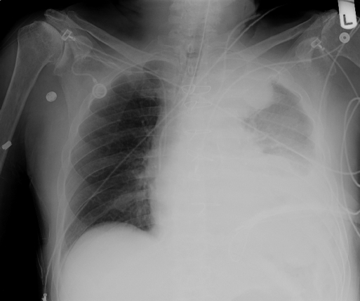
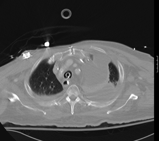
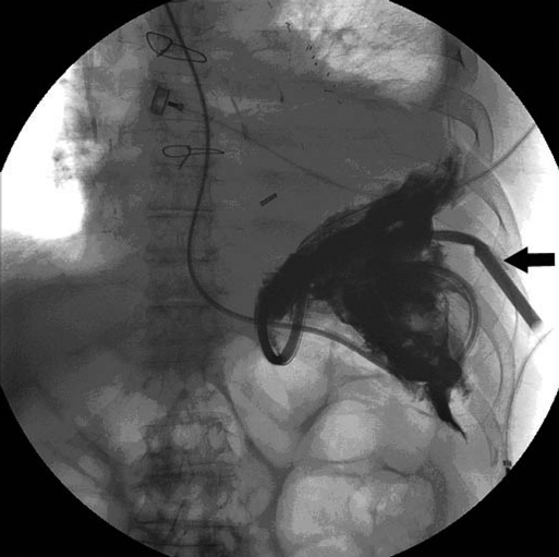
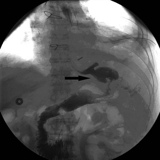
A 77-year-old man was admitted to the hospital with complaints of progressive lethargy and shortness of breath. He was found to be in septic shock with hypotension and hypoxia requiring ventilatory and vasopressor support. Tube thoracostomy was performed for a large left pleural effusion seen on the admission chest x-ray (Figure 1), revealing copious amounts of purulent fluid. His family reported a history significant for splenectomy and chemotherapy a year earlier for non-Hodgkin's B-cell lymphoma. Seven months later, tumor recurrence was treated with radiation therapy. Progressive dysphagia necessitated the initiation of home TPN. An esophagogastroduodenoscopy (EGD) was performed 2 weeks prior to this admission where a bleeding gastric ulcer was discovered and cauterized. None of the above interventions were done at our institution, and no outside records were available for review. A chest and abdominal computed tomography (CT) with contrast was eventually performed (Figure 2), raising the suspicion of oral contrast present within the chest tube (Figure 3). A subsequent upper gastrointestinal series (UGI) (Figure 4) and an EGD confirmed the presence of a gastropleural fistula (GPF).
Figure 1.
Admission chest x-ray showing large left pleural effusion.
Figure 2.
Chest computed tomographic scan confirming large amount of left pleural effusion.
Figure 3.
Gastrografin upper gastrointestinal series (via nasogastric tube) demonstrating the presence of oral contrast in the chest tube (arrow).
Figure 4.
Postoperative UGI showing slight narrowing at mid-body of stomach (arrow).
After maximum medical optimization, the patient was taken to the operating room while still on a ventilator. Diagnostic laparoscopy revealed a bulky tumor on the greater curvature of the stomach extending into the pancreas as well as the lateral abdominal wall and the thorax. A laparoscopic GPF takedown and partial sleeve gastrectomy were performed with the aid of intraoperative EGD. The fistulous tract was patched with a bovine pericardium mesh (Cook Surgical, Bloomington, IN). A closed suction drain and a feeding jejunostomy tube were also placed. The final pathology revealed malignant B-cell lymphoma. His immediate postoperative course was remarkable for a quick initial recovery, early extubation, and transfer out of the intensive care unit. Persistent anorexia and pulmonary complications, however, prolonged his hospital course. Failing conservative therapy, he eventually required a left thoracotomy with pulmonary decortication and debridement of an empyema. He was finally discharged home 45 days postoperatively where he died a short while later.
DISCUSSION
Gastropleural fistulas are uncommon causes of thoracic empyemas. They occur either as a consequence of prior pulmonary surgery, intrathoracic perforation of strangulated hiatal hernias, complication of peptic ulcer disease or its endoscopic treatment, intractable nausea and vomiting, trauma, and chemo/radiation therapy for gastric lymphomas,1–10 Our patient had multiple risk factors for developing GPF. The typical presentation is that of fever, cough, a persistent left-sided pneumonia or pleural effusion. The diagnosis is confirmed by the presence of oral contrast in the chest cavity following radiographic contrast examination. In case of gastric malignancy, the mechanism is thought to be secondary to perforation of the stomach, formation of an intraabdominal abscess, and its subsequent transdiaphragmatic erosion into the thoracic space.1–4 Depending on their underlying cause, GPFs generally carry a poor prognosis. Conservative management usually fails, requiring a laparotomy, with possible thoracotomy, and a partial or total gastrectomy.1–3 Our case, however, demonstrates the feasibility of a laparoscopic approach to this difficult problem. Laparoscopic treatment of GPF may be associated with less early morbidity in these often debilitated patients and should probably be considered as the initial procedure of choice.
Footnotes
No financial support was provided for this paper by any company or institution.
Contributor Information
Amir Mehran, Division of Minimally Invasive & Bariatric Surgery, Cleveland Clinic Florida, Weston, Florida, USA..
Andrew Ukleja, Department of Gastroenterology, Cleveland Clinic Florida, Weston, Florida, USA..
Samuel Szomstein, Division of Minimally Invasive & Bariatric Surgery, Cleveland Clinic Florida, Weston, Florida, USA..
Raul Rosenthal, Division of Minimally Invasive & Bariatric Surgery, Cleveland Clinic Florida, Weston, Florida, USA..
References:
- 1. Kellum JM, Jaffe BM, Calhoun TR, Ballinger WF. Gastric complications after radiotherapy for Hodgkin's disease and other lymphomas. Am J Surg. 1977;134(3):314–317 [DOI] [PubMed] [Google Scholar]
- 2. Warburton CJ, Calverley PM. Gastropleural fistula due to gastric lymphoma presenting as tension pneumothorax and empyema. Eur Resp J. 1997;10(7):1678–1679 [DOI] [PubMed] [Google Scholar]
- 3. Rotstein OD, Pruett TL, Simmons RL. Gastropleural fistula. Report of three cases and review of the literature. Am J Surg. 1985;150(3):392–396 [DOI] [PubMed] [Google Scholar]
- 4. Virlos I, Asimakopoulos G, Forrester C. Gastropleural fistula originating from the lesser curve: a recognised complication, an uncommon pathway of communication. Thorac Cardiovasc Surg. 2001;49(5):308–309 [DOI] [PubMed] [Google Scholar]
- 5. Tzeng JJ, et al. Gastropleural fistula caused by incarcerated diaphragmatic herniation of the stomach. Gastrointest Endosc. 2001;53(3):382–384 [DOI] [PubMed] [Google Scholar]
- 6. Mussi A, Lucchi M, Davini F, Angeletti CA. Gastropleural fistula as complication of postpneumonectomy empyema. J Cardiovasc Surg. (Torino) 2000;41(1):147–149 [PubMed] [Google Scholar]
- 7. Biswas IH, Raghavan C, Sevcik L. Gastropleural fistula: an unusual cause of intractable postoperative nausea and vomiting. Anesth Analg. 1996;83(1):186–188 [DOI] [PubMed] [Google Scholar]
- 8. Meredith HC, Seyumour EQ, Vujic I. Hiatal hernia complicated by gastric ulceration and perforation. Gastrointest Radiol. 1980;5(3):229–231 [DOI] [PubMed] [Google Scholar]
- 9. Schwab RJ, Jarvik JG. Tension pneumothorax secondary to a gastropleural fistula in traumatic diaphragmatic hernia. Chest. 1991;99(1):247–249 [DOI] [PubMed] [Google Scholar]
- 10. Okeefe PA, Goldstraw Gastropleural fistula following pulmonary resection. Thorax. 1993;48(12):1278–1279 [DOI] [PMC free article] [PubMed] [Google Scholar]


