Abstract
Colonic lipomas are infrequent lesions, yet they are the second most common benign lesions of the colon after benign adenomatous polyps. Their treatment ranges from observation to segmental colectomy and has been a matter of debate since Bauer first reported them in 1757. With the advent of new technologies, therapeutic options now include observation, endoscopic removal, laparoscopic removal, and traditional open surgery. We present a case of colonic lipoma presenting with indeterminate symptomatology, its workup, treatment outcome, and a review of the current literature.
Keywords: Lipoma, Colon, Endoscopy, Laparoscopy
CASE REPORT
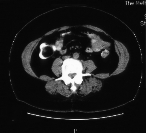
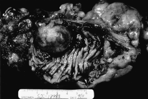
A 62-year-old female presented with a history of intermittent abdominal pain and bloating for 5 years. On physical examination, no abnormalities were found, and her stool was hemoccult negative. The laboratory workup was normal with no evidence of microcytic anemia. An abdominal computed tomographic (CT) scan with oral contrast was obtained revealing a round, well-demarcated, 3.8-cm homogeneous tumor with low-attenuation located in the colon near the hepatic flexure (Figure 1). A colonoscopy with tissue biopsies was performed, demonstrating pathology consistent with a benign colonic lipoma. A surgical consult was obtained and a laparoscopic colon resection was recommended. She tolerated this procedure well and was discharged home on postoperative day 3, tolerating a soft mechanical diet. Gross and microscopic pathologic examination confirmed the diagnosis of a benign colonic lipoma (Figure 2).
Figure 1.
Computed tomographic scan with oral contrast reveals a round, well-demarcated, 3.8-cm homogeneous tumor with low-attenuation located in the colon near the hepatic flexure.
Figure 2.
Gross and microscopic pathologic examination confirmed the diagnosis of a benign colonic lipoma.
DISCUSSION
Bauer reported the first case of a colonic lipoma in 1757.1 Lipomas are benign, nonepithelial tumors that can be found anywhere in the gastrointestinal tract, but most are located in the colon.1 As a benign lesion, they rank second in frequency only to benign adenomatous polyps. Pathologically, they are spherical deposits of adipose tissue in the bowel wall, in a submucosal, pedunculated, sessile, or very rarely annular position.2–4 When on a stalk, this psuedopedicle is felt to develop from continuous extrusion of the lipoma due to the peristalsis of the colon.3 Ninety percent of colonic lipomas lie in the submucosa; the remainder are subserosal.5 They have been noted to occur 1.5 to 2.0 times more frequently in women, and most patients are in their fifth or sixth decade of life.6 They are more frequently located on the right side, particularly in the cecum.1,7 They are multiple in up to 26% of cases.8 A review of over 10 000 colonoscopies found 16 lipomas with a distribution of 63% in the right colon and 37% in the left colon.9 Although they are the second most common benign colonic tumor, they are rare lesions with an incidence reported between 0.2 to 2.6%.1,7,10 A review of several major autopsy and clinical reports reveals an incidence of 0.26%.5,10
They are often found incidentally during colonoscopy or radiologic imaging. On contrast enema, lipomas appear circular, ovoid, well-demarcated, and smooth.11 A barium enema will show a radiolucent mass, and they may fluctuate in size and shape during the study.11 Radiolucency and the “squeeze sign” (change in size and shape during peristalsis) have been considered pathognomonic of colonic lipomas.12 On CT, the lipoma has a uniform appearance and density with absorption densities of -80 to -120 Hounsfield units to confirm the fatty composition. Meglumine diatrizoate (Gastrografin) should be administered as a dilute enema to maximize the imaging of the lipoma.13,14 A radiologic diagnosis can be made definitively in less than 20% of patients.11
Endoscopy, which locates a mass consistent with a lipoma, may reveal the “cushion sign” whereby a sponge-like impression is made as biopsy forceps are passed in the lesion and it then regains its original shape as they are withdrawn.11 Also the “naked fat sign” occurs after a biopsy is taken from the mucosa revealing fat protruding from the site.15 The specimen is also noted to float if extracted and placed in formalin. The mucosa will “tent” over the mass if it is grasped with forceps as it detaches from the lipomatous mass below it. Any of these findings are highly indicative of a benign lipoma.
Small colonic lipomas are rarely symptomatic, but when >2 cm, they can result in persistent or intermittent abdominal pain, bloating, changes in bowel habits, gastrointestinal bleeding, bowel obstruction, or intussusception.1,2,7,10,16 Among symptomatic patients, abdominal pain (23%) and rectal bleeding (20%) are the most common complaints with anemia, weight loss, nausea, vomiting, and abdominal distention being less commonly reported.1 Roughly 25% of all colonic lipomas are found to be symptomatic.17 Clinical proficiency helps distinguish this entity from other more common disease processes, such as chronic cholecystitis, diverticulosis, diverticulitis, colonic polyps, or malignancy, as its symptomatology can overlap significantly.2,10 Imaging with a contrast-enhanced abdominal CT scan can help delineate a benign colonic lipoma from other disease processes. Any deviation from the CT scan characteristics as described in this patient may indicate the presence of a malignancy. Colonoscopy is mandatory to locate, visualize, and biopsy the lesion. Endoscopic or surgical removal is indicated for symptomatic colonic lipomas or when malignancy is suspected or known.1,7,10 Although large colonic lipomas have been removed endoscopically, a greater risk of colon perforation exists when they are broad-based, intramural, or when they are >2 cm.1,7 Endoscopic ultrasound and injection of the base with epinephrine or saline is reported to help decrease this complication.2 Specifically, endoscopic ultrasonography can demonstrate whether the lipoma extends into the muscularis propria, a risk factor for perforation that should preclude endoscopic removal.16 Some lesions that are concealed behind mucosal folds or colonic flexures cannot be removed colonoscopically, and this worsens the risk of perforation after endoscopic removal.18,19 Recent literature to support the use of endoscopic ultrasound during excision has shown that lipomas can be removed safely by endoscopy if they are >2 cm; however, we recommend laparoscopic excision until this technique is more widely practiced.16
Surgical removal usually involves limited resection or colotomy with lipomectomy.20 Multiple operative techniques ranging from laparotomy with enucleation, colotomy, and segmental colonic resection have been described.20 Laparoscopic colon resection in the face of a known lipoma results in improved cosmesis, decreased length of stay, shorter disability, decreased adhesion formation, less postoperative pain, and faster return of bowel function. Segmental resection or colostomy with resection and closure are both feasible where indicated.21
We have adopted a clinical algorithm whereby symptomatology determines whether the patient will undergo radiologic or endoscopic evaluation. Radiologic identification leads to colonoscopic evaluation, and small lipomas <2 cm are removed endoscopically. If the mass is >2 cm or suspicious for malignancy, laparoscopic excision is performed with colotomy and closure or partial colectomy when indicated. For tumors of the rectosigmoid, transanal resection is possible.22
Footnotes
Funded in part by an educational grant from United States Surgical Corporation.
References:
- 1. Franc-Law JM, Begin LR, Vasilevsky CA, Gordan PH. The dramatic presentation of colonic lipomata: report of two cases and review of the literature. Am Surg. 2001;67(5):491–494 [PubMed] [Google Scholar]
- 2. Taylor BA, Wolff BG. Colonic lipomas: report of two unusual cases and review of the Mayo Clinic experience, 1976 –1985. Dis Colon Rectum. 1987;30:888–893 [DOI] [PubMed] [Google Scholar]
- 3. Fernandez MJ, Davis RP, Nora PF. Gastrointestinal lipomas. Arch Surg. 1983;118:1081–1083 [DOI] [PubMed] [Google Scholar]
- 4. Notaro JR, Masser PA. Annular colon lipoma: a case report and review of the literature. Surgery. 1991;110(3):570–572 [PubMed] [Google Scholar]
- 5. Haller JD, Roberst TW. Lipomas of the colon: a clinicopathologic study of 20 cases Surgery. 55:773–781, 1964 [PubMed] [Google Scholar]
- 6. Fazal K. Pedunculated lipoma of the colon: risks of endoscopic removal. South Med J. 1987;80:1176–1179 [DOI] [PubMed] [Google Scholar]
- 7. Rogers SO, Lee MC, Ashley SW. Giant colonic lipoma as lead point for intermittent colo-colonic intussusception. Surgery. 2002;131:687–688 [DOI] [PubMed] [Google Scholar]
- 8. Castro EB, Sterans MW. Lipoma of the large intestine: a review of 45 cases. Dis Colon Rectum. 1972;15:441–444 [DOI] [PubMed] [Google Scholar]
- 9. Chung YFA, HO YH, Nyam DCNK, Leong AFPK, SeowChoen F. Management of colonic lipomas. Aust N Z J Surg. 1998;68:133–135 [DOI] [PubMed] [Google Scholar]
- 10. Mayo CW, Pagtalunan RJG, Brown DJ. Lipoma of the alimentary tract. Surgery. 1963;53(5):598–603 [PubMed] [Google Scholar]
- 11. De Be, er RA, Shinya H. Colonic lipomas, an endoscopic analysis. Gastrointest Endosc. 1975;22:90–91 [DOI] [PubMed] [Google Scholar]
- 12. Kaplan P. Submucous lipoma of the colon. Int Surg. 1971; 56:113–117 [PubMed] [Google Scholar]
- 13. Megibow AM, Bosniak MA, Horowitz L. Diagnosis of gastrointestinal lipomas by CT. Am J Roentgenol. 1979;133:743–745 [DOI] [PubMed] [Google Scholar]
- 14. Heiken JP, Forde KA, Gold RP. Computerized tomography as a definite method for diagnosing gastrointestinal lipomas. Radiology. 1982;142:409–414 [DOI] [PubMed] [Google Scholar]
- 15. Messer J, Waye JD. The diagnosis of colonic lipoma-the naked fat sign. Gastrointest Endosc. 1982;28:186–188 [DOI] [PubMed] [Google Scholar]
- 16. Kim CY, Bandres D, Tio TL, Benjamin SB, Al-Kawas FH. Endoscopic removal of large colonic lipomas. Gastrointest Endosc. 2002;55(7):929–931 [DOI] [PubMed] [Google Scholar]
- 17. Rosario V, Ferrara M, Mosca F, Ignoto A, Latteri F. Lipomas of the large bowel. Eur J Surg. 1996;162:915–919 [PubMed] [Google Scholar]
- 18. Chase MP, Yarze JC. “Giant” colon-lipoma—To attempt endoscopic resection or not? Am J Gastroenterol. 2000;95:2143–2144 [DOI] [PubMed] [Google Scholar]
- 19. Bardaji M, Roset F, Camps R, Sant F, Fernandez-Layos MJ. Symptomatic colonic lipoma: differential diagnosis of large bowel tumors. Int J Colorectal Dis. 1998;13:1–2 [DOI] [PubMed] [Google Scholar]
- 20. Corman ML. Colon and Rectal Surgery. 4th ed. Philadelphia, PA: Lippincott-Raven; 1998;913–915 [Google Scholar]
- 21. Saclarides TJ, Ko ST, Airan M, Dillon C, Franklin J. Laparoscopic removal of a large colonic lipoma. Report of a case. Dis Colon Rectum. 1991;34:1027–1029 [DOI] [PubMed] [Google Scholar]
- 22. Tzilinis A, Fessenden JM, Ressler KM, Clarke LE. Transanal resection of a colonic lipoma mimicking rectal prolapse. Curr Surg. 2003;60(3):313–314 [DOI] [PubMed] [Google Scholar]


