Abstract
Gastric outlet obstruction as a result of gallstone (Bouveret's syndrome) is a rare but serious complication of cholelithiasis. Although patients present with persistent vomiting, colicky epigastric pain and dehydration, the clinical features of the Bouveret's syndrome are not pathognomonic. Due to its rarity, the diagnosis and treatment represent a challenge for the surgeon. In most of the reported cases, the diagnosis was made at the time of laparotomy. We report an unusual clinical presentation of Bouveret's syndrome with mild acute pancreatitis that was treated laparoscopically. To our knowledge, this is the first described case. Cause, clinical presentation, methods of diagnosis, and options for management of Bouveret's syndrome are also discussed.
Keywords: Bouveret's syndrome, Laparoscopy, Gastric outlet syndrome, Cholelithiasis
INTRODUCTION
In 1896, Bouveret reported gastric outlet obstruction due to gallstones with biliary-enteric fistula.1 Since then, approximately 300 cases of this proximal gallstone ileus have been described worldwide.2–5 Patients with Bouveret's syndrome present with persistent vomiting, colicky epigastric pain, and dehydration. Fever, hematemesis or jaundice may also be present. Previous biliary symptoms are often vague and occur in 50% to 70% of patients.6 The difficult and unpredictable nature of the pathological process may result in late diagnosis and treatment. As a matter of fact, in most of the reported cases, the diagnosis was made at the time of laparotomy. We report an unusual clinical presentation of Bouveret's syndrome with mild acute pancreatitis that was treated laparoscopically. To our knowledge, this is the first described case. Cause, clinical presentation, methods of diagnosis, and options for management of Bouveret's syndrome are also discussed.
CASE REPORT
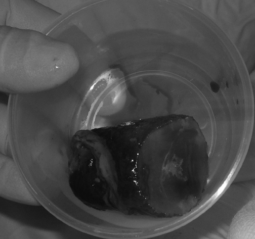
A 76-year-old female presented with a 3-day history of persistent nausea, vomiting, and upper abdominal pain. The physical examination was unremarkable apart from diffuse abdominal tenderness. Laboratory findings showed leukocytosis (white blood cell count >18,000), increased C-reactive protein (CRP158), increased serum amylasis (860 UI/L), lypasis (460 UI/L), and urea (80 mmol/L). Amylasuria was also present. The plain abdominal x-ray appeared normal. The patient was resuscitated and admitted with a diagnosis of acute pancreatitis. She remained stable over the next 48 hours but did not show any signs of clinical or biochemical improvement. She underwent a ultrasound of her abdomen, which showed intrahepatic pneumobilia in the absence of stones or biliary tract dilatation. The pancreas was not visualized. An abdominal computed tomographic scan with intravenous contrast confirmed intrahepatic pneumobilia and showed 2 large stones of 3 cm and 5 cm in the duodenum. Esophagogastroduodenoscopy (EGDS) confirmed the presence of 2 stones, and no attempts to dislodge the stones were made. Laparoscopy was performed, placing 4 trocars as in the standard cholecystectomy procedure. Calot's triangle was dissected, and a fistula between Hartmann's pouch, the cystic duct at its junction with the common bile duct (CBD), and the second portion of duodenum was visible. Cholecystitis and peri-cholecystitis were also present. The duodenum was dissected free, and the duodenal opening of the fistula was enlarged, the wedges excised, and 2 large stones extracted (Figure 1). The fistula was then closed with a single layer of running 3/0 absorbable suture. The anterior aspect of the CBD was involved in the fistula and was repaired with interrupted suture on a T-tube. A 20 F drain was left in the infrahepatic space. The gallbladder and the stones were retrieved separately by using 2 endo-bags. The umbilical incision was enlarged to extract the stones. The postoperative period was uneventful; the patient improved steadily and was discharged on postoperative day 8. Two weeks later, the T-tube was removed after a negative contrast study.
Figure 1.
One of the 2 retrieved stones causing Bouveret's syndrome.
DISCUSSION
The overall risk of gallstone ileus is 0.3% to 0.5%.5,7,8 It represents 1% to 4% of all cases of intestinal obstruction. However, this quote increases to 25% in people over 65 years of age.9 Gallstone ileus is due to a fistula between the gallbladder or the bile duct and the intestine. Chronic cholecystitis is the common underlying cause of the formation of the fistula. The most common type of fistula is cholecystoduodenal (60%), followed by cholecystocolic (17%), cholecystogastric (5%), and choledochoduodenal (5%).10–12 The destiny of the eroding stone(s) is variable depending on its size, the intestinal segment involved, and the presence of concomitant intestinal stenosis. It may be passed asymptomatically per rectum, may be vomited, or rarely, may cause intestinal obstruction that requires surgery (6% of cases).11 The obstruction usually occurs in the distal ileum (50% to 75%), occasionally in the proximal ileum or jejunum (20% to 40%), and less frequently in the colon.9 Seldom, large stones may impact in more proximal portions of the intestine (duodenum or stomach), causing gastric outlet symptoms, resembling Bouveret's syndrome.1
Mortality for gallstone ileus is high, occurring in up to 50% of patients (generally between 15% and 30%).13 The frequent old age of patients, the delay in diagnosis and treatment, and certainly the presence of the biliary-enteric fistula are the factors responsible for the high morbidity and mortality of this clinical condition.5 No separate mortality data are available for Bouveret's syndrome.
The clinical features of Bouveret's syndrome are not pathognomonic, and due to its rarity, the diagnosis and treatment represent a challenge for the surgeon. The typical patient is female, in the sixth to seventh decade of life. Abdominal pain, nausea, and vomiting are the symptoms more frequently reported and are variably associated with abdominal distension, constipation, and fever. Hematemesis is present in less than 10% of patients,14 while melena is very rare.15 Initial presentation with massive arterial bleeding from an eroded cystic artery has also been reported.16 Jaundice and abnormal liver function tests are present in about one third of patients,11 and previous biliary symptoms are present in up to two thirds of patients.6
A plain abdominal x-ray may show a distended stomach, extraluminal air in the right upper quadrant, and calcified shadows (Rigler's triad, less the 30% of the patients).17 Contrast studies of the upper gastrointestinal tract may identify both the course of the fistula and the level of the obstruction.5
Abdominal ultrasound may detect the presence of the stones in the duodenum, detect the fistula or the choledocholithiasis, or both.18 However, as observed in our patient, a distended abdomen or an “air filled” stomach can make this test unreliable.
Esophagogastroduodenoscopy (EGDS) allows visualization of the stones impacted in the duodenum and should always be performed to establish the diagnosis, especially if the gastric outlet obstruction is accompanied by ematemesis. Small stones can also be retrieved,19 but the procedure is often unsuccessful and may lead to further stone impaction,20 lacerations, and perforations.12
An abdominal CT scan facilitates the diagnosis of Bouveret's syndrome when the diagnosis is not straightforward. The stomach may be distended with deformation of the antrum or duodenum; cholelythiasis, cholecystitis, and the presence of air in the biliary tree are usually witnessed.21 In our case, a CT scan was performed with a working diagnosis of pancreatitis, and 2 large stones were visible in the duodenum. The presence of the 2 impacted stones was confirmed with EGDS, defining the anatomy and excluding other pathologies.
Surgery represents the treatment of choice. Less invasive procedures, such as endoscopic retrieval of the stones or extracorporeal shock wave lithotripsy with subsequent endoscopic extraction, have been proposed.22 However, in addition to potential complications previously described,12,20 endoscopic treatment does not correct the underling fistula and could not exclude the presence of additional stones distally.
Complete treatment aims to relieve the obstruction, to close the fistula, and to prevent relapses. Controversies exist regarding the use of simple enterolithotomy versus enterolithotomy associated with cholecystectomy and correction of the internal fistula as a 1- or 2-stage procedure. A 1-stage procedure involving the removal of the gallbladder with fistula closure is associated with a mortality of 20% to 30%, while a simple enterolithotomy has a mortality of <12%.7,9,19,23 However, the enterolithotomy alone is associated with an increased incidence of biliary complications (cholangitis, cholecystitis) and with a recurrence rate of 4.7%.9 Additionally, the risk of bleeding and gall-bladder carcinoma should be considered.12 Truncal vagotomy, antrectomy, gastric resection, or gastroenteric anastomosis have been described in association with cholecystectomy in the presence of large inflammatory masses.
From the literature, it is clear that extended operation time alone does not affect the outcome, while delay in diagnosis and treatment do.12 Our patient had a delayed diagnosis due to the confusing pancreatic sufferance, but because of her good performance status, she was fit enough for 1-stage surgery. We believe that whenever the physical conditions are satisfactory, extraction of the stone, closure of the fistula, and cholecystectomy should be performed to prevent further biliary complications.
No reports on laparoscopic treatment of Bouveret's syndrome, but our experience with this case, suggests the possibility of offering a trial of laparoscopic dissection. Both the fistula and the gallstones could be treated laparoscopically at the same time where the necessary expertise and equipment are available. It remains clear that the surgeon should adapt the treatment strategy according to the patient's age, general condition, and findings at surgery.
References:
- 1. Bouveret L. Stenose du pylore adherent a la vesicule. Rev Med (Paris). 1896;16:1–16 [Google Scholar]
- 2. Halaz NA. Gallstone obstruction of the duodenal bulb (Bouvert's syndrome). Am J Dig Dis. 1964;9:856–861 [DOI] [PubMed] [Google Scholar]
- 3. Togerson SA, Greening GK, Juniper K, et al. Gallstone obstruction of duodenal cap (Bouveret's Syndrome) diagnosed by endoscopy. Am J Gastroenterol. 1979;72:165–167 [PubMed] [Google Scholar]
- 4. Murthy GD. Bouveret's syndrome. Am J Gastroenterol. 1995; 90(4):638–639 [PubMed] [Google Scholar]
- 5. Ariche A, Czeiger D, Gortzak Y, Shaked G, Shelef I, Levy I. Gastric outlet obstruction by gallstone: Bouveret syndrome. Scand J Gastroenterol. 2000;35(7):781–783 [DOI] [PubMed] [Google Scholar]
- 6. Vidal O, Seco JL, Alvarez A, Trinanes JP, Serrano LP, Serrano SR. Bouveret's syndrome: 5 cases. Rev Esp Enferm Dig. 1994; 86(5):839–844 [PubMed] [Google Scholar]
- 7. Kasahara Y, Umemura H, Shiraha S, Kuyama T, Sakata K, Kubota H. Gallstone ileus. Review of 112 patients in the Japanese literature. Am J Surg. 1980;140(3):437–440 [DOI] [PubMed] [Google Scholar]
- 8. van Hi, llo M, van der Vliet JA, Wiggers T, Obertop H, Terpstra OT, Greep J. Gallstone obstruction of the intestine: an analysis of ten patients and a review of the literature. Surgery. 1987;101(3):273–276 [PubMed] [Google Scholar]
- 9. Reisner RM, Cohen JR. Gallstone ileus: a review of 1001 reported cases. Am Surg. 1994;60(6):441–446 [PubMed] [Google Scholar]
- 10. Clavien PA, Richon J, Burgan S, Rohner A. Gallstone ileus. Br J Surg. 1990;77(7):737–742 [DOI] [PubMed] [Google Scholar]
- 11. Thomas TL, Jaques PF, Weaver PC. Gallstone obstruction and perforation of the duodenal bulb. Br J Surg. 1976;63(2):131–132 [DOI] [PubMed] [Google Scholar]
- 12. Liew V, Layani L, Speakman D. Bouveret's syndrome in Melbourne. ANZ J Surg. 2002;72:161–163 [DOI] [PubMed] [Google Scholar]
- 13. Rodriguez-Sanjuan J, Cassado F, Fernandez MJ, et al. Cholecystectomy and fistula closure versus enterolithotomy alone in gallstone ileus. Br J Surg. 1997;84:634–637 [PubMed] [Google Scholar]
- 14. Al Ma, llah MH, Ibrahim M. Bouveret's syndrome, a rare cause of upper gastrointestinal bleeding and obstruction. Am J Gastroenterol. 2001;96(suppl S81):25411197270 [Google Scholar]
- 15. Mejia LE, Murthy R, Mapara S. Bouveret's syndrome presenting as upper GI bleed in a young female. Am J Gastroenterol 2003;98(suppl S131):386 [Google Scholar]
- 16. Heinrich D, Meier J, Wehrli H, Buhler H. Upper gastrointestinal hemorrhage preceding development of Bouveret's syndrome. Am J Gastroenterol. 1993;88(5):777–780 [PubMed] [Google Scholar]
- 17. Rigler LG, Borman CN, Noble JF. Gallstone obstruction. Pathogenesis and roentgen manifestations. JAMA. 1941;117:1753–1759 [Google Scholar]
- 18. Maglinte DD, Lappas JC, Ng AC. Sonography of Bouveret's syndrome. J Ultrasound Med. 1987;6:675–677 [DOI] [PubMed] [Google Scholar]
- 19. Lubbers H, Mahlke R, Lankisch PG. Gallstone ileus: endoscopic removal of a gallstone obstructing the upper jejunum. J Inter Med. 1999;246:593–597 [DOI] [PubMed] [Google Scholar]
- 20. Iuchtman M, Sternberg A, Alfici R, Sternberg E, Fireman T. Iatrogenic gallstone ileus as a new complication of Bouveret's syndrome [in Hebrew]. Harefuah 1999;136:122–124, 174. [PubMed] [Google Scholar]
- 21. Farman J, Goldstein DJ, Sugalski MT, Moazami N, Amory S. Bouveret's syndrome diagnosis by helical CT scan. Clin Imag. 1998;22:240–242 [DOI] [PubMed] [Google Scholar]
- 22. Holl J, Sackmann M, Hoffmann R, et al. Shock wave therapy of gastric outlet obstruction caused by a gallstone. Gastroenterology. 1989;97:472–474 [DOI] [PubMed] [Google Scholar]
- 23. Zuegel N, Hehl H, Lindemann F, Witte J. Advantages of one stage repair in case of gallstone ileus. Hepatogastroenterology. 1997;44:59–62 [PubMed] [Google Scholar]


