Abstract
Background and Objectives:
Meckel's diverticulum (MD) presents unique challenges for a pediatric surgeon, as it is prone to varied complications. This case series highlights the diverse presentations and laparoscopic management of MD in children.
Methods:
We performed a retrospective analysis of consecutive cases of laparoscopic-assisted transumbilical Meckel's diverticulectomy (LATUM) performed by the same surgeon for incidental as well as diverse Meckel's diverticular complications over 20 months.
Results:
Eight patients (5 males and 3 females) aged 3 years to 13 years (median, 12) underwent LATUM. Three patients had painless per-rectal bleeding and 1 presented with intestinal obstruction due to a mesodiverticular band and intestinal ischemia. Two patients had features masquerading as appendicitis; one had perforated MD with secondary inflammation of the appendix, and the other had a torsed, gangrenous MD. In 2 patients, incidental MD with a narrow base was noted at appendicectomy for appendicitis. All patients underwent successful LATUM along with appendicectomy in 4 patients. The operative duration was 72 minutes to 165 minutes (mean, 112.1±30.6). There were no operative complications, and no conversion to open surgery was required. The hospital stay was 4 days to 7 days (mean, 4.7±1.2). The patient with mesodiverticular band intestinal obstruction presented with adhesive intestinal obstruction 2 weeks after the surgery. Laparoscopic-assisted minilaparotomy was done to release the pelvic adhesions. There were no other complications during the follow-up (median, 11 months).
Conclusions:
LATUM is a simple, safe, and effective procedure with a better cosmetic outcome that can be performed for diverse manifestations of MD. The technique also allows palpation of the MD and avoids use of expensive staplers.
Keywords: Meckel's diverticulum, Complications, Children, Laparoscopy
INTRODUCTION
Laparoscopy has opened new avenues in the management of Meckel's diverticulum (MD), which poses different challenges to a clinician. Traditional investigative modalities like 99mTechnetium (99mTc) scintigraphy, radiographic contrast studies, ultrasonography, and computed tomography scan have many limitations in the accurate assessment of MD and its complications. Laparoscopy aids in the diagnosis and treatment of diverse complications associated with MD. Laparoscopic-assisted transumbilical Meckel's diverticulectomy (LATUM) is a simple, safe technique for the precise assessment of the complications of MD and performing the resection.1–4 Although laparoscopic intracorporeal Meckel's diverticulectomy can be accomplished with staplers5 or a pretied Endoloop,6 extracorporeal resection has the advantages of a simplified technique with a minimal number of ports and avoids the expensive staplers. This case series is unique as it is only the second series reported in the English language literature to depict the diverse forms of Meckel's diverticular complications successfully treated by LATUM.7
METHODS
A retrospective analysis of consecutive cases of LATUM performed by the same surgeon for incidental as well as diverse complications of MD during the study period from January 2004 and August 2005 was done. The presenting features, clinical signs, investigations, operative procedure, follow-up, and complications were noted and the safety and efficacy were analyzed.
Operative Procedure
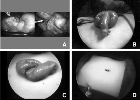
LATUM was performed through 3- or 2-port laparoscopy. A 10-mm port for the telescope was inserted through the umbilicus by an open Hasson's technique. Two 5-mm working ports were inserted in the suprapubic region and the left iliac fossa under vision after pneumoperitoneum was established. The second working port was omitted in cases of bleeding MD. Systematic exploration of the small intestine was performed in a retrograde fashion from the caecum. The intestinal loops were walked through evaluating the intestine on either side of the mesentery as cursory examination might overlook a small MD adherent to the mesentery. In the case of mesodiverticular band intestinal obstruction, the collapsed loops were walked towards the site of the obstruction with minimal handling of the proximal dilated intestinal loops. The MD was released from the mesentery after coagulating and dividing the feeding vessel. The MD was grasped with a Babcock forceps passed via the umbilical port through a reducer, visualizing with a 5-mm telescope through the working port. The umbilical incision was extended with a generous incision of the linea alba, but the skin incision remained within the umbilical cicatrix. The MD was brought out of the umbilical incision. Resection of the MD with a sleeve of ileum and hand-sewn single-layered endtoend anastomosis with interrupted 4–0 polyglactin sutures was performed extracorporeally. The anastomosed intestine was replaced back into the peritoneal cavity, and the umbilical incision was closed with few interrupted sutures of 2–0 polyglactin to approximate the linea alba (Figure 1).
Figure 1.
Stepwise depiction of laparoscopic-assisted transumbilical Meckel's diverticulectomy; A: laparoscopic view of incidental Meckel's diverticulum with acute appendicitis, B: Meckel's diverticulum brought out of umbilical incision, C: completed end-to-end anastomosis after resection of Meckel's diverticulum, D: Appearance of umbilical incision after laparoscopic-assisted transumbilical Meckel's diverticulectomy.
One case of torsed MD was handled with a modification of the technique. The narrow base of the torsed MD was ligated with a pretied loop suture (Vicryl Endoloop, Ethicon) and divided between the ligatures. The unruptured MD was retrieved through the umbilical incision avoiding spillage into the peritoneal cavity. Appendicectomy was accomplished simultaneously in 4 patients. The appendix was delineated and the mesentery was fulgurated with bipolar or monopolar hook diathermy and divided with scissors. The appendicular base was ligated with a single pretied Vicryl Endoloop suture. The appendix was divided between ligatures, the distal appendicular side of the ligature being tied using the same Endoloop suture with a slipknot tied manually. The appendix was retrieved through the umbilical port or the umbilical incision.
RESULTS
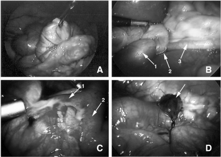
Eight patients (5 males and 3 females) age 3 years to 13 years (median, 12) underwent LATUM (Table 1). Three patients had painless per-rectal bleeding and 1 presented with intestinal obstruction due to a mesodiverticular band and intestinal congestion and ischemia. Two patients had features masquerading as appendicitis; one had perforated MD with secondary inflammation of the appendix, and the other had a torsed and gangrenous MD. In 2 patients, incidental MD with a narrow base was noted at appendicectomy for appendicitis (Figure 2).
Table 1.
Clinical Description of the LATUM Cases
| Case* | Age/Sex* | Symptoms* | Signs* | Investigations* | Surgery Findings* | Surgery* | Complications* |
|---|---|---|---|---|---|---|---|
| 1 | 5y6m/M | Abd pain, vomiting & fever for 3 days | Tender and guarded lower abd | Hb: 12.9 g/dL; TLC: 10.2×10⁹/L USG & CT scan abd: Inflammatory mass in the lower abdomen with interloop abscess Histo: Meckel's diverticulitis & secondary periappendicitis. No heterotopia | Meckel's diverticulitis with perforation and forming a mass with adjacent loops and appendix | LATUM and appendicectomy | Nil |
| 2 | 12y2m/F | Abd pain, vomiting and fever for 1 day | Tender RIF | Hb: 15.4 g/dL; TLC: 13.1×10⁹/L Histo: Acute appendicitis and MD (No heterotopia) | Appendicitis and incidental MD | LATUM and appendicectomy | Nil |
| 3 | 5y2m/F | PR bleeding and pallor for 1 day | Fresh PR bleeding, hypotension | Hb: 9.6 g/dL; TLC: 12×109/L Histo: MD with hemorrhagic peptic ulcer at base and gastric heterotopia | MD and blood filled distal intestinal loops | LATUM | Nil |
| 4 | 2y9m/M | PR bleeding, cold hands & feet and pallor for 1 day | Pallor, melenic stools on PR | Hb: 6.6 g/dL; TLC: 14.7×10⁹/L 99mTc scan: Consistent with MD Histo: MD with gastric & pancreatic heterotopia | MD and blood filled distal intestinal loops | LATUM | Nil |
| 5 | 12y2m/F | Abd pain, vomiting and abd distension for 4 days | Distended abd with lower abd tenderness | Hb: 14 g/dL; TLC: 11.5×10⁹/L AXR: Distended small bowel & air fluid levels Histo: MD with congestion and ischemic changes. No heterotopia | Meso-diverticular band intestinal obstruction with congestion of dilated proximal bowel | LATUM | Adhesive IO 2 weeks later |
| 6 | 13y5m/M | Abd pain, fever and blood streaked stools for 1 day | Tender RIF with rebound tenderness | Hb: 12.5 g/dL; TLC: 11.6×10⁹/L USG abd: Free fluid in right paracolic gutter Histo: Completely infarcted MD | Torsed MD with a narrow pedicle covered by omentum and adherent to parieties at RIF | Modified LATUM & appendicectomy | Nil |
| 7 | 11y10m/M | PR bleeding for 1 day | Pallor, melenic stools on PR | Hb: 10.8 g/dL; TLC: 17.2×10⁹/L 99mTc scan: Consistent with MD Histo: MD with gastric heterotopia and ulceration | MD and blood filled distal intestinal loops | LATUM | Nil |
| 8 | 12y8m/M | Abd pain for 1 day | Tender RIF | Hb: 13.2 g/dL; TLC: 16.4×10⁹/L Histo: Acute appendicitis; MD with gastric heterotopia, no ulcers | Appendicitis and incidental MD | LATUM and appendicectomy | Nil |
y=years, m=months, Abd=Abdomen, PR=Per-rectal, RIF=Right iliac fossa, Hb=Hemoglobin, TLC=Total leucocyte count, USG=Ultrasonogram, CT=Computed tomography, AXR=Abdominal radiograph, Histo=Histopathology, IO=Intestinal obstruction, MD=Meckel's diverticulectomy, LATUM=laparoscopic-assisted transumbilical Meckel's diverticulectomy.
Figure 2.
Laparoscopic view of various Meckel's diverticular complications; A: Bleeding Meckel's diverticulum, B: Meso-diverticular band intestinal obstruction (1-dilated & congested proximal ileum, 2- Meckel's band, 3-distal collapsed ileum), C: Meckel's diverticulitis & perforation forming a mass (1- Meckel's diverticulum encased in omentum, intestinal loops and appendix, 2-ileum) D: Torsed Meckel's diverticulum.
99mTc scintigraphy performed in 2 cases of per-rectal bleeding delineated the ectopic tracer uptake suggestive of MD. One patient with profuse lower gastrointestinal bleeding underwent laparoscopic exploration without 99mTc scintigraphy, after resuscitation with a blood transfusion. Abdominal radiograph detected features of intestinal obstruction in one patient who then underwent laparoscopic exploration. Incidental MD was detected, as it is our routine practice to walk the intestinal loops from the caecum proximally for a distance of to 2 feet to 3 feet, in all cases of laparoscopic appendicectomy.
All patients underwent successful LATUM along with laparoscopic appendicectomy in 4 patients. The operative duration was 72 minutes to 165 minutes (mean, 112.1±30.6). There were no operative complications and none required conversion to open surgery. The hospital stay was 4 days to 7 days (mean, 4.7±1.2). Ectopic gastric epithelium was found in all the 3 patients with bleeding MD and 1 patient with incidental MD. One of the bleeding MD also revealed ectopic pancreatic tissue. The patient with mesodiverticular band intestinal obstruction presented with adhesive intestinal obstruction 2 weeks after the surgery. Laparoscopic-assisted minilaparotomy through a suprapubic incision was done to release the pelvic adhesions. No other complications occurred during a median follow-up of 11 months.
DISCUSSION
The advances in laparoscopy in children have revolutionized surgical management, accomplishing surgical therapeutic goals with minimal somatic and psychological trauma. Laparoscopic application for Meckel's diverticular complications is reported as anecdotal case reports1,5,8–11 or short case series6,12–15 in the literature. Laparoscopic Meckel's diverticulectomy is also described as a few cases in a series of particular clinical condition, such as lower gastrointestinal bleeding,4,16 intestinal obstruction,17 or acute abdominal pain.3 We believe that our case series is distinctive as it is only the second case series in the English language literature to illustrate diverse complications of MD observed within a short period of time and successfully treated by LATUM.7
Systematic laparoscopic exploration aids in evaluating obscure cases of Meckel's diverticular complications in addition to uncovering incidental MD. Laparoscopy has been advocated as the first line procedure for evaluating cases of painless and profuse lower gastrointestinal bleeding in children as the 99mTc scan is not accurate.12 Laparoscopy has also been recommended ahead of colonoscopy or esophagogastroduodenoscopy in investigating cases of obscure lower gastrointestinal bleeding as the air insufflation with endoscopic procedures may complicate successful laparoscopic exploration under the same general anesthesia. In addition, laparoscopy would also aid in cases of negative exploration to determine the sequence of upper gastrointestinal endoscopy and colonoscopy, depending on the presence of blood in the small bowel or colon respectively.12
Laparoscopic exploration also serves in the diagnosis and release of intestinal obstruction due to various causes.9,10,17 In our patient with mesodiverticular band intestinal obstruction, the site of obstruction was detected by walking the collapsed loops from the ileocecal junction. The mesodiverticular band was defined and the tip was disconnected from the mesentery. The intestinal loops, which were congested, ischemic, and distended, showed improved vascularity with the relief of obstruction.
Laparoscopy has also proven beyond a doubt as a safe and effective tool in the management of patients with suspected appendicitis. Two of our patients presented with features masquerading as appendicitis. Meckel's diverticulitis with perforation and secondary appendicitis was detected in one and a torsed MD in the other. The computed tomographic scan in the first patient and the ultrasound scan in the second could only delineate an inflammatory lesion near the right iliac fossa region. Laparoscopy played a vital role not only in establishing the diagnosis but also in performing a definitive surgical procedure.
Another important aspect of laparoscopic exploration is the detection of incidental MD, the management of which is controversial as no clear guidelines are available in the literature. MD length more than 2 cm, presence of ectopic epithelium, and age less than 40 years are considered the risk factors for complications.18 Resection is indicated in cases of a narrow diverticular base, the presence of palpable thickening of the diverticulum consistent with ectopic mucosa, an association with vitellointestinal remnants, and a history of unexplained abdominal pain.18 Also, diverticulectomy for complications carries an operative mortality and morbidity of 2% and 12%, respectively with cumulative long-term complications of 7%, whereas incidental divertculectomies are safer with corresponding risks of 1%, 2%, and 2%.19 We believe that incidental MD in children should be resected if feasible except in cases associated with gastroschisis.
Laparoscopic Meckel's diverticulectomy can be performed either intracorporeally with staplers5 or pretied Endoloop, or extracorporeally with1,2 or without staplers.3,4 Attwood et al1 first described laparoscopic Meckel's diverticulectomy by extracorporeal transumbilical resection with transverse application of the stapler at the diverticular neck.1 This procedure had inherent drawbacks as Meckel's diverticulectomy without wedge resection or inclusion of a sleeve of ileum may leave behind the ectopic epithelium and the peptic ulcers. Ng et al2 illustrated the extracorporeal transumbilical Meckel's diverticulectomy with resection of a sleeve of adjacent ileum with a GIA stapler, to avert the complications of residual ectopic epithelium.2 They also stated that the procedure could be performed intracorporeally with the aid of an Endo GIA stapler, at the expense of peritoneal spillage, more wounds for insertion of bowel clamps, and a longer operating time. Altinli et al3 reported extracorporeal transumbilical resection of the MD with hand-sewn anastomosis.3 This procedure has the benefits of palpation of the base of the MD for ectopic epithelium and also avoids the use of staplers, which are expensive and difficult to procure during an emergency.
We utilized LATUM with hand-sewn anastomosis for our patients except for the case of torsed MD in which the narrow base was ligated with a pretied Endoloop suture. The procedure was performed through a 3- or 2-port laparoscopy. Bleeding MD was managed with a 10-mm umbilical camera port and 5-mm working port at the suprapubic region. The third port would be necessary in cases where 2 working ports are needed for dissection. The MD was brought out through the umbilical incision, and the end-to-end hand-sewn anastomosis was performed extracorporeally. Even though the umbilical incision remains within the umbilical cicatrix, the key technique is a generous incision in the linea alba, to allow easy replacement of the edematous and congested bowel following anastomosis. The procedure of LATUM for bleeding MD is also amenable through a solitary 12-mm umbilical port with channels for the telescope and the grasper.
There are recent reports stating that the external appearance of the MD may assist in the choice of the laparoscopic procedure21 The ectopic epithelium, a derivative of the totipotent cells from the extraembryonic portion of the vitellointestinal duct, is usually located in the distal part of the MD and spreads from the tip to the base of the diverticulum. Long MD with a height-to-diameter ratio (HD ratio) more than 1.6 have ectopic epithelium only in the distal part of the MD. This trend suggests that transverse resection at the neck of MD suffice for those with an HD ratio more than 1.6. However, a frozen section is mandatory to ensure that the stump is devoid of ectopic epithelium.21 All our patients underwent resection of MD along with a sleeve of ileum with end-to-end ileal anastomosis as this technique obviates the need for frozen section and also ensures resection of ectopic epithelium and peptic ulcers that are microscopic and occur in the adjacent normal ileum.
The most common complication following Meckel's diverticulectomy is the adhesive intestinal obstruction, which is seen in 5% to 10% of the patients.18 Our patient with mesodiverticular band obstruction presented with adhesive intestinal obstruction 2 weeks after surgery. The congested and ischemic bowel at the initial surgery however regained vascularity following relief of obstruction, and perhaps contributed to adhesions. Multiple adhesions between the intestinal loops were released with the same 3-port laparoscopy. Since the pelvic adhesions were dense, minilaparotomy through a more cosmetic suprapubic incision was performed to release the residual adhesions.
CONCLUSION
Laparoscopy aids in deciphering the complications of MD, which poses vivid challenges to a clinician. LATUM with hand-sewn anastomosis is a simple, safe, effective, and economic procedure that can be performed for diverse manifestations of MD. The technique allows palpation of the MD and avoids use of expensive staplers in addition to achieving a better cosmetic outcome.
Footnotes
Presented at the 14th International Congress and Endo Expo 2005, SLS Annual Meeting, San Diego, California, USA September 14 –17, 2005 and at IPEG's 14th Annual Congress for Endosurgery in Children, Venice Lido, Italy, June 1– 4, 2005
References:
- 1. Attwood SE, McGrath J, Hill AD, Stephens RB. Laparoscopic approach to Meckel's diverticulectomy. Br J Surg. 1992; 79: 211. [DOI] [PubMed] [Google Scholar]
- 2. Ng WT, Wong MK, Kong CK, Chan YT. Laparoscopic approach to Meckel's diverticulectomy. Br J Surg. 1992; 79: 973–974 [DOI] [PubMed] [Google Scholar]
- 3. Altinli E, Pekmezci S, Gorgun E, Sirin F. Laparoscopy-assisted resection of complicated Meckel's diverticulum in adults. Surg Laparosc Endosc Percutan Tech. 2002; 12: 190–194 [DOI] [PubMed] [Google Scholar]
- 4. Lee KH, Yeung CK, Tam YH, Ng WT, Yip KF. Laparoscopy for definitive diagnosis and treatment of gastrointestinal bleeding of obscure origin in children. J Pediatr Surg. 2000; 35: 1291–1293 [DOI] [PubMed] [Google Scholar]
- 5. Ruh J, Paul A, Dirsch O, Kaun M, Broelsch CE. Laparoscopic resection of perforated Meckel's diverticulum in a patient with clinical symptoms of acute appendicitis. Surg Endosc. 2002; 16: 1638–1639 [DOI] [PubMed] [Google Scholar]
- 6. Schier F, Hoffmann K, Waldschmidt J. Laparoscopic removal of Meckel's diverticula in children. Eur J Pediatr Surg. 1996; 6: 38–39 [DOI] [PubMed] [Google Scholar]
- 7. Shalaby RY, Soliman SM, Fawy M, Samaha A. Laparoscopic management of Meckel's diverticulum in children. J Pediatr Surg. 2005; 40: 562–567 [DOI] [PubMed] [Google Scholar]
- 8. Teitelbaum DH, Polley TZ, Jr, Obeid F. Laparoscopic diagnosis and excision of Meckel's diverticulum. J Pediatr Surg. 1994; 29: 495–497 [DOI] [PubMed] [Google Scholar]
- 9. Tashjian DB, Moriarty KP. Laparoscopy for treating a small bowel obstruction due to a Meckel's diverticulum. JSLS. 2003; 7: 253–255 [PMC free article] [PubMed] [Google Scholar]
- 10. Karahasanoglu T, Memisoglu K, Korman U, Tunckale A, Curgunlu A, Karter Y. Adult intussusception due to inverted Meckel's diverticulum: laparoscopic approach. Surg Laparosc Endosc Percutan Tech. 2003; 13: 39–41 [DOI] [PubMed] [Google Scholar]
- 11. Catarci M, Zaraca F, Scaccia M, Gossetti F, Negro P, Carboni M. Laparoscopic management of volvulated Meckel's diverticulum. Surg Laparosc Endosc. 1995; 5: 72–74 [PubMed] [Google Scholar]
- 12. Loh DL, Munro FD. The role of laparoscopy in the management of lower gastro-intestinal bleeding. Pediatr Surg Int. 2003; 19: 266–267 [DOI] [PubMed] [Google Scholar]
- 13. Huang CS, Lin LH. Laparoscopic Meckel's diverticulectomy in infants: report of three cases. J Pediatr Surg. 1993; 28: 1486–1489 [DOI] [PubMed] [Google Scholar]
- 14. Sarli L, Costi R. Laparoscopic resection of Meckel's diverticulum: report of two cases. Surg Today. 2001; 31: 823–825 [DOI] [PubMed] [Google Scholar]
- 15. Sanders LE. Laparoscopic treatment of Meckel's diverticulum. Obstruction and bleeding managed with minimal morbidity. Surg Endosc. 1995; 9: 724–727 [DOI] [PubMed] [Google Scholar]
- 16. Rivas H, Cacchione RN, Allen JW. Laparoscopic management of Meckel's diverticulum in adults. Surg Endosc. 2003; 17: 620–622 [DOI] [PubMed] [Google Scholar]
- 17. Strickland P, Lourie DJ, Suddleson EA, Blitz JB, Stain SC. Is laparoscopy safe and effective for treatment of acute small-bowel obstruction? Surg Endosc. 1999; 13: 695–698 [DOI] [PubMed] [Google Scholar]
- 18. Amoury RA, Snyder CL. Meckel's diverticulum. In: O'Neill JA, Rowe MI, Grosfeld JL, Fonkalsrud EW, Coran AG, eds. Pediatric Surgery. 5th ed. St. Louis, MO: Mosby; 1998; 1173–1184 [Google Scholar]
- 19. Cullen JJ, Kelly KA, Moir CR, Hodge DO, Zinsmeister AR, Melton LJ. 3rd. Surgical management of Meckel's diverticulum. An epidemiologic, population-based study. Ann Surg. 1994; 220: 564–568, discussion 568–569 [DOI] [PMC free article] [PubMed] [Google Scholar]
- 20. Prasad TR, Chui CH, Jacobsen AS. Laparoscopic resection of torted Meckel's diverticulum in a 13-year-old boy. J Laparoendosc. Adv Surg Tech A. In press [DOI] [PubMed] [Google Scholar]
- 21. Mukai M, Takamatsu H, Noguchi H, Fukushige T, Tahara H, Kaji T. Does the external appearance of a Meckel's diverticulum assist in choice of the laparoscopic procedure? Pediatr Surg Int. 2002; 18: 231–233 [DOI] [PubMed] [Google Scholar]


