Abstract
Objective:
Isolated torsion of the fallopian tube is an uncommon cause of acute lower abdominal pain. It is often found in reproductive-age women and is found less in prepubertal and perimenopausal women.
Methods:
We describe a 70-year-old postmenopausal woman who presented with lower abdominal pain and discomfort. Ultrasonography revealed a well-defined, echo-free cystic mass measuring 5.3 cm × 5.8 cm without septations. Laparoscopic examination showed a dark-red, round-shaped cystic lesion that twisted at the right infundibulo-pelvic ligament site in the right adnexa area with adhesion to the posterior uterine surface and separation from the atrophic ovary.
Results:
The pathology study of the excised tumor showed hydrosalpinx with torsion. The patient was asymptomatic after the procedure. Torsion of the hydro-salpinx is rare in postmenopausal women. In postmenopausal women presenting with low abdominal pain with an adnexal mass, the gynecologist should contemplate possible torsion of the hydrosalpinx.
Conclusions:
The case was unusual in the postmenopausal age group, making it a rare presentation of a rare entity. Laparoscopy could be a useful tool in diagnosing and treating isolated tubal torsion.
Keywords: Hydrosalpinx, Torsion, Laparoscopy, Ultra-sonography
INTRODUCTION
Isolated torsion of the fallopian tube is an uncommon cause of acute lower abdominal pain. The incidence is estimated to be 1 in 500 000 women.1 It is often found in reproductiveage women and is found less in prepubertal and perimenopausal women.2–4 Even if abdominal pain, nausea, and fever are accompanied by lesions, immediate diagnosis is sometimes difficult, especially in women without specific symptoms and signs. Due to lack of specific symptoms, specific imaging or laboratory characteristics make this entity difficult to diagnose preoperatively, which can delay surgical intervention. Introducing laparoscopy can be of great value not only by aiding accurate diagnosis but also by providing immediate successful management.
CASE REPORT
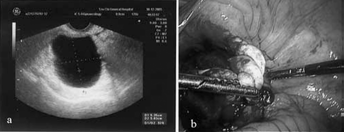
A 70-year-old postmenopausal woman presented with a 1-week history of lower abdominal pain and discomfort. She had undergone total knee replacement 1 year earlier. Her obstetric history was unremarkable, with no history of tubal sterilization. On examination, she was afebrile and normotensive. Her vaginal examination revealed a tense mass in the right adnexa. Ultrasound revealed a well-defined, echo-free cystic mass measuring 5.3 cm × 5.8 cm without septations (Figure 1a). Her blood count and erythrocyte sedimentation rate were normal. Serum markers of ovarian malignancy were obtained and found to be within normal limits.
Figure 1.
(a) Transvaginal ultrasound showed an echo-free cystic lesion, 5.3 cm × 5.8 cm without septations, located in the right adnexal area. (b) Laparoscopic examination revealed a dark-red, round-shaped cystic lesion that twisted at the infundibulo-pelvic ligament site with adhesion to the right posterior surface of uterus.
Laparoscopic surgery was performed due to suspicion of a right adnexa cystic lesion and possible torsion. Laparoscopic examination showed a dark-red, round-shaped cystic lesion that twisted at the right infundibulo-pelvic ligament site in the right adnexa area with adhesion to the posterior uterine surface with separation from the atrophic ovary (Figure 1b). Twisting at the right infundibulo-pelvic ligament site was noted. Right salpingo-oophorectomy by laparoscopy was smoothly performed, and the specimen was placed into a bag made from a glove and removed through the umbilical port site. Histological examination revealed tubal dilatation with epithelial flattening and foci of hemorrhage within the wall. The patient's hospital course was uneventful, and she was discharged 4 days after surgery. No special complaint was noted during 6-month follow-up.
DISCUSSION
The exact cause of fallopian tube torsion is unknown, and various theories have been postulated. Tubal abnormalities including previous tubal surgery, tubal ligation, tubal reconstruction, and inflammatory disease (hydrosalpinx, hematosalpinx) have been reported. Isolated torsion is rare in a normal fallopian tube, but it might occur in a premenarchal female with no identifiable risk factors. Only sporadic cases of fallopian tube torsion are reported each year. It rarely occurs during menopause.2
The most common symptom is pain located in the lower abdominal region or pelvis that may radiate to the flank or thigh. Sudden onset of cramping pain or intermittent pain is possible. Temperature, white blood cell count, and erythrocyte sedimentation rate may be normal or slightly elevated.2
Imaging findings are nonspecific in the preoperative diagnosis of torsed fallopian tubes. The ultrasound image associated with hydrosalpinx may reveal an elongated, convoluted cystic mass, tapering as it nears the uterine cornua and the ipsilateral ovary separate from the mass. Doppler evaluation could be helpful in a patient with a history of tubal ligation if high impedance or absence of flow in a tubular structure is noted. Computed tomography or magnetic resonance imaging is also reported to be helpful for diagnosis.5,6
Isolated tubal torsion can be managed with either detorsion or simple salpingectomy. Adnexal detorsion has an extremely low risk of thromboembolic events. However, it should be performed as early as possible to avoid irreversible damage to the tissue. The operative approach could be conventional exploratory laparotomy or laparoscopic surgery. Laparoscopic surgery serves not only as a diagnostic tool but is also an excellent therapeutic instrument except when contraindicated.3,7
CONCLUSION
This case is unusual in the postmenopausal age group, making it a rare presentation of a rare entity. Laparoscopy could be a useful tool in diagnosis and treatment of isolated tubal torsion. In postmenopausal women with lower abdominal pain accompanied by a cystic lesion in the adnexal region, a differential diagnosis of isolated hydro-salpinx torsion should be made.
References:
- 1. Hansen OH. Isolated torsion of the Fallopian tube. Acta Obstet Gynecol Scand. 1970;49(1):3–6 [DOI] [PubMed] [Google Scholar]
- 2. Shukla R. Isolated torsion of the hydrosalpinx: a rare presentation. Br J Radiol. 2004;77(921):784–786 [DOI] [PubMed] [Google Scholar]
- 3. Wang PH, Yuan CC, Chao HT, Shu LP, Lai CR. Isolated tubal torsion managed laparoscopically. J Am Assoc Gynecol Laparosc. 2000;7(3):423–427 [DOI] [PubMed] [Google Scholar]
- 4. Ullal A, Kollipara PJ. Torsion of a hydrosalpinx in an 18-year-old virgin. J Obstet Gynaecol. 1999;193:331. [DOI] [PubMed] [Google Scholar]
- 5. Ghossain MA, Buy JN, Bazot M, et al. CT in adnexal torsion with emphasis on tubal findings: correlation with US. J Comput Assist Tomogr. 1994;18(4):619–625 [DOI] [PubMed] [Google Scholar]
- 6. Terada Y, Murakami T, Nakamura S, et al. Isolated torsion of the distal part of the fallopian tube in a premenarcheal 12 year old girl: a case report. Tohoku J Exp Med. 2004;202(3):239–243 [DOI] [PubMed] [Google Scholar]
- 7. Ding DC, Chen SS. Conservative laparoscopic management of ovarian teratoma torsion in a young woman. J Chin Med Assoc. 2005;68(1):37–39 [DOI] [PubMed] [Google Scholar]


