Abstract
Introduction:
Injury to intraperitoneal organs is unusual during percutaneous renal surgery. We report a splenic injury during upper pole percutaneous renal access for nephrostolithotomy that was managed conservatively.
Methods:
A 52-year-old male with left upper pole renal stones associated with a narrow upper pole infundibulum underwent upper pole renal access prior to percutaneous nephrostolithotomy (PCNL). The access was performed in the 10th to 11th intercostal space, and the patient underwent PCNL with stone clearance. Plain film radiography after percutaneous access and PCNL revealed no pneumothorax or hydrothorax. The patient was discharged on postoperative day one with the nephrostomy tube in place.
Results:
On postoperative day 5, the patient was evaluated for persistent flank pain and bleeding from the nephrostomy tube. Computerized tomography revealed a transsplenic percutaneous renal access. The patient was admitted to the hospital, and the general surgery service was consulted. The patient was placed on strict bedrest. His hematocrit was within normal limits and remained stable. The nephrostomy tube was kept in place for 2 weeks. A pullback nephrostogram revealed no perirenal leak, and no evidence was present of acute bleeding. Follow-up computerized tomography on the same day revealed no evidence of acute bleeding. The patient was discharged without further complications and remains stone free at 1-year follow-up.
Conclusions:
A transsplenic renal access that was dilated and through which a successful left percutaneous nephrostolithotomy was performed is a highly unusual complication related to upper pole left renal access. We were able to manage this complication with conservative measures.
Keywords: Splenic injury, Percutaneous nephrostolithotomy
INTRODUCTION
Percutaneous nephrostolithotomy is the treatment of choice for most large renal stones, and the success of the procedure is critically dependent on obtaining an access with optimal angles for lithotripsy and stone removal. While thoracic entry and pneumothorax is a known complication with intercostal renal access, injury to intraperitoneal organs is unusual. We report the successful conservative management of a splenic injury resulting from transsplenic access and tract dilation in a patient undergoing PCNL.
CASE REPORT
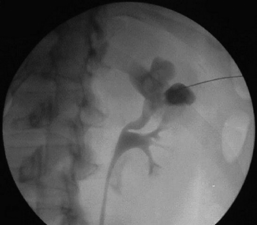
A healthy 52-year-old male presented with left upper pole renal stones associated with a narrow infundibulum (Figure 1). Due to stone location and related renal anatomy, the upper pole approach was preferred, and upper pole renal access through the 10th to 11th intercostal space was obtained in interventional radiology prior to PCNL. The access was successful, and the patient underwent balloon dilation of the tract and PCNL with complete stone clearance. Plain film radiography after PCNL revealed no pneumothorax or hydrothorax. The patient was discharged on postoperative day one with the nephrostomy tube in place.
Figure 1.
Nephrostogram during initial puncture demonstrates upper pole calculi with a narrow, tortuous infundibulum.
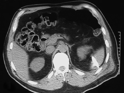
On postoperative day 3, the patient was evaluated in the emergency room due to flank pain and bleeding through the nephrostomy tube. His pain was successfully managed with narcotic analgesics. His blood work including hematocrit was within normal limits. He was discharged home. On postoperative day 5, he was reevaluated for persistent bleeding from the nephrostomy tube and flank pain. Computerized tomography of the abdomen and pelvis was obtained, revealing transsplenic percutaneous renal access (Figure 2). The patient was admitted to the hospital and placed on bedrest. His hematocrit was within normal limits and remained stable. In consultation with interventional radiology and general surgery, a decision was made to leave the nephrostomy in place for 2 weeks after surgery.
Figure 2.
Nephrostomy tube found to traverse the spleen on computerized tomography without hematoma.
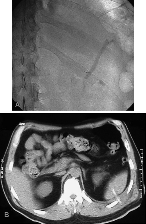
At that time, the patient underwent a pullback nephrostogram revealing no perirenal leak. The nephrostomy tube was removed without instillation of coagulants in the tract. No evidence was present of acute bleeding. Follow-up computerized tomography on the same day revealed no evidence of acute bleeding. The splenic tract was clearly demarcated with contrast (Figure 3). The patient was observed overnight and discharged without further complications.
Figure 3.
Splenic portion of the nephrostomy tract as seen by fluoroscopy (A) and computerized tomography (B).
DISCUSSION
The major complication rate for PCNL varies between 1.1% to 7%. Transfusion rates range from <1% to 10%.1 Most complications are related to percutaneous renal access, with bleeding and pneumothorax being most common. Renal anatomy and choice of access tract affect the rate of complications. When supracostal puncture is performed, the risk of pneumothorax or pleural effusion requiring drainage is 4% to 12%.2–6 Intraperitoneal visceral injury, particularly splenic laceration, is rare but has been reported.7,8 Splenic injury may require surgical management.9 However, conservative management with splenic preservation is feasible as demonstrated here.
The risk of splenic injury during PCNL has been estimated by Hopper and Yakes,10 who used CT to analyze the relationship of the kidney, spleen, and lower ribs. Their analysis noted that splenic injury is highly unlikely if an 11th or 12th rib supracostal approach is made during expiration. The risk increases to 13% if this approach is taken on inspiration and may be as high as 33% if a 10th to 11th approach is used for access.
Splenic injury in our patient was most likely due to supra-11th puncture at the skin level. Access was quite oblique with the needle directed caudally and, in retrospect, transperitoneally. No pleural injury occurred. The advantages of upper over lower pole access include direct access along the long axis of the kidney and to the ureteropelvic junction, usually allowing for less torque of the rigid nephroscope and less bleeding. We feel that the upper pole should have been accessed given the patient's anatomy. However, this may have been achieved at the 11th to 12th intercostal space, thus lessening the risk of transsplenic puncture and splenic injury.
Contributor Information
Robert I. Carey, University of Miami, Department of Urology, Miami, Florida, USA..
Farjaad M. Siddiq, University of Miami, Department of Urology, Miami, Florida, USA..
Jorge Guerra, University of Miami, Department of Radiology, Miami, Florida, USA..
Vincent G. Bird, University of Miami, Department of Urology, Miami, Florida, USA..
References:
- 1. Lingeman JE, Lifshitz DA, Evan AP. Surgical Management of Urinary Lithiasis. In: Walsh PC. ed. Cambell's Urology 8th Editon. Philadelphia, PA: Saunders; 2002:3361–3451 [Google Scholar]
- 2. Young AT, Hunter DW, Castaneda-Zuniga WR, et al. Percutaneous extraction of urinary calculi: Use of the intercostal approach. Radiology. 1985; 154: 633–638 [DOI] [PubMed] [Google Scholar]
- 3. Picus D, Weyman PJ, Clayman RV, et al. Intercostal-space nephrostomy for percutaneous stone removal. AJR Am J Roentgenol. 1986; 147: 393–397 [DOI] [PubMed] [Google Scholar]
- 4. Fuchs E, Forsyth MJ. Supracostal approach for percutaneous ultrasonic lithotripsy. Urol Clin North Am. 1990; 17: 99–102 [PubMed] [Google Scholar]
- 5. Narasimham DL, Jacobsson B, Vijayan P, Bhuyan BC, Nyman U, Holmquist B. Percutaneous nephrolithotomy through an intercostal approach. Acta Radiol. 1991; 32: 162–165 [PubMed] [Google Scholar]
- 6. Golijanin D, Katz R, Verstandig A, et al. The supracostal percutaneous nephrostomy for treatment of staghorn and complex kidney stones. J Endourol 1998; 12: 403–405 [DOI] [PubMed] [Google Scholar]
- 7. Santiago L, Bellman GC, Murphy J, et al. Small bowel and splenic injury during percutaneous renal surgery. J Urol. 1998; 159: 2071–2073 [DOI] [PubMed] [Google Scholar]
- 8. Goldberg SD, Gray RR, St. Louis EL, et al. Nonoperative management of complications of percutaneous renal nephrostomy. Can J Surg. 1989; 32: 192. [PubMed] [Google Scholar]
- 9. Kondas J, Szentgyorgyi E, Vaczi L, Kiss A. Splenic injury: a rare complication of percutaneous nephrolithotomy. Int Urol Nephrol. 1994; 26: 399–404 [DOI] [PubMed] [Google Scholar]
- 10. Hopper KD, Yakes WF. The posterior intercostal approach for percutaneous renal procedures: risk of puncturing the lung, spleen, and liver as determined by CT. AJR. 1990; 1541: 115–117 [DOI] [PubMed] [Google Scholar]


