Abstract
Schwannoma is a rare tumor of neural crest cell origin that is rarely seen arising from the adrenal gland. We report a case of an adrenal mass discovered incidentally in a 70-year-old man as part of a hematuria workup. Metabolic evaluation was unremarkable, and imaging studies did not meet strict imaging criteria for a typical adenoma. Following surgical excision and pathologic evaluation with confirmatory immunohistochemical staining, the mass was reported as a benign nerve sheath neoplasm.
Keywords: Adrenal schwannoma, Laparoscopy
INTRODUCTION
Schwannoma is a benign neoplasm originating from the myelin sheath of peripheral, motor, sensory, sympathetic, or cranial nerves. Visceral schwannomas are extremely rare and are usually discovered serendipitously. Adrenal schwannomas are equally rare with only 3 reports of 5 cases reported previously in the English literature.1–3 We report a case of adrenal schwannoma discovered incidentally on routine cross-sectional imaging performed as part of a hematuria workup.
CASE REPORT
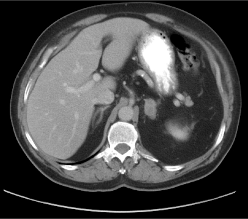
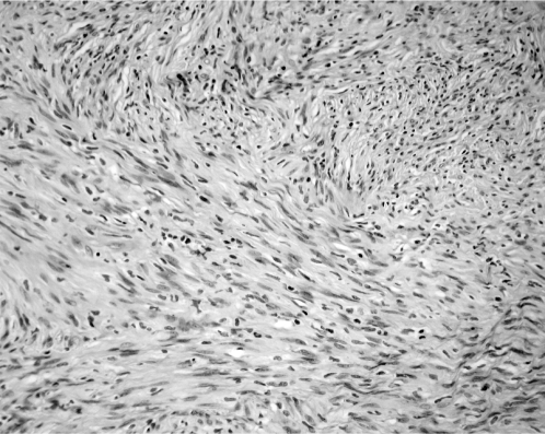
A 70-year-old male presented with a new onset of gross hematuria. As part of the evaluation, the patient underwent cystoscopy and urine cytology, both of which were unremarkable. Computed tomography of the abdomen and pelvis revealed a 2.6×1.8-cm heterogeneously enhancing soft tissue nodule in the left adrenal gland (Figure 1). Based on a density of 36 Hounsfield units, the mass did not meet strict noncontrast CT criteria for diagnosis of adenoma. Subsequent abdominal magnetic resonance imaging with intravenous administration of Gadolinium revealed a 3.5×1.8-cm lobular mass arising from the medial limb of the left adrenal gland. The mass demonstrated early peripheral enhancement with no significant washout. No retroperitoneal or upper abdominal lymphadenopathy was present. Metabolic workup, including serum electrolytes, cortisol, urinary metanephrine, and vanillylmandelic acid (VMA) were within normal range. A standard transperitoneal laparoscopic adrenalectomy was performed with no complications. Postoperative pathologic evaluation revealed a 65-g adrenal gland with a solitary tumor measuring 2.8×2.0×1.5-cm in overall dimensions that was grossly impinging on adrenal parenchyma. Histologically, the tumor exhibited characteristics of a benign nerve sheath neoplasm that appeared to arise from the adrenal medulla. Immunohistochemical analysis revealed cells that were uniformly S-100 positive, weakly vimentin positive, and were negative for CD34, CD177 (c-kit), desmin, and HMB-45 (Figure 2).
Figure 1.
Noncontrast-enhanced computed tomography scan showing a small mass in the left adrenal gland.
Figure 2.
100x power photomicrograph of hematoxylin and eosin stained section showing a spindle cell tumor with Verocay bodies.
DISCUSSION
Schwannoma is a benign, slow-growing, encapsulated neoplasm in which the principal component arises from neural crest cells and comprises differentiated Schwann cells in a poorly collagenized stroma. Schwannomas were first described by Verocay in 1908, with further sub-classification into 2 distinct histologic patterns performed by Antonini in 1920.4 This neoplasm commonly originates from the nerve sheath of peripheral, motor, sensory, sympathetic, or cranial nerves within the head, neck, and upper and lower extremities. Other sites of occurrence include the gastrointestinal tract5 and the retroperitoneum.6 Although the prevalence of clinically inapparent adrenal masses is about 2.1% and increases to 7% in patients older than 70 years,7 a schwannoma arising from adrenal medulla is a rare entity that has only been reported in the English literature 3 times,1–3 with several additional case reports in the world literature.8–10 It has been theorized that adrenal gland schwannomas originate from Schwann cells that insulate the nerve fibers innervating the adrenal medulla.3
Adrenal schwannomas are typically found incidentally; however, patients may present with clinical symptoms secondary to the mass effect of the tumor. Four of 8 cases reviewed in the literature were discovered incidentally, with abdominal discomfort being the most frequent presentation in the other 4 cases. Given the nonfunctional nature of schwannomas, only positive hormonal studies, such as elevated urine metanephrines in most cases of pheochromocytoma, can unequivocally rule out the diagnosis of schwannoma. On computed tomography, a schwannoma appears as a well-demarcated, round or oval mass that may be homogeneous, as in the case we present; however, other cases in the literature have shown prominent cystic degeneration and calcifications. With addition of contrast, schwannomas may demonstrate variable homogeneous or heterogeneous enhancement.11 Quite often, however, the diagnosis will remain unclear until after surgical intervention. With the advent of minimally invasive technology, laparoscopy provides a diagnostic and therapeutic modality with minimal morbidity. Histomorphological examination can provide the definitive diagnosis, demonstrating neoplastic cells that simulate the appearance of differentiated Schwann cells that are well circumscribed and composed of spindle cells organized as cellular areas with nuclear palisading (Antoni A) and paucicellular areas (Antoni B).12 Immunohistochemical staining can provide secondary confirmation of histology with diffusely positive staining for S-100 protein and vimentin. A small subset of schwannomas may be indistinguishable from neurofibromas, due to similar histologic appearance and positive staining for S-100 protein. Fine et al13 have previously demonstrated that in such instances, a positive stain for calretinin, a calcium-binding protein belonging to the same protein family as S-100, which is expressed in schwannoma, but not neurofibroma, will allow for discrimination of these 2 entities.
CONCLUSION
We report a case of an incidentally discovered schwannoma, arising from the adrenal medulla. Given the rarity of this tumor and lack of definitive nonhistologic diagnostic modalities, adrenal schwannoma remains a diagnosis of exclusion that nonetheless deserves a place on the differential of an incidentally discovered adrenal mass.
Contributor Information
Ruslan Korets, Montefiore Medical Center/Albert Einstein College of Medicine, Bronx, New York, USA.; Department of Urology, Montefiore Medical Center/Albert Einstein College of Medicine, Bronx, New York, USA.
Robert Berkenblit, Montefiore Medical Center/Albert Einstein College of Medicine, Bronx, New York, USA.; Department of Radiology, Montefiore Medical Center/Albert Einstein College of Medicine, Bronx, NY, USA.
Reza Ghavamian, Montefiore Medical Center/Albert Einstein College of Medicine, Bronx, New York, USA.; Department of Urology, Montefiore Medical Center/Albert Einstein College of Medicine, Bronx, New York, USA.
References:
- 1. Arena V, De Giorgio F, Drapeau CM, et al. Adrenal Schwannoma. Report of two cases. Folia Neuropathol. 2004;42:177–179 [PubMed] [Google Scholar]
- 2. Bedard YC, Horvath E, Kovacs K. Adrenal schwannoma with apparent uptake of immunoglobulins. Ultrastruct Pathol. 1986;10:505–513 [DOI] [PubMed] [Google Scholar]
- 3. Lau SK, Spagnolo DV, Weiss LM. Schwannoma of the adrenal gland: report of two cases. Am J Surg Pathol. 2006;30:630–634 [DOI] [PubMed] [Google Scholar]
- 4. Woodruff JM KH, Louis DN, Scheithauer BW: Schwannoma. Lyon France: IARC Press; 2000 [Google Scholar]
- 5. Prevot S, Bienvenu L, Vaillant JC, et al. Benign schwannoma of the digestive tract: a clinicopathologic and immunohistochemical study of five cases, including a case of esophageal tumor. Am J Surg Pathol. 1999;23:431–436 [DOI] [PubMed] [Google Scholar]
- 6. Daneshmand S, Youssefzadeh D, Chamie K, et al. Benign retroperitoneal schwannoma: a case series and review of the literature. Urology. 2003;62:993–997 [DOI] [PubMed] [Google Scholar]
- 7. Grumbach MM, Biller BM, Braunstein GD, et al. Management of the clinically inapparent adrenal mass (“incidentaloma”). Ann Intern Med. 2003;138:424–429 [DOI] [PubMed] [Google Scholar]
- 8. Gonzalez Gonzalez A, Perea R, Palacios Llopis S, et al. A benign adrenal schwannoma [in Spanish]. Med Clin (Barc). 2000;115:518–519 [DOI] [PubMed] [Google Scholar]
- 9. Igawa T, Hakariya H, Tomonaga M. Primary adrenal schwannoma [in Japanese]. Nippon Hinyokika Gakkai Zasshi. 1998;89:567–570 [DOI] [PubMed] [Google Scholar]
- 10. Ikemoto I, Yumoto T, Yoshino Y, et al. Schwannoma with purely cystic form originating from the adrenal area: a case report [in Japanese]. Hinyokika Kiyo. 2002;48:289–291 [PubMed] [Google Scholar]
- 11. Rha SE, Byun JY, Jung SE, et al. Neurogenic tumors in the abdomen: tumor types and imaging characteristics. Radiographics. 2003;23:29–43 [DOI] [PubMed] [Google Scholar]
- 12. Weiss SW GJ. Enzinger and Weiss's Soft Tissue Tumors. St Louis, MO: Mosby; 2001 [Google Scholar]
- 13. Fine SW, McClain SA, Li M. Immunohistochemical staining for calretinin is useful for differentiating schwannomas from neurofibromas. Am J Clin Pathol. 2004;122:552–559 [DOI] [PubMed] [Google Scholar]


