Abstract
Abdominal cerclage is necessary when the more commonly used transvaginal cerclage fails or anatomical abnormalities of the cervix preclude transvaginal placement. The disadvantage of an abdominal approach is that the patient can expect 2 laparotomies during her pregnancy, one for cerclage placement and the other associated with cesarean delivery. We report on an abdominal cerclage removed laparoscopically in the case of an intrauterine fetal death at 17 weeks. This minimally invasive surgical technique eliminates the need for laparotomy in response to a poor previable pregnancy outcome.
Keywords: Cerclage, Abdominal, Laparoscopy
INTRODUCTION
The original abdominal cerclage was described by Benson and Durfee1 in 1965. The majority of patients suffering a recurrent second trimester pregnancy loss due to an incompetent cervix, now termed cervical insufficiency, can be treated successfully with a transvaginal cerclage. A select group of patients who suffer recurrent loss despite a transvaginal cerclage or have an anatomically deformed cervix may benefit from the transabdominal approach.2–4
The disadvantage of the transabdominal approach has been the necessity for 2 laparotomies, one associated with placement of the cerclage and the other with cesarean delivery.5 Occasionally, disorders of pregnancy in the second trimester, prior to fetal viability, warrant delivery. These women require a laparotomy to remove the cerclage and allow vaginal delivery or a classical hysterotomy to deliver the infant leaving the cerclage in situ. We describe the successful laparoscopic removal of an abdominal cerclage in a patient experiencing an intrauterine fetal death at 17 weeks.
CASE REPORT
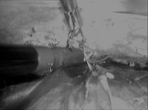
The patient is a 32-year-old, G 6 P 0 A 5, with a history of 1 first trimester loss and 3 second trimester losses felt to be due to cervical insufficiency. A transvaginal cerclage was placed at 13 weeks gestation during the fourth pregnancy, but progressive effacement and dilatation with another second trimester pregnancy loss occurred. The patient presented to the MUSC Prenatal Wellness Center at 13 weeks gestation and was scheduled for a laparotomy and placement of an abdominal cerclage using a Mersilene band. Postoperatively, she developed premature, preterm rupture of membranes. Subsequently, oligohydramnios was noted and fetal death occurred at 17+ weeks. The patient was counseled regarding her options for delivery, laparotomy with cerclage removal, and vaginal delivery or laparotomy and classical hysterotomy for delivery of the dead fetus. We felt that we would be able to release the cerclage laparoscopically. After informed consent, the patient was taken to the operating room where she was prepped and draped accordingly in the lithotomy position with Allen Stirrups. Sequential compression devices were placed prior to induction of anesthesia. A Foley catheter was placed into the patient's bladder. Attention was turned to the patient's abdomen where a vertical intraumbilical incision with Hasson trocar placement was carried out. Intraabdominal CO2 pressure was set at 14 mm Hg and insufflation was begun. Adequate pneumoperitoneum was obtained. A brief survey of the abdomen revealed a large pregnant uterus approximately 3 cm below the umbilicus, soft in consistency. Bilateral 5-mm trocars were placed under direct visualization after first placing a 20-gauge needle with a 10-mL finger control syringe filled with 0.25% bupivacaine with epinephrine to confirm an avascular placement and decrease postoperative pain. Both were placed laterally at the level of the umbilicus yet medial to the epigastric vessels. A suprapubic 5-mm trocar was placed as well. Through these ports, a laparoscopic grasper and a blunt probe were inserted to obtain visualization of the Mersilene band. Using the probe to displace the gravid uterus posteriorly, the scissors were used to redevelop the bladder flap, under which the Mersilene knot and suture was situated. The knot was grasped and elevated away from the uterine surface (Figure 1). The right portion of the Mersilene suture just lateral to the knot was transected with the scissors allowing for the entire Mersilene cerclage to be gently removed. We were careful not to cut both sides of the knot to prevent the band from retracting into the operative site and making removal more difficult. The suture was brought out through the Hasson port. The cervical isthmus and lower uterine segment were noted to be hemostatic. The remaining procedure of port removal and incision closure was performed in a standard manner. While the patient was anesthetized, laminaria were passed into her cervix and the vagina was packed with gauze. The patient tolerated the procedure well and was transferred to the recovery room. She underwent successful dilatation and evacuation of a fetus weighing 150 grams.
Figure 1.
Transecting Mersilene suture after dissecting bladder flap.
DISCUSSION
The continued development of minimally invasive surgical skills allows women new options resulting in less morbidity and more prompt recovery. Scarantino et al6 and McComiskey et al7 were the earliest to report that operative laparoscopy is a viable alternative to laparotomy in those women needing abdominal cerclage removal in the second trimester. We confirm the notion that operative laparoscopy is a viable alternative for removing a cerclage previously placed by laparotomy. We wish to emphasize the importance of excellent communication with our obstetrical colleagues.
Important technical aspects of the case revolve around placement of the trocars.8,9 The large uterus can block visualization when the camera and telescope are placed through the infraumbilical port. In our case, we were able to displace the uterus posteriorly enough to be able to develop the bladder flap and incise the knot. If this had not been possible, we would have considered moving the camera to one of our lateral ports. Likewise, placement of the lateral ports relatively high at the level of the umbilicus is important in being able to manipulate an oversized yet soft uterus. Again, we were successful in our approach but we would not have hesitated to place 2 additional 5-mm ports, one on each side, several centimeters inferior to our more superior accessory probes to gain access to the cervical isthmus and the bladder flap.
One final consideration for abdominal cerclage removal by operative laparoscopy involves the posterior approach. Although difficult, the enlarged uterus could have been displaced either laterally or anteriorly, allowing us to incise the band as it crosses the cervical isthmus above the level of the uterosacral ligament. Although this would not allow band removal, incision of the band should allow subsequent cervical dilatation and vaginal delivery. Although not reported in the literature, we have heard of anecdotal reports of band incision via a colpotomy approach.
CONCLUSION
Operative laparoscopy with abdominal cerclage removal is a viable alternative for women with second trimester fetal loss or diagnoses necessitating midtrimester vaginal delivery.
References:
- 1. Benson RC, Durfee RB. Transabdominal cervicouterine cerclage during pregnancy for treatment of cervical incompetency. Obstet Gynecol. 1965; 25: 145–155 [PubMed] [Google Scholar]
- 2. Novy MJ. Transabdominal cervicoisthmic cerclage: a reappraisal 25 years after its introduction. Am J Obstet Gynecol. 1991; 164: 1635–1642 [DOI] [PubMed] [Google Scholar]
- 3. Cammarano CL, Herron MA, Parer JT. Validity of indications for transabdominal cerclage for cervical incompetence. Am J Obstet Gynecol. 1995; 172: 1871–1875 [DOI] [PubMed] [Google Scholar]
- 4. Leiman G, Harrison NA, Rubin A. Pregnancy following conization of the cervix. Complications related to cone size. Am J Obstet Gynecol. 1980; 136: 14–18 [DOI] [PubMed] [Google Scholar]
- 5. Lesser KB, Childers JM, Surwit EA. Transabdominal cerclage: a laparoscopic approach. Obstet Gynecol. 1998; 91: 855–856 [DOI] [PubMed] [Google Scholar]
- 6. Scarantino SE, Reilly JG, Moretti ML, Pillari VT. Laparoscopic removal of a transabdominal cervical cerclage. Am J Obstet Gynecol. 2000; 182 (5): 1086–1088 [DOI] [PubMed] [Google Scholar]
- 7. McComiskey M, Dornan JC, Hunter D. Removal of transabdominal cerclage. Ulster Med J. 2006; 75 (3): 228. [PMC free article] [PubMed] [Google Scholar]
- 8. Carter JF, Soper DE. Laparoscopy in pregnancy. JSLS. 2004; 8: 57–60 [PMC free article] [PubMed] [Google Scholar]
- 9. Carter JF, Soper DE. Laparoscopic abdominal cerclage: a case report. JSLS. 2005; 9: 491–493 [PMC free article] [PubMed] [Google Scholar]


