Abstract
Background and Objectives:
We report a case of appendicitis presenting in an incarcerated femoral hernia, otherwise known as de Garengeot hernia. This rare hernia usually presents with both diagnostic and therapeutic dilemmas. We wish to underline the usefulness of laparoscopy in both the diagnosis and treatment of de Garengeot hernias.
Methods:
A diagnostic laparoscopy was performed initially. The appendix was seen to disappear into the hernia sac. A laparoscopic appendectomy was then performed prior to open exploration of the groin.
Results:
We were able to obtain a correct diagnosis and perform an appendectomy prior to making a groin incision. Operative findings included an incarcerated, inflamed appendix within a femoral hernia.
Conclusions:
Diagnostic laparoscopy could be a valuable tool in the correct diagnosis and management of unusual presentations of incarcerated groin hernias.
Keywords: Hernia, Appendicitis, de Garengeot, Diagnostic laparoscopy
INTRODUCTION
We report the successful management of a 77-year-old lady who presented with acute onset of a right groin mass and tenderness, which turned out to be an incarcerated femoral hernia containing an inflamed appendix.
CASE REPORT
A 77-year-old lady presented to the emergency room with a right groin mass that had appeared suddenly 4 days before and then gradually increased in size. She also complained of increasing pain and redness in the right groin. Her past medical history was significant for hypertension, gastroesophageal reflux disease (GERD), and right inguinal hernia repair 30 years before. She denied fevers, nausea, vomiting, change in bowel habits, or any urinary symptoms and did not have any recent trauma.
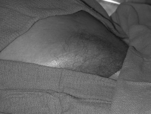
On admission, the patient was afebrile with age-appropriate vital signs. Abdominal examination revealed a soft, obese, and nondistended abdomen with a large, tender mass in the right inguinal region, measuring 18 cm × 10 cm. The mass was firm to palpation and nonreducible, with erythema of the overlying skin. No peritoneal signs were elicited, and the remainder of the patient's physical examination was unremarkable. Laboratory data showed a significant white blood cell count of 19,100/mm3 but were otherwise within normal range.
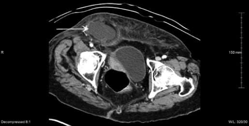
A recurrent, incarcerated inguinal hernia was suspected, but the lack of systemic or abdominal symptoms and the absence of findings suggesting bowel obstruction was concerning. The differential diagnosis of a localized abscess of uncertain origin was also considered. A computed tomographic (CT) scan of the abdomen and pelvis was obtained to help differentiate between intraabdominal and localized pathology and to plan our surgical approach. A 3 cm × 6 cm lobulated fluid collection in the anterior abdominal wall in the right inguinal region was seen. Stranding and thickening of the adjacent anterior abdominal wall fat was also seen. The primary concern was for an incarcerated, recurrent inguinal hernia resulting in tissue necrosis and abscess formation. We decided to do a diagnostic laparoscopy prior to exploration of the groin.
Figure 1.
External view of the patient's right groin swelling.
Figure 2.
CT scan of the abdomen showing the abscess-like collection (white arrow) in the right lower quadrant.
Figure 3.
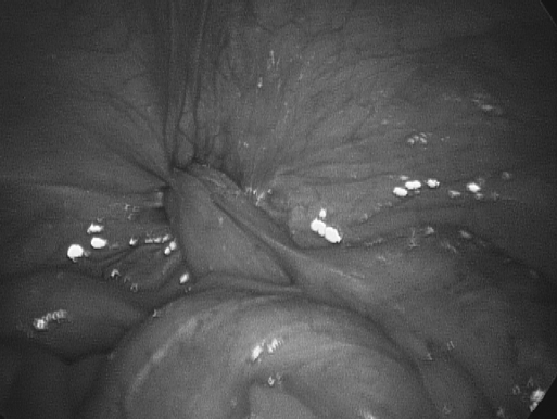
Laparoscopic view of the femoral hernia with the incarcerated appendix.
Figure 4.
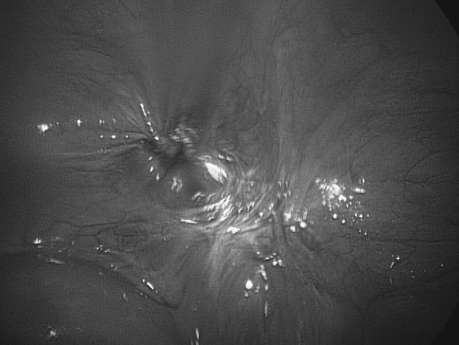
Laparoscopic view of the hernia orifice after repair.
Operative Technique
An infraumbilical incision was made, and pneumoperitoneum was obtained by using Hasson's open technique. A blunt port was then inserted through the incision and a 10-mm, 30-degree scope was used to examine the abdominal cavity. A major portion of the appendix was seen to pass through a defect adjacent to the inguinal ligament. Two 5-mm abdominal ports were placed, one in the left lower quadrant and the other in the right upper quadrant. The mesoappendix and the base of the appendix were then stapled and transected separately.
We then turned our attention to the right inguinal swelling. An incision was made externally over the right groin swelling and then carried down to the hernia sac, which appeared to be a femoral hernia. The sac was opened and seropurulent fluid was evacuated. The appendix was then isolated and removed through the external incision. Dissection was carried down to the neck of the sac, which was then ligated, followed by excision of the sac. The inferior portion of the inguinal ligament and the pectineus fascia were then approximated by using 0 polypropylene sutures. A Penrose drain was placed over this layer. The rest of the wound was closed in layers, by using absorbable sutures for deep subcutaneous layer and staples for skin.
The patient had an uneventful postoperative period and was discharged to her home on the second day after surgery. She was seen twice afterwards as an outpatient, once for removal of the drain and a second time for removal of staples.
DISCUSSION
The appendix being present within a femoral hernia is an unusual incidental finding at surgery, occurring in 0.8% to 1% of all femoral hernias.1,2 This interesting condition was first described in 1731 by the French surgeon Rene Jacques Croissant de Garengeot.3 Even more rare is the presence of acute appendicitis within a femoral hernia, and Hevin in 1785 first described an appendectomy in one such case. Fewer than 80 such cases have been reported to date.4,5 Complications such as rupture and abscess formation have been described in a handful of cases. A review of the literature indicates the incidence of de Garengeot hernias to be greater in women, paralleling the sex-related incidence of femoral hernias.5 The diagnosis is a difficult one to make preoperatively, and only 2 reports of a positive CT diagnosis are available in literature.2,6,7
The patient is usually an elderly female with a few days' history of a painful groin swelling, suggestive of an incarcerated hernia or a groin abscess. These patients seldom develop signs of peritonitis, as the inflamed appendix is isolated from the peritoneal cavity by the tight neck of the hernia sac. Frequently, the inflamed or ruptured appendix is a surprise finding when the groin swelling is explored.
No standard approach to treatment of de Garengeot hernias has been described, possibly due to the rarity of this condition. Various authors have suggested different surgical options ranging from initial open drainage and interval appendectomy and hernia repair, to initial appendectomy followed by interval hernia repair.8–10 In previously reported cases when the groin was explored first, it was often not possible to reach the base of the appendix, and a formal laparotomy became necessary.
In our patient, the unusual presentation of the hernia prompted us to do a diagnostic laparoscopy first, during which the appendix was seen entering the hernia sac. We were therefore able to promptly perform an appendectomy laparoscopically, eliminating the need for laparotomy and peritoneal contamination. We were also able to anticipate the contents of the hernia before opening the sac. The same approach can be applied to acute appendicitis occurring within an inguinal hernia (also known as Amyand hernia).
We therefore recommend initial diagnostic laparoscopy in the treatment of groin hernias with atypical presentations and when the contents of the hernia cannot be determined either clinically or radiologically. To the best of our knowledge, this approach has never been described before.
CONCLUSION
De Garengeot hernia is a rare condition in which appendicitis occurs within a femoral hernia. Initial diagnostic laparoscopy can be an invaluable adjunct in both diagnosis and treatment of atypical hernias.
References:
- 1.Wise L, Tanner N. Strangulated femoral hernia appendix with perforated sigmoid diverticulitis. Proc R Soc Med. 1963;56:1105. [DOI] [PMC free article] [PubMed] [Google Scholar]
- 2.Wakely CPG. Hernia of the vermiform appendix. In Maingot R. ed. Abdominal Operations. New York, NY: Appleton Century Crofts; 1969;1288 [Google Scholar]
- 3.De Garengeot RJC. Traite des operations de chuirgie. 2nd ed. Paris: Huart; 1731:369–371 [Google Scholar]
- 4.Akopian G, Alexander M. De Garengeot Hernia. Appendicitis within a Femoral Hernia. Am Surg. 2005;71(6):526–527 [PubMed] [Google Scholar]
- 5.Unguent ET, Komenaka IK. Strangulated femoral hernia containing a perforated appendix. Can J Surg. 2004;47(1):69. [PMC free article] [PubMed] [Google Scholar]
- 6.Fukukura Y, Chang SD. Acute appendicitis within a femoral hernia: multidetector CT findings. Abdom Imaging. 2005;30(5):620–622 [DOI] [PubMed] [Google Scholar]
- 7.Zissin R, Brautbar O, Shapiro-Feinburg M. CT diagnosis of acute appendicitis in a femoral hernia. Br J Radiol. 2000;73:1013–1014 [DOI] [PubMed] [Google Scholar]
- 8.Watkins RM. Appendix abscess in a femoral hernia sac – case report and review of the literature. Postgrad Med J. 1981;57:306–307 [DOI] [PMC free article] [PubMed] [Google Scholar]
- 9.Voitk AJ, Macfarlane JK, Estrada RL. Ruptured appendicitis in femoral hernias: Report of two cases and review of the literature. Ann Surg. 1974;179:24–26 [DOI] [PMC free article] [PubMed] [Google Scholar]
- 10.Cuotolo LC, Wasserman I, Pinck RL, et al. Acute suppurative appendicitis occurring within femoral hernia: Report of a case. Dis Colon Rectum. 1978;21:203–204 [DOI] [PubMed] [Google Scholar]


