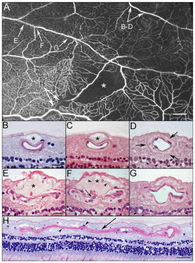
Figure 2.

Area of ADPase labeled retina embedded in JB-4 polymer showing an arteriolar occlusion (arrow), capillary dropout (asterisk) and a ribbon-like appearance of a vein (paired arrows). This region was serially sectioned and the arrows correspond to areas shown in panels A–H below.
Panels B and C demonstrate a dome-shaped serous–like material located internal to the artery just below the internal limiting membrane (asterisks). These bleb-like features were common but intermittent along the course of this artery. Even when these bleb-like structures were absent, the wall of the artery appeared thickened with an amorphous material (arrows) as shown in “D”. The spaces in the inner nuclear layer (arrowheads) probably represent a postmortem artifact. Cross sections in panels E–F taken through a portion of the venous segment demonstrate a serous–like material located internal to the vessel just below the internal limiting membrane (asterisks in E–F). The lumen in some areas appears to be compressed or possibly collapsed. The wall of the vein is thick and contains an amorphous material. Cell processes extend into the site (arrowheads in F) and endothelial cell nuclei appear quite large with pale basophilic staining (arrow in F). A more distal segment of the vein is less affected (G), however, the wall is abnormally thickened. The spaces in the inner nuclear layer (arrowheads) probably represent a postmortem artifact. An old arteriolar occlusion in longitudinal section (H). Some smooth muscle cells remain within the collapsed basement membrane matrix (arrowheads in H). Scale bar = 1mm in A, 20 microns in B–G, 40 microns in H [B and H. Periodic acid Schiff’s (PAS) and hematoxylin; C. hematoxylin and eosin; D–G. toluidine blue/basic fuchsin]
