

Introduction
An 8-year-old child, daughter of Omani parents, presented to the pediatric ophthalmology clinic of Sultan Qaboos University Hospital, Oman, with defective vision, photophobia, and rapid movements of both eyes since birth. The child was otherwise healthy. Both parents were healthy and the marriage was first degree consanguineous. There was a positive family history of similar problems in the patient’s younger brother and paternal aunt. Clinical examination concluded with the diagnosis of oculocutaneous albinism. The patient’s sibling was also examined and diagnosed with the same condition.
Comment
Albinism (origin - Latin word albus which means white) refers to a group of hereditary disorders with an abnormality of melanin synthesis or distribution. The incidence of oculocutaneous albinism in Oman is 1 in 30,000 live births.[1] There are various types of albinism [Table 1]. The main clinical features are hypopigmentation of hair, skin, and eyes. Ophthalmic manifestations include photophobia, nystagmus, defective vision, and squint. The visual acuity in patients with albinism usually is poor and ranges from 20/60 to 20/400. Visual prognosis is usually guarded. However, the children with albinism grow and develop normally and reach normal intelligence levels.[2] The visual-evoked potential test (VEP) is a sensitive diagnostic modality for confirming the diagnosis of albinism. Patients with albinism show an asymmetry of VEP between the two eyes secondary to misrouting of optic pathways.[3] Optical coherence tomography (OCT) is useful in identifying foveal hypoplasia in patients with albinism.[4]
Table 1.
Common types of albinism with brief description of characteristics
| Types of albinism | Subdivision | Subdivision | Etiological factor | Manifestations | Signs and symptoms | Inheritance/trait |
|---|---|---|---|---|---|---|
| Oculo-cutaneous | OCA1* | OCA1A | Tyrosinase negative | Absence of skin | 1. Skin, hair and eye discoloration [Figure 1] | Autosomal recessive disorder |
| or ocular pigment | 2. Tendency to sunburn easily | (affects both sexes equally) | ||||
| 3. Photophobia due to pigmentary | ||||||
| abnormalities of iris and retina | ||||||
| 4. defective vision | ||||||
| 5. Pendular nystagmus | ||||||
| 6. Strabismus, mostly esotropia | ||||||
| 7. Refractive error | ||||||
| 8. Red pupil [Figure 2) | ||||||
| 9. Iris trans- illumination [Figure 3] | ||||||
| 10. Foveal hypoplasia [Figure 4] | ||||||
| 11. Optic nerve hypoplasia | ||||||
| 12. Abnormal decussation of the optic | ||||||
| nerve fi bers | ||||||
| OCA1B | Tyrosinase positive | Can develop some | ||||
| pigment during | ||||||
| the growth | ||||||
| OCA2* | Defect in the P protein | Minimal amount | ||||
| of pigment | ||||||
| OCA3* | Defect in TYRP1 protein | Substantial pigment | ||||
| OCA4* | Defect in SLC45A2 | Minimal amount | ||||
| protein | of pigment | |||||
| Syndromic albinism | Hermansky– | Other genes | Hypo pigmentation, | Signs and symptoms 1 to 12 | ||
| Pudlak | involvement | bleeding problems and, | as above and systemic features | |||
| syndrome | cellular storage disorders | |||||
| Chediak – | Other genes | Hypo pigmentation and | ||||
| Higashi | involvement | defect in white blood cells, | ||||
| syndrome | prone to infections | |||||
| Ocular albinism | OA 1* | Defect of the | Absence of ocular pigment | Only ocular symptoms and | X-linked recessive (affects | |
| GPR143 gene | signs (3 to 12 as above) and | males) Autosomal recessive | ||||
| mosaic fundal pigmentation | (affects both sexes equally) | |||||
| OA 2* | Not exactly known | Absence of ocular | Only ocular symptoms and | |||
| pigment | signs (3 to 12 as above) | |||||
| and color blindness | ||||||
| AROA* | Defect in | Absence of | Only ocular symptoms and | |||
| chromosome 6 | ocular pigment |
OCA - Oculocutaneous albinism
OA - Ocular albinism
AROA - Autosomal Recessive Ocular Albinism,
There is no medical cure for albinism. Supportive care includes: 1] Periodic examination of patients to monitor their visual development and to assess the status of their refractive error and/or strabismus. 2] Vision rehabilitation by correction of refractive errors, use of filter/tinted glasses and caps/visors and prescription of low visual aids and vision enhancing tips. 3] Skin care: There is an increased risk of skin cancer in these patients. They should be advised to use skin-tanning lotion with Sun Protection Factor of 15 or greater and proper clothing for protection against exposure to sunlight. 4] Referral to appropriate sub-specialists for systemic evaluation. 5] Genetic testing and counseling for patients and their families. 6] Counseling by a social worker for social support and to dispel existing myths about albinism.
Figure 1.
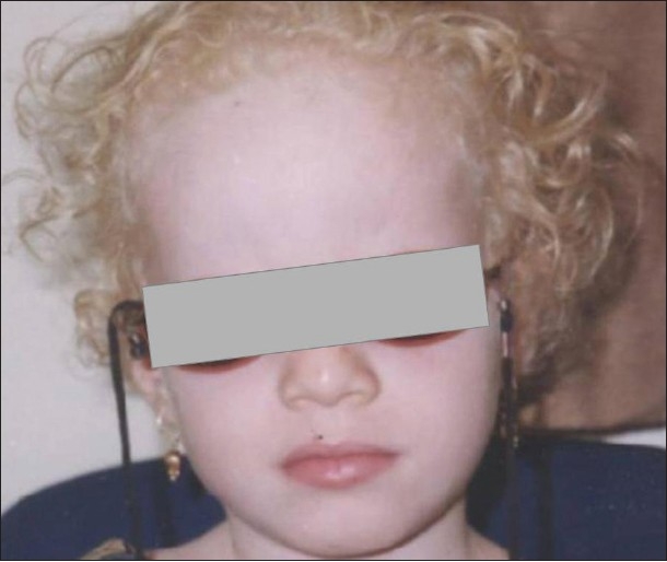
Face photo of an Omani girl shows external features of oculocutaneous albinism. Note blond hair and white skin
Figure 2.
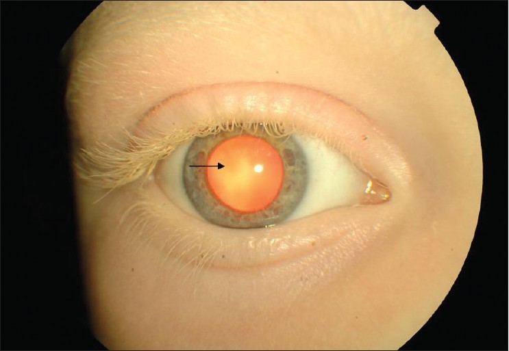
Red pupil (arrow) due to the poor absorption of light from hypo-pigmented retina. Also note hypo-pigmented eye lashes, eye brows, and light iris
Figure 3.
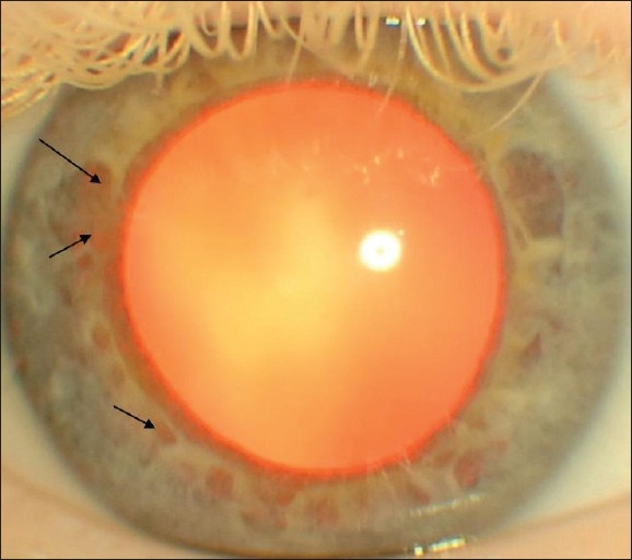
Iris transillumination (arrows) due to escape of refl ected light from the retina through the iris
Figure 4.
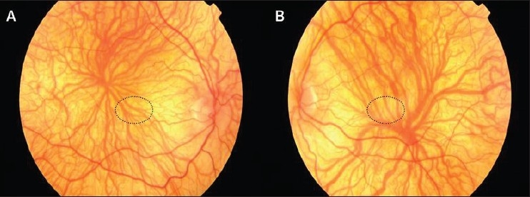
Fundus photo of right eye (A) and left eye (B); shows clear view of choroidal vasculature due to the hypo-pigmentation of retinal pigment epithelium, pale retina, foveal hypoplasia, and indistinct optic disc margin. Dotted circle shows the location of normal fovea.
Acknowledgments
We thank Dr. Nadia Al-Kharousi, Head of the Department of Ophthalmology, Sultan Qaboos University Hospital, Oman for her support in the preparation of this manuscript.
Footnotes
Source of Support: Nil
Conflict of Interest: None
References
- 1.Rajab A, Bappal B, Al-Shaikh H, Al-Khusaibi S, Mohammed AJ. Common autosomal recessive diseases in Oman derived from a hospital-based registry. Community Genet. 2005;8:27–30. doi: 10.1159/000083334. [DOI] [PubMed] [Google Scholar]
- 2.Kutzbach BR, Summers CG, Holleschau AM, MacDonald JT. Neurodevelopment in children with albinism. Ophthalmology. 2008;115:1805–8. doi: 10.1016/j.ophtha.2008.03.006. [DOI] [PubMed] [Google Scholar]
- 3.Hoffmann MB, Schmidtborn LC, Morland AB. Abnormal representations in the visual cortex of patients with albinism: Diagnostic aid and model for the investigation of the visual cortex. Ophthalmology. 2007;104:666–73. doi: 10.1007/s00347-007-1589-7. [DOI] [PubMed] [Google Scholar]
- 4.Izquierdo NJ, Emanuelli A, Izquierdo JC, García M, Cadilla C, Berrocal MH. Foveal thickness and macular volume in patients with oculocutaneous albinism. Retina. 2007;27:1227–30. doi: 10.1097/IAE.0b013e3180592b48. [DOI] [PubMed] [Google Scholar]
- 5.Scheinfeld NS. Syndromic albinism: A review of genetics and phenotypes. Dermatol Online J. 2003;95:5. [PubMed] [Google Scholar]
- 6.The national organization for albinism and hypopigmentation. 2007. Available from: http://www.albinism.org. [PubMed]