Abstract
Background
Hip arthroscopy is an evolving procedure. One small study suggested that a low modified Harris hip score and arthritis at the time of surgery were predictors of poor prognosis.
Questions/purposes
We therefore intended to confirm those findings with a large patient cohort to (1) determine the long-term nonarthritic hip score; (2) determine survivorship; (3) identify risk factors that increase the likelihood of THA; and (4) use those factors to create a usable risk assessment algorithm.
Patients and Methods
We retrospectively reviewed 324 patients (340 hips) who underwent arthroscopy for pain and/or catching. Of these, 106 patients (111 hips or 33%) had a minimum followup of 10 years (mean, 13 years; range, 10–20 years). The average age was 39 years (± 13) with 47 men and 59 women. We recorded patient age, gender, acetabular and femoral Outerbridge grade at surgery, and the presence of a labral tear. Followup consisted of a nonarthritic hip score or the date of a subsequent THA. We determined survivorship with the end point of THA for the acetabular and femoral Outerbridge grades.
Results
Overall survivorship among the 111 hips was 63% at 10 years. The average nonarthritic hip score for non-THA patients was 87.3 (± 12.1). Survivorship was greater for acetabular and femoral Outerbridge grades normal through II. Age at arthroscopy and Outerbridge grades independently predicted eventual THA. Gender and the presence of a labral tear did not influence long-term survivorship.
Conclusions
The long-term survivorship of labral tears with low-grade cartilage damage indicates hip arthroscopy is reasonable for treating labral tears.
Level of Evidence
Level IV, therapeutic study. See Guidelines for Authors for a complete description of levels of evidence.
Introduction
Hip arthroscopy is a rapidly evolving, minimally invasive procedure. Historically, surgery on this joint lagged behind other arthroscopically accessed joints such as the knee and shoulder. The anatomic constraints, inelasticity of the periarticular ligaments, proximity of neurovascular structures, and lack of dedicated hip-specific detractors and instruments contributed to the lack of experience in this area. Improvements in arthroscopic optical capabilities and surgical tools, including the introduction of hip-specific distractors in the late 1980s [26], spurred the use of arthroscopic hip surgery. Since then, hip arthroscopy has been used to treat a variety of problems, including labral tears, chondral lesions, loose bodies, and synovitis.
Long-term followup of surgical procedures drives innovation through specific outcome data. Such followup is limited for hip arthroscopy with the only long-term study reporting arthritis as an indicator of poor prognosis despite an average improvement in modified Harris hip score (mHHS) [4]. However, this study lacked any statistical analysis as a result of its small sample size (50 patients) and did not address survivorship. Although long-term followup studies exist for knee arthroscopy for meniscectomies and meniscal repairs [8, 9, 17, 25], the conclusions from the knee cannot be extrapolated to the hip. Furthermore, results from short- and long-term studies of both hip and knee arthroscopy have resulted in conflicting conclusions [17]. In one study, at an average followup of 5 years after hip arthroscopy, chondromalacia and osteoarthritis did not predict eventual THA or poor post-operative mHHS [15], whereas another study at 2 years followup, and the previously discussed study at 10 years followup [4], showed a clear influence of preoperative arthritis on postoperative surveys or the mHHS [4, 10]. Improved mHHS, patient satisfaction, and no subsequent reoperation or THA at 1.5 years are associated with a diagnosis of labral pathology [1], left-sided surgery, higher preoperative activity level, and longer preoperative duration of symptoms [15], whereas lower improvements in mHHS and other questionnaires examining pain and function are associated with older age at surgery [1, 2, 15]. The 10-year followup study [4] showed an increase in mHHS after arthroscopy treatment for labral tears, loose bodies, synovitis, and chondral injury as long as those indications were not also associated with arthritis or avascular necrosis. Although these studies suggest links between patient factors and various outcome measures, long-term studies with large patient populations are needed to establish factors that influence the long-term success of hip arthroscopy so it can be used more effectively.
Therefore, we (1) determined the ability of hip arthroscopy to relieve pain and restore function; (2) determined survivorship for patients grouped based on patient factors with failure defined as eventual THA; (3) identified patient risk factors that predicted the likelihood of eventual THA; and (4) used those risk factors to create a usable risk assessment algorithm.
Patients and Materials
We retrospectively identified 324 patients (340 hips) who underwent hip arthroscopy for pain and/or catching between 1989 and 1997 and were at least 10 years postoperative. The indications for surgery included anterior, inguinal, trochanteric, or buttock pain with or without a painful catch, lock, or buckle. Further preoperative indications for surgery were a positive McCarthy sign [23] or inguinal pain on a resisted straight leg raise. All patients were chosen for surgery using MRI, arthrography, or if they had clinical findings and failed at least 3 months of nonoperative therapy. In the entire cohort, there were 140 men (43%) and 184 women (57%) with an average age of 36 years (± 13) at the time of surgery. Four patients (five hips) had died, 43 patients (44 hips) were lost to followup, five patients refused contact, and 166 patients (175 hips) did not respond to mailings. We obtained followup from 106 (111 hips) of the 324 patients (33%) at an average followup of 13 years (range, 10–20 years) (Table 1). In our followup cohort, there were 47 men (44%) and 59 women (56%) with an average age of 39 years (± 13) at the time of surgery. These 106 patients (111 hips) were similar in age and gender to the entire study cohort (Table 2). No other patient demographics were available for the entire cohort or followup study group. The followup cohort contained 30% to 55% of patients in each operative Outerbridge grade category (Table 1). Results presented are based only on patients with known followup.
Table 1.
Hips with followup by Outerbridge grade at surgery
| Outerbridge grade at surgery | Number of hips (percent of 340 total) | Received followup | Percent obtained followup |
|---|---|---|---|
| Acetabular | |||
| Normal | 165 (49%) | 51 | 31% |
| Grade I | 45 (13%) | 13 | 29% |
| Grade II | 45 (13%) | 15 | 33% |
| Grade III | 29 (9%) | 10 | 35% |
| Grade IV | 56 (16%) | 22 | 39% |
| Femoral | |||
| Normal | 197 (58%) | 56 | 28% |
| Grade I | 50 (15%) | 16 | 32% |
| Grade II | 36 (11%) | 12 | 33% |
| Grade III | 25 (7%) | 10 | 40% |
| Grade IV | 32 (9%) | 17 | 53% |
| Both acetabular and femoral grades at least | |||
| Normal | 227 (67%) | 68 | 30% |
| Grade I | 39 (11%) | 12 | 31% |
| Grade II | 31 (9%) | 13 | 42% |
| Grade III | 21 (6%) | 7 | 33% |
| Grade IV | 22 (6%) | 11 | 50% |
| Either acetabular or femoral grades at least | |||
| Normal | 135 (40%) | 39 | 29% |
| Grade I | 56 (16%) | 17 | 30% |
| Grade II | 50 (15%) | 14 | 28% |
| Grade III | 33 (10%) | 13 | 39% |
| Grade IV | 66 (19%) | 28 | 42% |
| Total | 340 | 111 | 33% |
Table 2.
Demographic and presurgical clinical variables for the study cohort
| Variable | Followup group (N = 111) | Nonfollowup group (N = 229) | Entire cohort (N = 340) | |
|---|---|---|---|---|
| Age at arthroscopy (years) | Mean ± SD | 38.3 ± 12.6 | 35.0 ± 12.8 | 36.1 ± 12.9 |
| Range | 16–72 | 12–82 | 12–82 | |
| Gender | Male | 47 (42%) | 101 (44%) | 148 (44%) |
| Female | 64 (58%) | 128 (56%) | 192 (56%) | |
| Acetabular Outerbridge grade | Normal | 51 (46%) | 114 (50%) | 165 (49%) |
| Grade I | 13 (12%) | 32 (14%) | 45 (13%) | |
| Grade II | 15 (14%) | 30 (13%) | 45 (13%) | |
| Grade III | 10 (9%) | 19 (8%) | 29 (9%) | |
| Grade IV | 22 (20%) | 34 (15%) | 56 (16%) | |
| Femoral Outerbridge grade | Normal | 56 (50%) | 141 (62%) | 197 (58%) |
| Grade I | 16 (14%) | 34 (15%) | 50 (15%) | |
| Grade II | 12 (11%) | 24 (10%) | 36 (11%) | |
| Grade III | 10 (9%) | 15 (7%) | 25 (7%) | |
| Grade IV | 17 (15%) | 15 (7%) | 32 (9%) | |
| Labral tear | No | 51 (46%) | 102 (45%) | 153 (45%) |
| Yes | 60 (54%) | 127 (55%) | 187 (55%) | |
Percentages represent the portion of each cohort.
The lateral decubitus position with a dedicated hip distractor at 7 mm to 10 mm of distraction was used for all patients. Average surgical time was 38 minutes (range, 21–84 minutes). All arthroscopies were performed as outpatient procedures. Labral tears were treated with débridement with a low-frequency thermal tool or curved shaver and minimal resection of white zone lesions, leaving the capsular attachment. Chondral lesions with full-thickness damage were treated with microfracture surgery, and partial-thickness lesions were minimally resected to a stable base. Loose bodies were removed and the synovial lining was resected in the case of impingement. At the time of surgery, the presence of a labral tear and chondral lesions was recorded along with the type, location, and Outerbridge grade [27] of the lesion. The Outerbridge grades are: normal cartilage; Grade I with softening and swelling; Grade II with a partial-thickness defect with fissures on the surface that do not reach subchondral bone or exceed 1.5 cm in diameter; Grade III with fissuring to the level of subchondral bone in an area with a diameter more than 1.5 cm; and Grade IV with exposed subchondral bone. Observations at the time of surgery showed that of the 340 hips, half had normal chondral surfaces, whereas the other half had anterior chondral lesions in the femur or acetabulum (Table 1). We found labral tears in 102 (30.1%) hips, all of which were anteriorly located and in the white zone. Chondral lesions were also present in 72 of these hips with labral tears (69%), of which 18 were Grade I, 16 were Grade II, 13 were Grade III, and 25 were Grade IV.
All procedures were performed as outpatient procedures with patients on crutches immediately and weightbearing as tolerated. Most patients were off crutches in 3 to 4 days. Recommended postoperative activities included walking, stationary biking, and pool activities. Running, high-impact sports, walking on a treadmill, squats, and leg presses were restricted for the first 6 weeks. Physical therapy was not used for the first 6 weeks. After 6 weeks, depending on the patient’s comfort level and a clinical examination (full ROM without pain, negative McCarthy sign [23] indicating a painful hip click and mechanical symptoms of locking, signs of impingement, and negative resisted straight leg raise), the patient could progress as tolerated or begin physical therapy based on predetermined protocols [22].
Patients were seen at 1 week, 6 weeks, 12 weeks, 6 months, 1 year, 2 years, 5 years, and 10 years postoperatively. The intervals may have been shortened depending on the patient’s progress. The 1-week visit was for suture removal and review of operative findings. All other visits consisted of gait evaluation, radiographs (including AP pelvis and a false profile), and a hip examination including ROM, checking for McCarthy and impingement signs, and resisted straight leg raises. Furthermore, we obtained an MRI arthrogram if there was a recurrence of mechanical symptoms: catching, locking, or buckling.
Followup data were obtained through phone calls from a research assistant (39 hips), office visits with the operating surgeon (15), and mailed letters that were returned (57 hips). Patients who were contacted by phone and/or mailing were done so in groups alphabetically based on their last name. Information collected during a phone call included whether the patient had undergone a subsequent THA on their surgical hip. If so, the date of the THA was recorded. If not, the date of the phone call was used as the followup date. Office visits and returned mailings provided the same information as a phone call as well as a nonarthritic hip score [5] measuring pain, mechanical symptoms, physical function, and level of activity. Survey scores were attained through validated, patient-administered paper surveys. Nonarthritic hip scores were obtained either during office visits or from returned mailings for 62 of the 111 hips with followup (56%) who had not undergone THA.
We constructed a Kaplan-Meier survivorship curve [13] with 95% confidence intervals for 111 hips with follow-up with THA used as an the end point. The inclusion of the patients with no follow-up in the Kaplan-Meier survivorship is not warranted because there is no information about the timing of a specific outcome, and we therefore concluded it was inappropriate and unrealistic to include them and merely assume that all were failures, especially given that we would have no idea when these failures occurred. Survivorship was further determined for groups of acetabular and femoral Outerbridge grades divided into low-grade (normal through Grade II) and high-grade (Grades III or IV) groups with subgroups compared by the log-rank test. To identify correlations between factors and eventual need for THA, a univariable crosstabulation was completed with THA or no THA as a binary outcome and the following factors: age at arthroscopy, gender, acetabular and femoral Outerbridge grades, and the presence of a labral tear. To determine predictors of the eventual need for a THA, a stepwise multivariable logistic regression analysis was performed. To simplify this analysis, the three categories of age, acetabular Outerbridge grades, and femoral Outerbridge grades were each divided into two groups. The two age groups were at or below and above the age of 40 years at surgery to simplify and add meaning to the analysis based on age. This was done because discrete groups are needed to calculate odds ratios of increased risk as well as a table of risk factors for eventual THA. Forty years was chosen because of its proximity to the average and median age of our study population and because it allowed for a clear cut-off to be used in the clinical algorithm. For the multivariable analysis, Outerbridge grades were divided into groups of low-grade (normal to II) and high-grade (Grades III–IV) based on the presence of damage involving the full thickness of the cartilage in Grades III and IV. Two-way interactions between age, gender, grade, and presence or absence of a labral tear were examined in the multivariable logistic models. Odds ratios were calculated for all significant independent predictors. Odds ratios for all possible combinations of significant predictors were used to create a table to be used for risk assessment. Statistical analysis was performed using the SPSS statistical package (Version 18.0; SPSS Inc/IBM, Chicago, IL). Two-tailed p < 0.05 was considered statistically significant.
Results
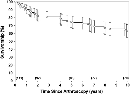
The average of the nonarthritic hip score, based on the 62 hips that did not go on to have a THA, was 87.3 (± 12.1) (Table 3). For patients who underwent THA, the average time to THA was 4.8 years (± 4.4 years; range, 0.1–15.7 years). The Kaplan-Meier estimated survivorship of the entire population with THA as an end point was 91% at 1 year, 75% at 5 years, and 63% at 10 years (Fig. 1).
Table 3.
Nonarthritic hip scores for hips not resulting in THA
| Outerbridge grade at surgery | Average ± SD | Median | Range |
|---|---|---|---|
| Acetabular grade | |||
| Normal (n = 43) | 90.2 ± 9.8 | 94 | 60–100 |
| I (n = 8) | 87.3 ± 12.2 | 81 | 56–100 |
| II (n = 8) | 73.1 ± 14.5 | 73 | 56–95 |
| III (n = 0) | — | — | — |
| IV (n = 3) | 83.8 ± 14.7 | 80 | 71–100 |
| Femoral grade | |||
| Normal (n = 44) | 88.2 ± 11.8 | 94 | 56–100 |
| I (n = 12) | 85.1 ± 14.5 | 81 | 59–100 |
| II (n = 5) | 85.5 ± 11.5 | 73 | 69–95 |
| III (n = 1) | — | 84 | — |
| IV (n = 0) | — | — | — |
| Total (n = 62) | 87.3 ± 12.1 | 91 | 56–100 |
Fig. 1.
The Kaplan-Meier estimated survivorship for the subset (n = 111) of the entire cohort (324) using THA as an end point was 91% (86%–96%) at 1 year, 75% (68%–82%) at 5 years, and 63% (55%–71%) at 10 years. Numbers in parentheses at the bottom are patients who did not have THA and were still being followed.
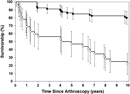
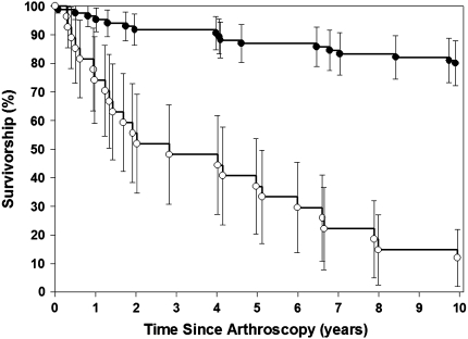
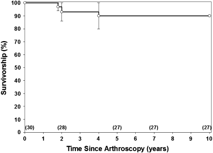
When divided by acetabular Outerbridge grades, 10-year survivorship was 80% for low grades and 22% for high grades (Fig. 2). Based on femoral Outerbridge grades, the 10-year survivorship was 80% for low grades and 12% for high grades (Fig. 3). A decrease (p < 0.001) was found in 10-year survivorship for the high-grade groups compared with the low-grade groups for both the acetabular and femoral Outerbridge grades. For hips with a labral tear and a low-grade chondral lesion (n = 30), the 5-year and 10-year survivorships were 90% (Fig. 4).
Fig. 2.
The Kaplan-Meier estimated survivorship for the followup cohort divided into low-grade (n = 79, 20 THAs) and high-grade (n = 32, 29 THAs) groups based on acetabular Outerbridge grade using THA as an end point showed a decrease in 10-year survivorship for the high grades (22% [9%–35%]) compared with the low grades (80% [71%–89%], p < 0.001, log-rank test = 59.75).
Fig. 3.
The Kaplan-Meier estimated survivorship for the followup cohort divided into low-grade (n = 84, 23 THAs) and high-grade (n = 27, 26 THAs) groups based on femoral Outerbridge grade using THA as an end point showed a decrease in 10-year survivorship for the high grades (12% [2%–22%]) compared with the low grades (80% [72%–88%], p < 0.001, log-rank test = 72.72).
Fig. 4.
The Kaplan-Meier estimated survivorship for a subgroup of the followup cohort, which had a labral tear and a low Outerbridge grade (normal to Grade II) on both the femoral and acetabular side using THA as an end point showed a 90% (80%–100%) survivorship at 5 and 10 years postoperatively. Numbers in parentheses at the bottom are patients who did not have THA and were still being followed.
The univariable analysis indicated possible predictors of age and Outerbridge grades (Table 4) and showed a clear trend toward hips with greater Outerbridge grades resulting in eventual THA. In the multivariable analysis, age group at surgery (p = 0.024) and grouped femoral and acetabular Outerbridge grades (p < 0.001) were confirmed to be independent predictors of eventually having a THA (Table 5). We observed no two-way interactions found in the multivariable analysis (all p > 0.50). Odds ratios showed patients older than 40 years were 3.6 times more likely to require a THA and those with Outerbridge grades of III or IV were 20 to 58 times more likely to eventually require a THA (Table 5). A labral tear did not predict eventual THA.
Table 4.
Univariate analysis according to need for THA
| Variable | THA group (N = 49) | No THA group (N = 62) | p | |
|---|---|---|---|---|
| Age at arthroscopy (years) | Mean ± SD | 43.2 ± 12.5 | 34.5 ± 11.4 | < 0.001* |
| Range | 17–72 | 16–58 | ||
| Age Group | ≤ 40 years | 22 (32%) | 46 (68%) | 0.002* |
| > 40 years | 27 (63%) | 16 (37%) | ||
| Gender | Male | 24 (51%) | 23 (49%) | 0.208 |
| Female | 25 (39%) | 39 (61%) | ||
| Acetabular Outerbridge grade | Normal | 8 (16%) | 43 (84%) | < 0.0001* |
| Grade I | 5 (38%) | 8 (62%) | ||
| Grade II | 7 (47%) | 8 (53%) | ||
| Grade III | 10 (100%) | 0 (0%) | ||
| Grade IV | 19 (86%) | 3 (14%) | ||
| Femoral Outerbridge grade | Normal | 12 (21%) | 44 (79%) | < 0.0001* |
| Grade I | 4 (25%) | 12 (75%) | ||
| Grade II | 7 (58%) | 5 (42%) | ||
| Grade III | 9 (90%) | 1 (10%) | ||
| Grade IV | 17 (100%) | 0 (0%) | ||
| Labral tear | No | 26 (43%) | 34 (57%) | 0.852 |
| Yes | 23 (45%) | 28 (55%) | ||
* Statistically significant.
Table 5.
Multivariate logistic regression analysis with increased odds of needing THA
| Variable | Odds ratio | 95% Confidence interval | p |
|---|---|---|---|
| Age at arthroscopy | 3.6 | 1.2–11.5 | 0.024* |
| > 40 versus ≤ 40 years | |||
| Gender (male versus female) | — | — | 0.134 |
| Acetabular Outerbridge grade | 20 | 4.6–87.0 | < 0.0001* |
| Grades III–IV versus Normal–II | |||
| Femoral head Outerbridge grade | 58.1 | 6.4–528 | < 0.0001* |
| Grades III–IV versus Normal to II | |||
| Presence of a labral tear | — | — | 0.312 |
* Statistically significant.
Based on these odds ratios, the probability of needing a THA by 10 years postoperatively was calculated for all possible risk factors and ranged from 10% for a patient younger than 40 with a Grade 0 to II femoral and acetabular Outerbridge grade to 99% for a patient over 40 with a Grade III or IV femoral and acetabular Outerbridge grade (Table 6).
Table 6.
Probability of THA based on combinations of predictors from multivariate analysis
| Age at arthroscopy (years) | Femoral head Outerbridge grade | Acetabular Outerbridge grade | Probability of THA (%) | 95% Confidence interval |
|---|---|---|---|---|
| 40 or younger | 0–II | 0–II | 10 | 5–22 |
| 40 or younger | 0–II | III–IV | 70 | 40–90 |
| 40 or younger | III–IV | 0–II | 88 | 47–98 |
| 40 or younger | III–IV | III–IV | 99 | 93–99 |
| Older than 40 | 0–II | 0–II | 30 | 15–50 |
| Older than 40 | 0–II | III–IV | 90 | 70–97 |
| Older than 40 | III–IV | 0–II | 96 | 74–99 |
| Older than 40 | III–IV | III–IV | 99 | 98–100 |
Discussion
Long-term followup in the field of arthroscopy is essential because most arthroscopic patients are young and active, and the ultimate goal of these elective procedures is to relieve symptoms and preserve the biologic joint for as long as possible. Long-term studies of knee arthroscopy exist [7, 8, 18, 30], but the conclusions of these studies cannot necessarily be extended to the hip. Furthermore, mid- and long-term studies of hip arthroscopy have sometimes yielded conflicting conclusions [4, 15], and the previously published long-term study is limited by a small sample size and lack of statistically supported conclusions [4]. Therefore, we designed this study to assess the efficacy of the hip arthroscopy procedure, determine the long-term survivorship based on patient factors, identify patient risk factors for eventual THA, and create a usable risk assessment algorithm based on those risk factors.
Our study has a number of limitations. First, we had difficulty obtaining followup for a young population of patients who have relocated and/or changed their contact information since surgery. Although we are very aware that a followup of only 33% of the original patient base could have a large effect on our results, we believe it important to present our findings of the indicators of eventual need for THA in the group we were able to obtain followup. We also observed similarities in both the demographics and presurgical clinical variables between our followup and overall cohorts. Thus, although we realize the status of the patients from whom we did not obtain followup could vary, we speculate that our finding that femoral and acetabular Outerbridge grades predicted eventual THA can be, at least partially, extrapolated to the entire patient cohort based on the previous long-term study presenting similar findings and, importantly, the high level of significance of these variables. Second, in looking at these demographic similarities, there was a small difference in the average age of patients eligible for the study compared with the eventual study cohort (36 versus 39 years, respectively). Because age at surgery has been previously reported [2, 4, 21] as a predictor of “good” or “excellent” postoperative questionnaire scores (improvement of at least 25 points in the mHHS [4], 61% showing improvement of their preoperative scores assessing satisfaction, pain, and mobility [2], and 58% showing “excellent outcomes” [21]), or eventual failure of the arthroscopic procedure, our average postoperative score could be slightly lower, and we could have observed a slightly higher rate of patients eventually requiring THA as a result of the older age of our followup cohort. Third, there was an overrepresentation of patients whose Outerbridge grades at surgery were Grades III or IV (35% to 53% versus 28% to 33% for normal to Grade II). Based on our data indicating higher grades are more likely to result in a THA, this suggests our followup cohort was biased toward a worse disease status, but we cannot say whether they had worse long-term results than the study population. Fourth, this is a single-surgeon series at a tertiary care hospital in a large metropolitan area, which could affect the methods of treatment and results. Fifth, we have no preoperative scores available as a result of the lack of a validated outcome measure at the time of surgery; however, we were still able to compare our postoperative scores with previous literature. Sixth, the treatment of this patient cohort preceded the current knowledge base regarding femoral acetabular impingement [11]. Seventh, we were unable to obtain patient demographics such as body mass index and duration of symptoms preoperatively in our model and our definition of failure was eventual THA without considering reduced function or pain as possible failure modes. However, this clear definition of failure allows us to provide risk factors for a specific, relevant negative outcome: THA. Lastly, the size and location of the cartilage lesions was not recorded, a factor that could affect our results. However, we believe the Outerbridge classification is a sufficient assessment of the severity of the cartilage damage to provide clinical guidance in treating these lesions in the future.
We assessed the effectiveness of hip arthroscopy using the nonarthritic hip score for patients who did not undergo THA. Patients who did undergo THA were considered a failed outcome because the main goal of the arthroscopy was preservation of the natural joint with return to pain-free function. The average postoperative nonarthritic score of 87.3 for our cohort was slightly higher than postoperative scores from previous reports in the literature, which ranged from 74 to 86 from 6 months to 4.8 years followup [3, 14, 19, 29], most likely as a result of the long-term nature of this study. There is currently only one long-term study using the nonarthritic hip score as its validation [5]. To examine the overall success of hip arthroscopy, failure was defined as an eventual THA, and the 5- and 10-year survivorship of our study population were 75% and 63%, respectively. These are comparable, yet slightly lower than previously reported survivorships of 70% to 90% for midterm [2, 10, 15, 20, 28] and 73% for long-term [4] followup of hip arthroscopy (Table 7).
Table 7.
Review of current literature
| Author | Followup (range) | Number of hips | Outcome measures | Restoration of function | Factors affecting outcome | Survivorship (TKA as end point) | |
|---|---|---|---|---|---|---|---|
| Positively | Negatively | ||||||
| Awan and Murray [1] | 1.5 years (1–3.7) | 22 | Questionnaire (pain, mechanical symptoms, activity levels, sporting activities) | Preoperative: 67.7 median, postoperative: 84 median | Mechanical symptoms with confirmed labral pathology | ||
| Boyer and Dorfman [2] | 6.5 years (1–16) | 111 | Questionnaire (satisfaction, pain, mobility), telephone, or office visit | 56.7% had “excellent or good outcomes” | Older age | 83% | |
| Byrd and Jones [4] | 10 years | 52 | mHHS (pain, function) | Preoperative: 56 median, postoperative: 81 median | Symptom duration < 18 months, normal center-edge angles, traumatic onset of symptoms | Arthritis (major predictor), older age | 73% |
| Farjo et al. [10] | 2.8 years (1–8.3) | 28 | Questionnaire (pain, mechanical symptoms, activity level, daily living) versus office visit or telephone | 46% had “good to excellent results” (71% and 21% in nonarthritis and arthritis groups, respectively) | Presence of arthritis (radiographic confirmation), or acetabular or femoral chondromalacia (arthroscopic confirmation) | 71% (86% without arthritis, 57% with arthritis) | |
| Kamath et al. [15] | 4.8 years | 52 | mHHS, clinical outcomes | Preoperative: 56.8 average, postoperative: 80.4 average, 56% had “good or excellent outcomes” | Left side, high preoperative activity, symptom duration > 18 months | Smoking, secondary gain issues (not chondromalacia and arthritis) | 94% |
| Kim et al. [17] | 4.2 years | 43 | Japanese Orthopaedic Association pain score | Arthritis group: 0.76 preoperative to 2.38 postoperative, nonarthritis group: 0.75 preoperative to 1.9 postoperative | Femoroacetabular impingement | ||
| Londers and Van Melkebeek [20] | 6 years (5–10) | 56 | 80.4% showed improvement, 80% would undergo procedure again | Grade of articular damage | 88% | ||
| Margheritini and Villar [21] | 1.5 years | 133 | mHHS | 61% showed improvement, 36% had “good or excellent results” | Older age, severity of arthritis | ||
| Philippon et al. [28] | 2.3 years (2–2.9) | 112 | mHHS | Preoperative: 58 average, postoperative: 84 average, median patient satisfaction 9/10 | Preoperative mHHS, joint space narrowing ≥ 2 mm, repair of labral pathology (versus débridement) | 91% | |
| Streich et al. [31] | 2.8 years (2–4) | 50 | VAS, mHHS, Larson hip score | No degenerative changes of the articular cartilage surface | Articular cartilage lesions | ||
| Walton et al. [32] | 70 | Modified Farjo and Glick classification | Chondral degradation (at arthroscopy), Evidence of degenerative changes on plain radiographs | ||||
| McCarthy et al. [current study] | 13 years (10–20) | 111 | Eventual THA date or Nonarthritic hip score from mailings, telephone, or office visits | Average postoperative: 87.3 | Older age and older age group (≥ 40 years), femoral Outerbridge grade, acetabular Outerbridge grade | 91% at 1 year, 75% at 5 years, 63% at 10 years | |
mHHS = modified Harris hip score; VAS = visual analog scale.
Ten-year survivorship broken down by acetabular and femoral Outerbridge Grades III and IV, compared with Grades 0 to II, showed a decrease for the higher grades of damage from 80% to between 12% and 20%. This decrease in survivorship associated with cartilage damage is consistent with a previous report of decreased survivorship from 86% to 57% for nonarthritis and arthritis groups, respectively [10]. The 10-year survivorship of the arthroscopy procedure for labral tears was 90% as long as the acetabular and femoral Outerbridge grades were low (0 to II). Studies suggest the labrum minimizes the risk of premature arthritis by maintaining proper biomechanical stability of the hip with the purpose of a labral repair to restore this function [6, 12, 16, 24]. Our findings that a labral tear without articular lesions can be successfully treated with arthroscopy and maintain long-term survivorship of the natural hip provides strong support for the early detection and treatment of a labral tear with arthroscopy before the resulting instability causes further cartilage damage leading to worse long-term outcomes.
The need for treatment before cartilage damage occurs was further supported by our finding that femoral and acetabular Outerbridge grades were independent predictors of failure of the arthroscopic procedure defined as eventual THA. Although the previous long-term study identified only a trend between lower mHHS and preoperative diagnoses of osteoarthritis or avascular necrosis [4], our data show higher acetabular and femoral Outerbridge grades of III and IV predict eventual need for a THA. Also, previously published short- and midterm studies have indicated the presence and severity of arthritis or chondral degradation were predictors of worse questionnaire scores, mHHS, modified Farjo and Glick classifications, and Larson hip scores (Table 7) [4, 10, 21, 31, 32]. Because the treatment for articular lesions in the hip, namely judicious removal or microfracture surgery, is similar to that in the knee, this finding is not surprising given similar conclusions [8]. However, our study challenges the conclusions from a 5-year followup study by Kamath et al. [15], which reported postoperative mHHS after hip arthroscopy for labral tear repair was not affected by the presence of arthritis. Although previous works discussed the influence of cartilage changes on outcome, none of these studies has broken down the cartilage changes by acetabular and femoral severity. Our results indicate although cartilage changes in both the acetabular socket and femoral head are important, the risk of failure for hips with femoral head cartilage changes is over two times as high as the risk associated with acetabular changes. The knowledge that patients with cartilage damage are 20 to 60 times more likely to require an eventual THA can be combined with a high-resolution arthro-MRI scan to more effectively screen patients who will benefit the most from hip arthroscopy. In addition to identifying damage on the acetabular or femoral chondral surfaces as a predictor of eventual need for THA, older age was also indicated as increasing the risk of failure by 3.6 times. This is consistent with previous findings from short-, mid-, and long-term studies showing a negative effect of older age on postoperative surveys (Table 7) [2, 4, 21]. Gender did not predict eventual need for THA. This finding is contradictory to the gender relationship seen in knee arthroscopy procedures in which females are at greater risk for failure in both short-term recovery and in long-term studies up to 20 years [7, 9, 25], and no current hip arthroscopy studies report gender as a predictor of outcome. Importantly, our study supplements the observed trend that arthritis and older age negatively affect mHHS at 10 years followup [4] with the conclusion that patients with cartilage damage of Outerbridge grades of III or I to V as well as patients older than 40 years have increased risk for eventual THA after arthroscopy.
Combining the predictors of eventual THA, a chart was made showing the probability that a patient will require a THA by 10 years postoperatively based on the various combinations of risk factors. This is previously unreported and can serve as a guide for physicians and patients during the decision-making process about whether to proceed with the arthroscopy. The intended use of this probability algorithm is to aid in clinical decisions while serving as a benchmark to compare with future long-term studies.
This is the only long-term study of hip arthroscopy large enough to report statistically relevant conclusions. Our results suggest caution should be taken when treating older individuals with articular cartilage damage using this technique but that long-term preservation of a well-functioning hip is possible in patients with labral tears and/or minimal cartilage damage. Additionally, the 90% survivorship 10 years after treatment of labral tears without associated cartilage damage indicates long-term preservation of a natural hip can be achieved with treatment using hip arthroscopy.
Acknowledgments
We thank David Zurakowski, PhD, Department of Orthopedic Surgery, Children’s Hospital, for his assistance with the statistical analysis.
Footnotes
Each author certifies that he or she has no commercial associations (eg, consultancies, stock ownership, equity interest, patent/licensing arrangements, etc) that might pose a conflict of interest in connection with the submitted article.
Each author certifies that his or her institution approved the human protocol for this investigation, that all investigations were conducted in conformity with ethical principles of research, and that informed consent for participation in the study was obtained.
This work was performed at Massachusetts General Hospital, Boston, MA, USA.
References
- 1.Awan N, Murray P. Role of hip arthroscopy in the diagnosis and treatment of hip joint pathology. Arthroscopy. 2006;22:215–218. doi: 10.1016/j.arthro.2005.12.005. [DOI] [PubMed] [Google Scholar]
- 2.Boyer T, Dorfmann H. Arthroscopy in primary synovial chondromatosis of the hip: description and outcome of treatment. J Bone Joint Surg Br. 2008;90:314–318. doi: 10.1302/0301-620X.90B3.19664. [DOI] [PubMed] [Google Scholar]
- 3.Brunner A, Horisberger M, Herzog RF. Sports and recreation activity of patients with femoroacetabular impingement before and after arthroscopic osteoplasty. Am J Sports Med. 2009;37:917–922. doi: 10.1177/0363546508330144. [DOI] [PubMed] [Google Scholar]
- 4.Byrd JW, Jones KS. Prospective analysis of hip arthroscopy with 10-year followup. Clin Orthop Relat Res. 2010;468:741–746. doi: 10.1007/s11999-009-0841-7. [DOI] [PMC free article] [PubMed] [Google Scholar]
- 5.Christensen CP, Althausen PL, Mittleman MA, Lee JA, McCarthy JC. The nonarthritic hip score: reliable and validated. Clin Orthop Relat Res. 2003;406:75–83. doi: 10.1097/00003086-200301000-00013. [DOI] [PubMed] [Google Scholar]
- 6.Crawford MJ, Dy CJ, Alexander JW, Thompson M, Schroder SJ, Vega CE, Patel RV, Miller AR, McCarthy JC, Lowe WR, Noble PC. The 2007 Frank Stinchfield Award. The biomechanics of the hip labrum and the stability of the hip. Clin Orthop Relat Res. 2007;465:16–22. doi: 10.1097/BLO.0b013e31815b181f. [DOI] [PubMed] [Google Scholar]
- 7.Englund M, Lohmander LS. Risk factors for symptomatic knee osteoarthritis fifteen to twenty-two years after meniscectomy. Arthritis Rheum. 2004;50:2811–2819. doi: 10.1002/art.20489. [DOI] [PubMed] [Google Scholar]
- 8.Fabricant PD, Jokl P. Surgical outcomes after arthroscopic partial meniscectomy. J Am Acad Orthop Surg. 2007;15:647–653. doi: 10.5435/00124635-200711000-00003. [DOI] [PubMed] [Google Scholar]
- 9.Fabricant PD, Rosenberger PH, Jokl P, Ickovics JR. Predictors of short-term recovery differ from those of long-term outcome after arthroscopic partial meniscectomy. Arthroscopy. 2008;24:769–778. doi: 10.1016/j.arthro.2008.02.015. [DOI] [PMC free article] [PubMed] [Google Scholar]
- 10.Farjo LA, Glick JM, Sampson TG. Hip arthroscopy for acetabular labral tears. Arthroscopy. 1999;15:132–137. doi: 10.1053/ar.1999.v15.015013. [DOI] [PubMed] [Google Scholar]
- 11.Ganz R, Parvizi J, Beck M, Leunig M, Notzli H, Siebenrock KA. Femoroacetabular impingement: a cause for osteoarthritis of the hip. Clin Orthop Relat Res. 2003;417:112–120. doi: 10.1097/01.blo.0000096804.78689.c2. [DOI] [PubMed] [Google Scholar]
- 12.Groh MM, Herrera J. A comprehensive review of hip labral tears. Curr Rev Musculoskelet Med. 2009;2:105–117. doi: 10.1007/s12178-009-9052-9. [DOI] [PMC free article] [PubMed] [Google Scholar]
- 13.Harris EK, Albert A. Survivorship Analysis for Clinical Studies. New York, NY: Marcel Dekker; 1991. pp. 27–49. [Google Scholar]
- 14.Horisberger M, Brunner A, Herzog RF. Arthroscopic treatment of femoroacetabular impingement of the hip: a new technique to access the joint. Clin Orthop Relat Res. 2010;468:182–190. doi: 10.1007/s11999-009-1005-5. [DOI] [PMC free article] [PubMed] [Google Scholar]
- 15.Kamath AF, Componovo R, Baldwin K, Israelite CL, Nelson CL. Hip arthroscopy for labral tears: review of clinical outcomes with 4.8-year mean follow-up. Am J Sports Med. 2009;37:1721–1727. doi: 10.1177/0363546509333078. [DOI] [PubMed] [Google Scholar]
- 16.Kelly BT, Weiland DE, Schenker ML, Philippon MJ. Arthroscopic labral repair in the hip: surgical technique and review of the literature. Arthroscopy. 2005;21:1496–1504. doi: 10.1016/j.arthro.2005.08.013. [DOI] [PubMed] [Google Scholar]
- 17.Kim SJ, Chun YM, Jeong JH, Ryu SW, Oh KS, Lubis AM. Effects of arthroscopic meniscectomy on the long-term prognosis for the discoid lateral meniscus. Knee Surg Sports Traumatol Arthrosc. 2007;15:1315–1320. doi: 10.1007/s00167-007-0391-z. [DOI] [PubMed] [Google Scholar]
- 18.Kimura M, Shirakura K, Higuchi H, Kobayashi Y, Takagishi K. Eight- to 14-year followup of arthroscopic meniscal repair. Clin Orthop Relat Res. 2004;421:175–180. doi: 10.1097/01.blo.0000119461.83244.69. [DOI] [PubMed] [Google Scholar]
- 19.Laude F, Sariali E, Nogier A. Femoroacetabular impingement treatment using arthroscopy and anterior approach. Clin Orthop Relat Res. 2009;467:747–752. doi: 10.1007/s11999-008-0656-y. [DOI] [PMC free article] [PubMed] [Google Scholar]
- 20.Londers J, Melkebeek J. Hip arthroscopy: outcome and patient satisfaction after 5 to 10 years. Acta Orthop Belg. 2007;73:478–483. [PubMed] [Google Scholar]
- 21.Margheritini F, Villar RN. The efficacy of arthroscopy in the treatment of hip osteoarthritis. Chir Organi Mov. 1999;84:257–261. [PubMed] [Google Scholar]
- 22.McCarthy JC. Early Hip Disease: Advances in Detection and Minimally Invasive Treatment. New York, NY: Springer-Verlag; 2003. pp. 175–190. [Google Scholar]
- 23.McCarthy JC, Lee JA. Acetabular dysplasia: a paradigm of arthroscopic examination of chondral injuries. Clin Orthop Relat Res. 2002;405:122–128. doi: 10.1097/00003086-200212000-00014. [DOI] [PubMed] [Google Scholar]
- 24.McCarthy JC, Noble PC, Schuck MR, Wright J, Lee J. The Otto E. Aufranc Award: The role of labral lesions to development of early degenerative hip disease. Clin Orthop Relat Res. 2001;393:25–37. [DOI] [PubMed]
- 25.Meredith DS, Losina E, Mahomed NN, Wright J, Katz JN. Factors predicting functional and radiographic outcomes after arthroscopic partial meniscectomy: a review of the literature. Arthroscopy. 2005;21:211–223. doi: 10.1016/j.arthro.2004.10.003. [DOI] [PubMed] [Google Scholar]
- 26.Norman-Taylor FH, Villar RN. Arthroscopic surgery of the hip: current status. Knee Surg Sports Traumatol Arthrosc. 1994;2:255–258. doi: 10.1007/BF01845599. [DOI] [PubMed] [Google Scholar]
- 27.Outerbridge RE. The etiology of chondromalacia patellae. J Bone Joint Surg Br. 1961;43:752–757. doi: 10.1302/0301-620X.43B4.752. [DOI] [PubMed] [Google Scholar]
- 28.Philippon MJ, Briggs KK, Yen YM, Kuppersmith DA. Outcomes following hip arthroscopy for femoroacetabular impingement with associated chondrolabral dysfunction: minimum two-year follow-up. J Bone Joint Surg Br. 2009;91:16–23. doi: 10.1302/0301-620X.91B1.21329. [DOI] [PubMed] [Google Scholar]
- 29.Stahelin L, Stahelin T, Jolles BM, Herzog RF. Arthroscopic offset restoration in femoroacetabular cam impingement: accuracy and early clinical outcome. Arthroscopy. 2008;24:51–57.e51. [DOI] [PubMed]
- 30.Steenbrugge F, Verdonk R, Verstraete K. Long-term assessment of arthroscopic meniscus repair: a 13-year follow-up study. Knee. 2002;9:181–187. doi: 10.1016/S0968-0160(02)00017-0. [DOI] [PubMed] [Google Scholar]
- 31.Streich NA, Gotterbarm T, Barie A, Schmitt H. Prognostic value of chondral defects on the outcome after arthroscopic treatment of acetabular labral tears. Knee Surg Sports Traumatol Arthrosc. 2009;17:1257–1263. doi: 10.1007/s00167-009-0833-x. [DOI] [PubMed] [Google Scholar]
- 32.Walton NP, Jahromi I, Lewis PL. Chondral degeneration and therapeutic hip arthroscopy. Int Orthop. 2004;28:354–356. doi: 10.1007/s00264-004-0585-7. [DOI] [PMC free article] [PubMed] [Google Scholar]


