Introduction
Pilon fractures, or fractures of the tibial plafond, range from low- to high-energy axial-loading injuries. This relatively rare injury (< 10% of lower extremity fractures) usually occurs in adults (aged 30s to 40s) owing to a fall from height or a motor vehicle crash [6]. Because of the often high energy involved and the limited soft tissue envelope that surrounds the distal tibia, these fractures require careful attention to the soft tissue envelope for timing and method of surgical fixation. Complications are common after fixation, and posttraumatic arthritis occurs in a high percentage of patients even with adequate restoration of the joint surface [1].
Structure and Function
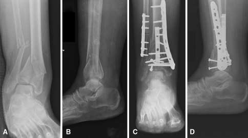
The plafond (or roof) is the distal articular and load-bearing surface of the tibia. An axial load drives the talar dome into the distal tibia (Fig. 1A) with the ankle position influencing the pattern of injury (eg, dorsiflexion tends to cause anterior injury) (Fig. 1B). The fibula, fractured in the majority of cases, is important in understanding the mechanism of injury and as a reference during surgical fixation to help determine correct limb length, alignment, and rotation.
Fig. 1A–D.
(A) AP and (B) lateral radiographs show a comminuted intraarticular pilon fracture with concurrent fibular fracture and valgus malalignment. (C) AP and (D) lateral radiographs after open reduction and internal fixation with correction of limb alignment show near anatomic restoration of the articular surface.
The ankle is a ginglymus joint that is stabilized by strong ligamentous connections between the distal tibia and fibula, calcaneus, talus, and navicular. Common fracture fragments seen in plafond fractures are defined by these ligamentous attachments: for example, the posterolateral (Volkmann’s), anterolateral (Chaput), and medial fragments. Reduction of the articular surface after fracture is essential to decreasing the peak contact pressures realized in the joint and possibly decreasing the incidence of posttraumatic osteoarthritis [5].
The soft tissue envelope around the distal tibia is thin and constrained and the majority of the blood supply is supported by an anastamotic network of extraosseous vessels from the posterior tibial and anterior tibial arteries [13]. These characteristics underscore the importance of meticulous soft tissue management during fixation for optimal soft tissue and bony healing.
Diagnosis and Classification
Plain radiographs, obtained at the time of injury and after provisional reduction, and a CT scan after initial stabilization, are essential to fully understand the fracture and form an adequate preoperative plan. The soft tissues should be inspected carefully; substantial swelling, soft tissue damage, and open injuries are common.
The Rüedi and Allgöwer classification system [11] is based on the degree of articular comminution. Type I fractures are nondisplaced cleavage fractures. Type II fractures have displacement of the fracture fragments but there is minimal comminution. Finally, Type III fractures are those with a high degree of comminution and displacement. Although there are only three categories of fractures, there is low interobserver reliability and debate still exists regarding whether the system has prognostic value [7].
The AO/OTA classification system is an alternative way to describe fractures of the distal tibia [13]. The distal tibia is designated in the system as 43; the tibia being designated as 4 and the distal aspect designated as 3. The system then describes fractures as type A, which are nonarticular fractures; type B, which are partial articular; and type C, which are total articular fractures. In each category, the fracture then is broken into three groups based on the degree of comminution, and then each group is broken again into three subgroups based on different characteristics of the fracture pattern or lines [8]. Despite being a thorough and possibly guiding treatment and prognosis, the system has inadequate observer reliability beyond the basic categories of type A, B, or C [14].
Treatment
Nonoperative management, such as cast immobilization, is reserved only for nondisplaced articular fractures, patients who have surgical contraindications because of medical comorbidities, or patients with low demand such as those who are nonambulatory. Definitive use of external fixation, without articular reduction or fixation, has been used in highly comminuted or open pilon fractures to maintain length, alignment, and rotation [2]. The indications for open reduction and internal fixation include inability to achieve or maintain adequate limb alignment and/or articular reduction. There are no strict guidelines regarding the amount of articular step-off which can be tolerated, but as a general rule, if incongruity can be seen on plain radiographs, operative intervention should be considered. Two major challenges for fixation in displaced fractures include restoration of the articular surface and preservation of the soft tissue envelope to optimize osseous healing. Fractures often are treated in two stages with initial external fixator application followed by definitive fixation at a later time (10 to 14 days or later) [12].
Once the soft tissue edema and swelling have resolved around the extremity, operative intervention can take place. This can be determined subjectively by evaluating the soft tissues and/or checking whether the skin around the zone of injury can wrinkle. The distal tibia can be approached through different surgical intervals and which surgical exposure is used often depends on an open wound, residual fracture blister, or the location of key fracture fragments. The anteromedial, anterolateral, anterior, lateral, posteromedial, and posterolateral approaches have been described [9]. There is no single surgical technique for fractures of the tibial plafond but step-wise reduction of fracture fragments (including attempted restoration of the joint surface) with wires or pins, followed by stable internal fixation is typical. For unstable fracture patterns, plates often are applied to the fibula and tibia (sometimes in multiples) in a direct or indirect (submuscular) fashion.
In achieving fixation goals, observation and reduction of the articular surface (Fig. 1C–D) and minimization of soft tissue trauma are of utmost importance. Bone graft should be used to fill osseous voids during fracture fixation to substitute for bone loss [13]. In select instances where comminution makes articular reconstruction untenable, primary ankle fusion can be considered [3].
Outcomes
The outcomes of surgically treated pilon fractures frequently depend on the associated injury to, and management of, the soft tissues surrounding the injury and accuracy of the articular reduction. Pollak et al. reported in their retrospective study of patients who had sustained fractures of the tibial plafond, after 3 years, more than 1/3 of all patients had substantial ankle stiffness and pain, with more than 1/4 reporting persistent swelling [10]. Rates of delayed union and nonunion increase with fracture severity and soft tissue injury, especially open fractures, but are quoted to be near 5% for all intraarticular distal tibia fractures [6]. An intact fibula can lead to varus malunion after fixation of complete articular plafond fractures. Late stiffness may be related to osteophyte formation, arthrofibrosis, or pain. A correlation exists between the severity of the fracture, the development of arthritis, and an overall outcome [4].
Pearls
Respect the soft tissues: do not operate too early or through compromised skin (open injury, blisters, ecchymotic/attenuated tissue, tight swelling); instead maintain length, alignment, and rotation with an external fixator; practice meticulous surgical technique and minimize surgical trauma to the soft tissue envelope.
Understand the fracture completely before planning surgery (obtain adequate radiographs, CT scan, and radiographs of the uninjured limb as needed).
Restoration of the articular surface and reestablishing its relationship to the tibial shaft is a primary goal of treatment.
For highly comminuted injuries with unreconstructable articular surface, primary ankle fusion is an alternative to ORIF.
Postoperative pain, swelling, and stiffness are the most common sequelae of high-energy tibial plafond injuries and seen in nearly 1/3 of all patients.
Footnotes
Each author certifies that he has no commercial association (eg, consultancies, stock ownership, equity interest, patent, licensing arrangements, etc) that might pose a conflict of interest in connection with the submitted article.
References
- 1.Bonar SK, Marsh JL. Tibial plafond fractures: changing principles of treatment. J Am Acad Orthop Surg. 1994;2:297–305. doi: 10.5435/00124635-199411000-00001. [DOI] [PubMed] [Google Scholar]
- 2.Bone L, Stegemann P, McNamara K, Seibel R. External fixation of severely comminuted and open tibial pilon fractures. Clin Orthop Relat Res. 1993;292:101–107. [PubMed] [Google Scholar]
- 3.Bozic V, Thordarson DB, Hertz J. Ankle fusion for definitive management of non-reconstructable pilon fractures. Foot Ankle Int. 2008;29:914–918. doi: 10.3113/FAI.2008.0914. [DOI] [PubMed] [Google Scholar]
- 4.Egol KA, Wolinsky P, Koval KJ. Open reduction and internal fixation of tibial pilon fractures. Foot Ankle Clin. 2000;5:873–885. [PubMed] [Google Scholar]
- 5.Li W, Anderson DD, Goldsworthy JK, Marsh JL, Brown TD. Patient-specific finite element analysis of chronic contact stress exposure after intraarticular fracture of the tibial plafond. J Orthop Res. 2008;26:1039–1045. doi: 10.1002/jor.20642. [DOI] [PMC free article] [PubMed] [Google Scholar]
- 6.Marsh JL, Saltzman CL. Axial-loading injuries: tibial plafond fractures. In: Bucholz RW, Heckman JD, Court-Brown CM, eds. Fracturesin Adults. Ed 6. Vol 2. Philadelphia, PA: JB Lippincott; 2006:2203–2234.
- 7.Martin JS, Marsh JL, Bonar SK, DeCoster TA, Found EM, Brandser EA. Assessment of the AO/ASIF fracture classification for the distal tibia. J Orthop Trauma. 1997;11:477–483. doi: 10.1097/00005131-199710000-00004. [DOI] [PubMed] [Google Scholar]
- 8.Müller ME, Nazarian S, Koch P, Schatzker J, eds. The Comprehensive Classification of Fractures of Long Bones. Berlin, Germany: Springer-Verlag; 1990:170–179.
- 9.Nork SE. Distal tibia fractures. In: Stannard JP, Schmidt AH, Kregor PJ, eds. Surgical Treatment of Orthopaedic Trauma. New York, NY: Thieme; 2007:767–791.
- 10.Pollak AN, McCarthy ML, Bess RS, Agel J, Swiontkowski MF. Outcomes after treatment of high-energy tibial plafond fractures. J Bone Joint Surg Am. 2003;85:1893–1900. doi: 10.2106/00004623-200310000-00005. [DOI] [PubMed] [Google Scholar]
- 11.Rüedi TP, Allgöwer M. The operative treatment of intra-articular fractures of the lower end of the tibia. Clin Orthop Relat Res. 1979;138:105–110. [PubMed] [Google Scholar]
- 12.Sirkin M, Sanders R, DiPasquale T, Herscovici D., Jr A staged protocol for soft tissue management in the treatment of complex pilon fractures. J Orthop Trauma. 1999;13:78–84. doi: 10.1097/00005131-199902000-00002. [DOI] [PubMed] [Google Scholar]
- 13.Sommer C, Rüedi TP. Tibia distal (pilon). In Rüedi TP, Murphy WM, eds. AO Principles of Fracture Management. New York, NY: Thieme; 2000:543–560.
- 14.Swiontkowski MF, Sands AK, Agel J, Diab M, Schwappach JR, Kreder HJ. Interobserver variation in the AO/OTA fracture classification system for pilon fractures: is there a problem? J Orthop Trauma. 1997;11:467–470. doi: 10.1097/00005131-199710000-00002. [DOI] [PubMed] [Google Scholar]


