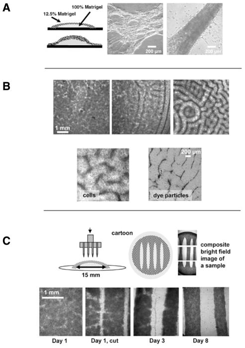
Figure 1. Spontaneous and imposed formation of cardiac fibers within Matrigel pillows.
(A) Cartoon of a pillow and bright field images of live unstained samples that show the extreme examples of very thin and very thick fibers that form spontaneously in these samples. (B) In the upper panel, three examples of low, intermediate, and high degree of patterning observable on different coverslips within the same cell preparation (cell seeding density: 5 × 10⁶/mL, concentration of Matrigel in the top layer: 12.5%, Day 1 after cell plating). In the bottom panel, close-up of the patterns obtained when either myocytes or dye particles were included into the top Matrigel layer. (C) Freshly isolated cardiomyocytes were plated according to the standard Matrigel pillow protocol. The next day a razor stamp was applied to the surface of the Matrigel pillow. The stamp cuts both the newly formed cell layer and the top layer of the Matrigel matrix, creating fibers with defined geometry. The bottom row of bright field images shows the development of these macrofibers as they are maintained in standard cell culture conditions.

