Abstract
Mechanosensory transduction underlies touch, hearing and proprioception and requires mechanosensitive channels that are directly gated by forces; however, the molecular identities of these channels remain largely elusive. A new study has identified Piezo1 and Piezo2 as a novel class of mechanosensitive channels.
The activity of mechanosensitive channels has been detected in nearly every organism [1]. These channels are directly gated by forces to convert mechanical stimuli into electrical signals and thus function as the force transducer in mechanosensory transduction [1,2]. They are also called mechanotransduction channels or mechanically activated channels. Mechanosensitive channels open very rapidly with short latency, usually less than 5 milliseconds [2], which makes it unlikely that second messengers are involved in channel gating [2]. It has also been argued that mechanical stimuli may not always result in direct gating of ion channels by forces, but instead may trigger second-messenger signaling that leads to activation of downstream ion channels [3]. In this case, the ion channels are mechanically sensitive but not mechanically gated. Nevertheless, it is generally believed that the three common mechanical sensory modalities — touch, hearing and proprioception — are mediated by mechanosensitive channels that are directly gated by forces [1]. The molecular identities of these channels, however, remain largely elusive, particularly in mammals. A new study by Coste et al. [4], published recently in Science, has now shed light on this enigma.
The best characterized mechanosensitive channels are the bacterial Msc proteins [5], but the quest for mechanosensitive channels in the animal kingdom has turned out to be rather difficult for several reasons [6]. First, the expression level of mechanosensitive channels is typically low, making it difficult to identify them through biochemical approaches [6]. Second, it is relatively difficult to functionally express mechanosensitive channels in heterologous systems. Unlike voltage-, ligand-, or temperature-gated channels, the proper function of many mechanosensitive channels may require tethering of the channel to the cytoskeleton and/or extracellular matrix and may also depend on auxiliary subunits, a setting that is difficult to recapitulate in heterologous systems [1,6]. Third, the biophysical properties of mechanosensitive channels recorded from different cell types show large variation, suggesting that the molecular nature of mechanosensitive channels is highly heterogeneous [6].
The first breakthrough came from studies in the genetic model organism Caenorhabditis elegans. Using genetic and electrophysiological approaches, Chalfie and colleagues have identified a mechanosensitive channel complex comprising MEC-4, MEC-10, MEC-2 and MEC-6 that senses gentle body touch in C. elegans [6–9]. In this complex, MEC-4 and MEC-10 form the channel pore, while MEC-2 and MEC-6 are the auxiliary subunits that link the channel to the cytoskeleton and extracellular matrix, respectively [6,9]. MEC-4 and MEC-10 belong to the ENaC/DEG family of sodium channels that are conserved from worms to humans (Figure 1) [6,9].
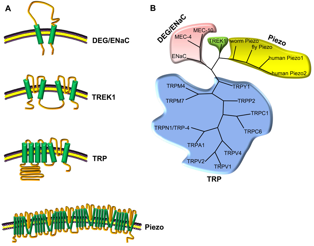
Figure 1. Mechanosensitive channels in eukaryotes.
(A) Schematics of mechanosensitive channels in eukaryotes. Only one subunit is shown for each channel. The membrane topology of Piezo is unclear, and one possibility is shown here. (B) A dendrogram plot of different classes of putative mechanosensitive channels. In the case of TRP family channels, only those that have been implicated in mechanosensation are included, amongst which TRPN1 is the only TRP protein that has been demonstrated to function as a mechanosensitive channel that is mechanically gated [12].
TRP family channels have recently emerged as another class of leading candidates for mechanosensitive channels (Figure 1) [2]. These channels are found in nearly all eukaryotes [10]. Among the seven TRP subfamilies (TRPC, TRPV, TRPM, TRPN, TRPA, TRPP, and TRPML), nearly every subfamily has members that have been implicated in mechanosensation [2]. However, it has also been suggested that TRP channels are not mechanically gated and may merely play indirect roles in mechanosensation by modulating/amplifying the activity of mechanosensitive channels of unknown molecular identity [11]. But more recent work in C. elegans shows that TRP family proteins can function as mechanosensitive channels that are mechanically gated. In this work, Kang et al. [12] demonstrated that the C. elegans TRPN1 channel TRP-4 forms the pore of a mechanically gated channel that senses touch in the worm nose. Interestingly, this channel also mediates proprioception in both C. elegans and Drosophila [13,14].
Work in model organisms such as worms and flies raises the possibility that ENaC/DEG and TRP family genes encode the mechanosensitive channels sensing touch, sound and gravity in mammals, although this has not yet been confirmed, at least at the genetic level [2]. A second, but not mutually exclusive, possibility is that mechanosensitive channels in mammals are encoded by completely different types of genes. Indeed, the two-pore-domain K+ channel TREK1 has been reported to form a mechanosensitive channel in mammals [15], but, given that the opening of this K+ channel hyperpolarizes rather than depolarizes a neuron, it cannot be the primary channel mediating touch, hearing and proprioception in mammals.
In the new work, Patapoutian and colleagues [4] have now identified a novel class of mechanosensitive channels in mammals. They took a reverse genetic approach by screening for channel-like genes that, when knocked down by RNA interference (RNAi), result in suppression of mechanosensitive currents in cell lines. This tour de force effort began with the mouse neuroblastoma cell line Neuro2A (N2A), which expresses endogenous rapidly-adapting mechanosensitive channels. Two protocols were used to evoke mechanosensitive currents in these cells — membrane touch and membrane stretch. As a first step, the authors carried out a microarray analysis of enriched transcripts in N2A cells and selected 73 candidates that contained at least two transmembrane segments. RNAi-mediated knockdown of these candidates identified a single gene — Fam38A — that is required for the mechanosensitive currents in N2A cells. They renamed this gene piezo1, from the Greek ‘πíεση’ (píesi) meaning ‘pressure’. Another piezo gene, piezo2 (Fam38B), was identified and found to be present in all vertebrates, like piezo1. Overexpression of Piezo1 or Piezo2 in multiple cell lines (i.e. N2A, HEK293T, and C2C12) generated robust mechanosensitive cation currents that are non-selective, exhibit a linear current–voltage relationship, and are sensitive to ruthenium red and gadolinium, two known inhibitors of many mechanosensitive channels. Although the response latency of Piezo channels has not yet been determined, it is probably in the millisecond range according to the traces presented in the paper. Piezo1 can be detected at the plasma membrane in transfected HEK293T cells. These data together provide convincing evidence that Piezo proteins can form mechanosensitive channels in vitro.
But are Piezo proteins required for mechanosensation in vivo? Both Piezo1 and Piezo2 are expressed in multiple tissues, such as bladder, colon and lung. In addition, Piezo2 is enriched in dorsal root ganglion (DRG) neurons, suggesting a role for Piezo2 in mechanosensation. Indeed, RNAi-mediated knockdown of Piezo2 in cultured mouse DRG neurons caused a specific suppression of the rapidly-adapting, but not the intermediately- or slowly-adapting, mechanosensitive currents [4]. This provides strong evidence that Piezo2 is an essential subunit of a native mechanosensitive channel in a group of DRG neurons.
piezo genes are evolutionarily conserved and can be found in most eukaryotic organisms, including plants, nematodes, insects and vertebrates, but appear to be absent in yeast. At the sequence level, Piezos show little homology to any other known ion channels. These proteins contain 24–36 putative transmembrane segments, reminiscent of the structure of voltage-gated sodium and calcium channels that comprise fourfold repeats of six transmembrane segments. Piezo1 and Piezo2 clearly represent a new class of membrane proteins.
The identification of Piezo1 and Piezo2 raises many interesting questions about the role of these proteins in mechanosensation. First, do Piezo proteins form the pore of a mechanosensitive channel(s)? The lack of homology in Piezos to known channel proteins and the presence of dozens of transmembrane segments make it a daunting challenge to pinpoint the channel pore. Nevertheless, the fact that overexpression of Piezos in heterologous cells can largely recapitulate the properties of endogenous mechanosensitive currents makes it highly likely that Piezos line the channel pore. It also suggests that Piezos can function largely on their own without a special requirement for auxiliary subunits. Second, Piezo2 appears to be specifically required for the rapidly-adapting mechanosensitive conductance in DRG neurons. So which channels are responsible for the intermediately- and slowly-adapting mechanosensitive currents in these neurons? Are they mediated by Piezo1 or by ENaC/DEG and TRP family channels? It will also be interesting to examine the phenotypes associated with Piezo knockout mice. Third, if Piezos are expressed in hair cells, do they contribute to the formation of the long-sought mechanotransduction channels that detect sound waves in the inner ear? Finally, the cloning of Piezos highlights the power of RNAi-based screening in identifying mechanosensitive channels. Similar approaches could be applied to other cell lines that express distinct types of mechanosensitive conductance. The work by Patapoutian and colleagues [4] heralds a new era in the study of mechanosensation.
References
- 1.Gillespie PG, Walker RG. Molecular basis of mechanosensory transduction. Nature. 2001;413:194–202. doi: 10.1038/35093011. [DOI] [PubMed] [Google Scholar]
- 2.Christensen AP, Corey DP. TRP channels in mechanosensation: direct or indirect activation? Nat. Rev. Neurosci. 2007;8:510–521. doi: 10.1038/nrn2149. [DOI] [PubMed] [Google Scholar]
- 3.Patel A, Sharif-Naeini R, Folgering JR, Bichet D, Duprat F, Honore E. Canonical TRP channels and mechanotransduction: from physiology to disease states. Pflugers Arch. 2010;460:571–581. doi: 10.1007/s00424-010-0847-8. [DOI] [PubMed] [Google Scholar]
- 4.Coste B, Mathur J, Schmidt M, Earley TJ, Ranade S, Petrus MJ, Dubin AE, Patapoutian A. Piezo1 and Piezo2 are essential components of distinct mechanically activated cation channels. Science. 2010 doi: 10.1126/science.1193270. doi: 2010.1126/science.1193270. [DOI] [PMC free article] [PubMed] [Google Scholar]
- 5.Kung C, Martinac B, Sukharev S. Mechanosensitive channels in microbes. Annu. Rev. Microbiol. 2010;64:313–329. doi: 10.1146/annurev.micro.112408.134106. [DOI] [PubMed] [Google Scholar]
- 6.Arnadottir J, Chalfie M. Eukaryotic mechanosensitive channels. Annu. Rev. Biophys. 2010;39:111–137. doi: 10.1146/annurev.biophys.37.032807.125836. [DOI] [PubMed] [Google Scholar]
- 7.Driscoll M, Chalfie M. The mec-4 gene is a member of a family of Caenorhabditis elegans genes that can mutate to induce neuronal degeneration. Nature. 1991;349:588–593. doi: 10.1038/349588a0. [DOI] [PubMed] [Google Scholar]
- 8.O'Hagan R, Chalfie M, Goodman MB. The MEC-4 DEG/ENaC channel of Caenorhabditis elegans touch receptor neurons transduces mechanical signals. Nat. Neurosci. 2005;8:43–50. doi: 10.1038/nn1362. [DOI] [PubMed] [Google Scholar]
- 9.Goodman MB. Mechanosensation. WormBook; 2006. pp. 1–14. [DOI] [PMC free article] [PubMed] [Google Scholar]
- 10.Xiao R, Xu XZS. Function and regulation of TRP family channels in C. elegans. Pflugers Arch. 2009;458:851–860. doi: 10.1007/s00424-009-0678-7. [DOI] [PMC free article] [PubMed] [Google Scholar]
- 11.Sharif-Naeini R, Dedman A, Folgering JH, Duprat F, Patel A, Nilius B, Honore E. TRP channels and mechanosensory transduction: insights into the arterial myogenic response. Pflugers Arch. 2008;456:529–540. doi: 10.1007/s00424-007-0432-y. [DOI] [PubMed] [Google Scholar]
- 12.Kang L, Gao J, Schafer WR, Xie Z, Xu XZS. C. elegans TRP family protein TRP-4 is a pore-forming subunit of a native mechanotransduction channel. Neuron. 2010;67:381–391. doi: 10.1016/j.neuron.2010.06.032. [DOI] [PMC free article] [PubMed] [Google Scholar]
- 13.Li W, Feng Z, Sternberg PW, Xu XZS. A C. elegans stretch receptor neuron revealed by a mechanosensitive TRP channel homologue. Nature. 2006;440:684–687. doi: 10.1038/nature04538. [DOI] [PMC free article] [PubMed] [Google Scholar]
- 14.Cheng LE, Song W, Looger LL, Jan LY, Jan YN. The role of the TRP channel NompC in Drosophila larval and adult locomotion. Neuron. 2010;67:373–380. doi: 10.1016/j.neuron.2010.07.004. [DOI] [PMC free article] [PubMed] [Google Scholar]
- 15.Dedman A, Sharif-Naeini R, Folgering JH, Duprat F, Patel A, Honore E. The mechano-gated K(2P) channel TREK-1. Eur. Biophys. J. 2009;38:293–303. doi: 10.1007/s00249-008-0318-8. [DOI] [PubMed] [Google Scholar]


