Neuro Oncol. 2010 October;12(10):1014.
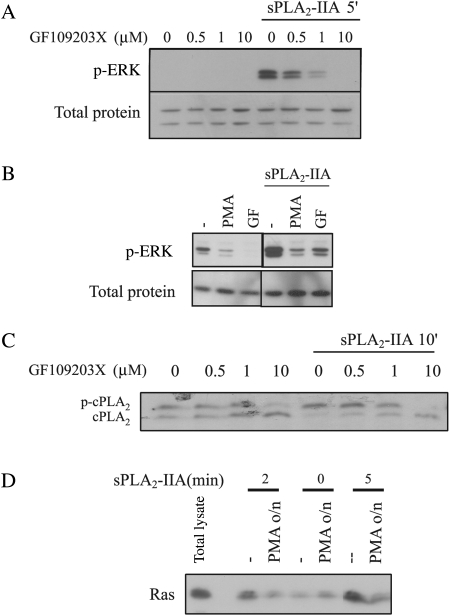
Figure 2A and 2C's GF109203X doses should be micromolar, not millimolar. The author regrets this error.
Fig. 2.
PKC participation in sPLA2-IIA–induced activation of ERK, cPLA2, and Ras. (A and C) 1321N1 cells were incubated with the indicated doses of GF109203X for 30 minutes prior to stimulation with sPLA2-IIA. Western blots of total lysates were incubated with either phospho-ERK (upper A) or actin (lower A) antibodies or cPLA2 antibody (C). (B) 1321N1 cells were incubated with 1-μM PMA overnight or with 1-μM GF109203X for 30 minutes, prior to the addition of sPLA2-IIA for 5 minutes. Western blots of the lysates were incubated either with phospho-ERK (upper) or with actin antibody (lower). Samples shown belong to the same experiment and were run simultaneously but not consecutively; therefore, a lane represents the gap. (D) 1321N1 cells were incubated with 1-μM PMA overnight and then with sPLA2-IIA for the indicated times, and RasGTP was determined using the RBD pull-down assay. The first lane shows a sample of total lysate; the other lanes are Ras bound to the RBD, which represents RasGTP. In all cases, cells were serum starved overnight and the experiments shown are representative of the results obtained in at least 3 trials for each condition.


