Abstract
Talon cusp is a rare developmental extra cusp-like projection on the cingulum area of affected anterior teeth that may cause various functional and aesthetic problems. The present report describes a case of bilateral palatal talon cusps on permanent maxillary incisors and the treatment procedure to overcome the clinical problems associated with talon cusps.
Keywords: Talon cusp, Maxillary lateral incisor, Root canal therapy
INTRODUCTION
Talon cusp is a rare dental anomaly in which a cusp-like mass of hard tissue protrudes from the cingulum area of maxillary or mandibular anterior teeth. The typical appearance of this projection is conical and resembles an eagle’s talon. Although its reported incidence is higher in permanent dentition, talon cusp can also be observed in primary dentition.1–3 The predominantly affected teeth are maxillary lateral incisors,1–3 with a higher incidence in men than women.1
As seen with other dental anomalies that affect teeth, talon cusp occurs during early odontogenesis. Although both genetic and environmental factors are involved, the aetiology of such an extraordinary formation has not been elucidated. Nevertheless, this anomaly has been reported to be associated with parental consanguinity.4
The presence of a talon cusp is more than just an aesthetic problem. The prominent bulge of a talon cusp may cause interference with occlusion, irritation of soft tissues and tongue, or accidental cusp fractures causing pulpal exposure.1 The developmental groove where the cusp joins the lingual surface of the crown has a tendency to accumulate plaque, which makes the tooth susceptible to caries.1 In the literature, conservative or radical treatment modalities have been advocated to manage talon cusps. Treatment should be as conservative as possible and be planned depending on the shape and size of the talon cusp.5,6 The talon cusp is composed of enamel and dentin.7 However, the possibility of pulp tissue involvement should not be overlooked during treatment planning.8
This case report highlights the clinical and radiographic characteristics and management of bilateral talon cusps on both maxillary lateral incisors with a combination of endodontic and restorative approaches.
CASE REPORT
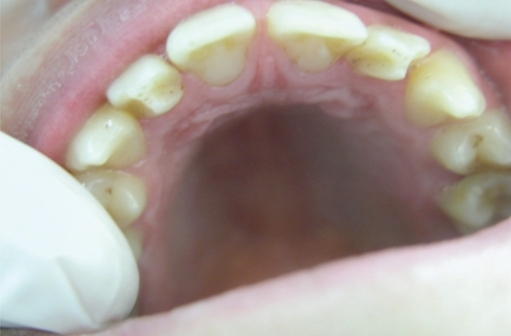
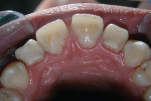
A 19-year-old male patient was referred by a general practitioner to the Department of Endodontics of Hacettepe University for consultation and further management of bilateral cusp-like projections from the cingulum area of maxillary lateral incisors, causing premature contact and plaque accumulation (Figure 1).
Figure 1.
Intraoral view of the palatal talon cusps on the maxillary lateral incisors.
Both malformed lateral incisors exhibited extra cusp-like projections in the palatal region, forming a ‘T-shaped’ incisal edge of the crown. The patient’s medical history was non-contributory. No other member of the family was affected by similar dental anomalies. Intraoral examination revealed a class 2 molar occlusal relationship, and the prominent cusps prevented an ideal overbite/overjet relationship. No significant soft-tissue abnormality was observed, and the taloned lateral incisor teeth were caries-free. The teeth responded normally to thermal and percussion tests.
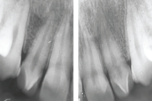
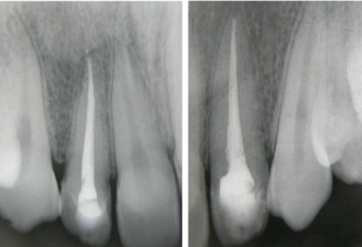
Radiographic examination showed a typical, V-shaped radio-opaque structure superimposed over the image of the affected crowns, with the point of V toward the incisal edges (Figure 2).
Figure 2.
Initial intraoral radiographs demonstrate the presence of inverted V-shaped talon cusps on the maxillary lateral incisors.
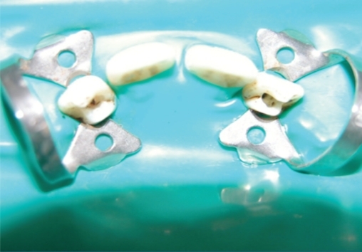
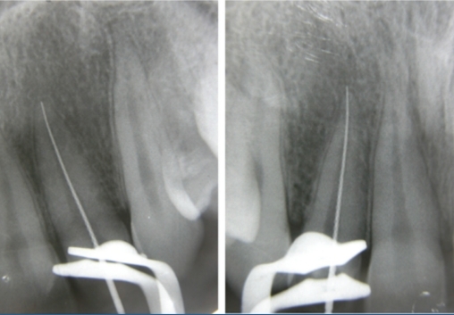
After orthodontic consultation, it was decided to reduce the talon cusps completely to avoid occlusal interferences. As pulp exposure was expected, root canal therapy was initiated before reducing the talon cusps. The teeth were anaesthetized, and an adequate endodontic access preparation was made under rubber dam isolation (Figure 3). After removal of the pulp tissues, working lengths were determined with a radiograph (Figure 4). Working lengths were also confirmed with the use of an electronic apex locator (ProPex™ Apex Locator; Dentsply Maillefer, Ballaigues, Switzerland). The root canals were prepared using Pro-Taper (Dentsply-Maillefer, Switzerland) rotary instruments and RC Prep (Premier, Norristown, PA, USA) as a lubricant. Copious irrigation with 2% sodium hypochlorite solution was employed throughout the procedure. Obturation of the root canals was performed using gutta-percha and AH 26 (Dentsply Maillefer) root canal sealer with lateral condensation (Figure 5), and the talon cusps were reduced using a water-cooled diamond bur in a high-speed handpiece. Following this procedure, access cavities were restored with bonded composite resin material (TPH Spectrum, Dentsply) (Figure 6).
Figure 3.
Access cavities of the maxillary lateral incisors.
Figure 4.
Root length determination radiographs.
Figure 5.
Final periapical radiographs taken after completion of endodontic treatment.
Figure 6.
Clinical appearance after selective cuspal grinding and composite restoration followed by root canal treatment.
DISCUSSION
Talon cusp has been reported as a relatively rare odontogenic anomaly that is most frequently observed on the cingulum area of incisors, and treatment may be required if occlusion is compromised.8 Normal enamel covers the cusp and fuses with the lingual aspect of the tooth. The cusp may or may not contain pulp horns of varying extensions.9
The morphological characteristics of talon cusps were classified by Hattab et al10 into three types on the basis of the degree of cusp formation and extension
Type 1. Talon: a morphologically well-delineated additional cusp that prominently projects from the palatal surface of a primary or permanent anterior tooth and extends at least half the distance from the cemento-enamel junction to the incisal edge.
Type 2. Semi talon: an additional cusp of 1 mm or more, but extending less than half the distance from the cemento-enamel junction to the incisal edge. It may blend with the palatal surface or project away from the rest of the crown.
Type 3. Trace talon: an enlarged or prominent cingulum in any of its variant forms (i.e. conical, bifid, or tubercle-like) originating from the cervical third of the root.
According to this classification, the anomalous cusps on the maxillary lateral incisors in our case are type 3 bifid cingula or trace talon cusps.
The possible clinical problems caused by the presence of a talon cusp are poor aesthetics, occlusal trauma, occlusal interferences, displacement of the affected tooth, attrition of the opposing tooth, caries, pulp infection and pulp necrosis related to pulp extension or irritation of soft tissues, periapical pathosis, changes in the alveolar bone and periodontal connective tissue due to excessive occlusal forces, and irritation of soft tissues during speech and mastication.8,10
A tooth with talon cusp is commonly thicker faciolingually and wider mesiodistally than the normal contralateral tooth. The talon cusp prevents the tooth from being orthodontically repositioned into an acceptable overbite/overjet relationship. Therefore, the talon cusp is reduced partially or completely to allow the establishment of a proper overbite/overjet relationship.1
Previous reports have demonstrated an association of talon cusp with a number of syndromes including Mohr syndrome, Sturge-Weber syndrome, Rubinstein-Taybi syndrome, ıncontinentia pigmenti achromians, and Ellis-van Creveld syndrome.10–12 However, in the present case, the patient had none of the above-mentioned syndromes.
In the literature, different treatment modalities have been advocated to manage talon cusps.6 It has been reported that the presence of a talon cusp is not an indication for dental treatment unless it causes clinical problems.6,8 Furthermore, early diagnosis avoids future complications.5,13 Depending on the size and configuration of the talon cusp, the treatment should be planned individually after clinical evaluation. The presence or absence of pulp tissue in the talon cusp can be indicative of different treatment alternatives. These alternatives include a periodic grinding procedure with a desensitising agent such as fluoride varnish or a single-visit reduction of the cusp with or without endodontic therapy.1,10
In the present case, the cusp was prominent, sharply defined, and projected from the cervical region to the incisal edge of the tooth. If both talon cusps were grinded completely, pulpal exposure would occur. Additionally, since the reduction of talon cusps is done gradually on consecutive visits, the patient preferred a single-visit root canal therapy as a radical treatment option. Therefore, grinding of both talon cusps and endodontic treatment were chosen as the treatment option to prevent occlusal problems and to achieve normal tooth structure, which would help maintain proper hygiene.
CONCLUSIONS
This case illustrates the endodontic treatment of talon cusps involving both upper maxillary lateral incisors. Exposure of the pulp was a concern during complete cuspal reduction. Considering our experience with this case, we think that in such cases where exposure of the pulp is a concern, endodontic therapy could be the treatment of choice when treatment by protective measures is not possible.
REFERENCES
- 1.Hattab FN, Yassin OM, al-Nimri KS. Talon cusp—clinical significance and management: case reports. Quintessence Int. 1995;26:115–120. [PubMed] [Google Scholar]
- 2.Sumer AP, Zengin AZ. An unusual presentation of talon cusp: a case report. Br Dent J. 2005;199:429–430. doi: 10.1038/sj.bdj.4812741. [DOI] [PubMed] [Google Scholar]
- 3.Bolan M, Gerent Petry Nunes AC, de Carvalho Rocha MJ, De Luca Canto G. Talon cusp: report of a case. Quintessence Int. 2006;37:509–514. [PubMed] [Google Scholar]
- 4.Davis PJ, Brook AH. The presentation of talon cusp: diagnosis, clinical features, associations and possible aetiology. Br Dent J. 1986;160:84–88. doi: 10.1038/sj.bdj.4805774. [DOI] [PubMed] [Google Scholar]
- 5.Maroto M, Barbería E, Arenas M, Lucavechi T. Displacement and pulpal involvement of a maxillary incisor associated with a talon cusp: report of a case. Dent Traumatol. 2006;22:160–164. doi: 10.1111/j.1600-9657.2006.00343.x. [DOI] [PubMed] [Google Scholar]
- 6.al-Omari MA, Hattab FN, Darwazeh AM, Dummer PM. Clinical problems associated with unusual cases of talon cusp. Int Endod J. 1999;32:183–190. doi: 10.1046/j.1365-2591.1999.00212.x. [DOI] [PubMed] [Google Scholar]
- 7.Mellor JK, Ripa LW. Talon cusp: a clinically significant anomaly. Oral Surg Oral Med Oral Pathol. 1970;29:225–228. doi: 10.1016/0030-4220(70)90089-7. [DOI] [PubMed] [Google Scholar]
- 8.Hattab FN, Hazza’a AM. An unusual case of talon cusp on geminated tooth. J Can Dent Assoc. 2001;67:263–266. [PubMed] [Google Scholar]
- 9.Segura-Egea JJ, Jimenez-Rubio A, Velasco-Ortega E, Ríos-Santos JV. Talon cusp causing occlusal trauma and acute apical periodontitis: report of a case. Dent Traumatol. 2003;19:55–59. doi: 10.1034/j.1600-9657.2003.00110.x. [DOI] [PubMed] [Google Scholar]
- 10.Hattab FN, Yassin OM, al-Nimri KS. Talon cusp in permanent dentition associated with other dental anomalies: review of literature and report of seven cases. ASDC J Dent Child. 1996;63:368–376. [PubMed] [Google Scholar]
- 11.Chen RJ, Chen HS. Talon cusp in primary dentition. Oral Surg Oral Med Oral Pathol. 1986;62:67–72. doi: 10.1016/0030-4220(86)90072-1. [DOI] [PubMed] [Google Scholar]
- 12.Hattab FN, Yassin OM, Sasa IS. Oral manifestations of Ellisvan Creveld syndrome: report of two siblings with unusual dental anomalies. J Clin Pediatr Dent. 1998;22:159–165. [PubMed] [Google Scholar]
- 13.Dash JK, Sahoo PK, Das SN. Talon cusp associated with other dental anomalies: a case report. Int J Paediatr Dent. 2004;14:295–300. doi: 10.1111/j.1365-263X.2004.00558.x. [DOI] [PubMed] [Google Scholar]


