Abstract
The Bunyaviridae family includes pathogens of medical and veterinary importance. Rift Valley fever virus (RVFV), a member in the Phlebovirus genus of the family Bunyaviridae, is endemic to sub-Saharan Africa and causes a mosquito-borne disease in ruminants and humans. Viruses in the family Bunyaviridae carry a tripartite, single-stranded, negative-sense RNA genome composed of L, M, and S RNAs. Little is known about how the three genomic RNA segments are copackaged to generate infectious bunyaviruses. We explored the mechanism that governs the copackaging of the three genomic RNAs into RVFV particles. The expression of viral structural proteins along with replicating S and M RNAs resulted in the copackaging of both RNAs into RVFV-like particles, while replacing M RNA with M1 RNA, lacking a part of the M RNA 5′ UTR, abrogated the RNA copackaging. L RNA was efficiently packaged into virus particles released from cells supporting the replication of L, M, and S RNAs, and replacing M RNA with M1 RNA abolished the packaging of L RNA. Detailed analyses using various combinations of replicating viral RNAs suggest that M RNA alone or a coordinated function of M and S RNAs exerted efficient L RNA packaging either directly or indirectly. Collectively, these data are consistent with the possibility that specific intermolecular interactions among the three viral RNAs drive the copackaging of these RNAs to produce infectious RVFV.
Keywords: hemorrhagic fever, segmented RNA virus
Rift Valley fever virus (RVFV) (genus Phlebovirus, family Bunyaviridae) causes a mosquito-borne disease characterized by a febrile illness resulting in a high rate of abortions in ruminants and an acute febrile illness followed by fatal hemorrhagic fever, encephalitis, or ocular diseases in humans (1, 2). RVFV is endemic in sub-Saharan African countries although outbreaks also occur in countries outside the endemic area, including Egypt, Saudi Arabia, and Yemen (3). RVFV has the potential to spread to other areas of the world, including North America by naturally occurring mosquito populations (4); large numbers of competent mosquito vectors and vertebrate hosts are all that are needed for RVFV epidemics. Currently, there are neither licensed RVFV vaccines nor antivirals for use in humans or in animals.
RVFV has a single-stranded, tripartite RNA genome composed of L, M, and S segments. Both L and M RNA segments are of negative polarities. The antiviral-sense L RNA encodes the RNA-dependent RNA polymerase (L protein) and the antiviral-sense M RNA encodes two envelope glycoproteins, Gn and Gc, and two accessory proteins, NSm and the 78-kDa protein. The S segment uses an ambisense strategy for gene expression; a nonstructural protein, NSs, is translated from the mRNA that is transcribed from the antiviral-sense S segment, whereas N protein is produced from the mRNA that is transcribed from the viral-sense S segment (5). N and L proteins are essential for viral RNA synthesis (6–11). RVFV RNA replication takes place in cytoplasm and the viral assembly occurs on the Golgi complex (5). Released RVFV particles are morphologically highly homogenous; moreover, the structure of Phleboviruses is highly ordered, and the virus surface is arranged in an icosahedral lattice with T = 12 quasisymmetry (12–14).
An understanding of the mechanisms that govern the copackaging of segmented RNA genomes into virus particles to generate infectious virus has been one of the important research areas in virology. Past studies of the genomic RNA packaging mechanism in influenza A virus showed the presence of an RNA packaging signal in each viral RNA segment (15–22), a hierarchy among virus RNA segments for incorporation into virions (18), and the possible intersegment association of viral RNAs during virus assembly (18, 23). For the production of infectious bunyaviruses, the virus must carry all three RNA segments but the mechanism that controls the copackaging of the three RNA segments is unknown. In Bunyamwera virus, which belongs to the genus Orthobunyavirus, low particle-to-pfu ratios of 2.6–7.2 are observed (24), suggesting an efficient copackaging of the three viral RNA segments in bunyaviruses. Minigenome RNAs, which carry UTRs of bunyavirus RNA segments and encode a reporter gene, have been used to study the mechanism of bunyavirus RNA packaging (24–27). However, the minigenome-based system is not ideal for addressing the mechanism of viral RNA copackaging because infectious virus is used as a helper virus for minigenome RNA packaging. Habjan et al. developed a system to generate virus-like particles (VLPs) of RVFV (28); the coexpression of the minigenome RNA, carrying M RNA UTRs and a reporter gene and L, N, and Gn/Gc proteins results in minigenome RNA amplification and the generation of VLPs carrying the minigenome RNA. The VLPs resemble authentic RVFV particles and are able to infect new cells; in particular, after infection, the VLP-associated nucleocapsids autonomously undergo a primary round of transcription resulting in the expression of low levels of reporter gene (28). Furthermore, the coexpression of L and N proteins in VLP-infected cells promotes the subsequent replication and transcription of RNA leading to high levels of reporter gene expression (28). These data suggest that the mechanism driving the production of RVFV VLP is similar to the one used in the production of infectious RVFV. This helper virus-independent minigenome RNA packaging system into VLPs can be used to explore bunyavirus RNA copackaging mechanisms.
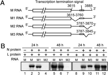
Both the 5′ and 3′ ends of bunyaviral genomic RNA segments have UTRs, which contain cis-acting RNA replication signals of ∼30 nt at their termini (5). RVFV M RNA has a 271-nt-long 5′ UTR, which includes a ∼26 nt-long M mRNA transcription termination signal (Fig. 1A) (29) and is substantially longer than the 110-nt-long L RNA 5′ UTR and the 38-nt-long S RNA 5′ UTR. Our studies on the biological significance of the long 5′ UTR in M RNA led to the discovery that this region played critical roles in the copackaging of the three viral RNA segments into virus particles. Our data implied that specific intermolecular interactions among the three viral RNA segments drive the viral RNA copackaging to produce infectious RVFV.
Fig. 1.
Replication competencies of M, M1, M2, and M3 RNAs. (A) Schematic diagram of antiviral-sense M, M1, M2, and M3 RNAs. The M gene ORF is shown in boxes. The numbers in M RNA represent the location of the 5′ UTR (in viral-sense RNA) from the 5′ end of antiviral-sense M RNA and those in each M RNA mutant represent the deletion site. (B) Cells were cotransfected with plasmids encoding N protein, L protein, and antiviral-sense M RNA or one of the M RNA mutants. The plasmid expressing N protein was omitted in samples shown in lanes 1 and 8. Intracellular RNAs were extracted at 24 h or 48 h posttransfection and subjected to Northern blot analysis using an RNA probe that selectively binds to viral-sense M RNA.
Results
Effects of Deletions Within the 5′ UTR of M RNA on the Recovery of Infectious Viruses.
To explore the biological functions of the 5′ UTR of RVFV M RNA, we constructed three plasmids expressing the antiviral-sense M RNA mutants, M1, M2, and M3 RNAs, each carrying a deletion at different sites within the 5′ UTR (Fig. 1A). Using the RVFV reverse genetics system (30), we recovered 1.0 × 10⁷ pfu/mL of infectious virus carrying M RNA and 3.5 × 10⁶ pfu/mL of virus carrying M3 RNA at 5-d-postplasmid tranfection, demonstrating that the deletion site in M3 RNA was not essential for RVFV replication in cell culture. We recovered 2.0 × 10⁴ pfu/mL of virus carrying M1 RNA (M1 virus) in one out of three independent attempts, whereas infectious virus carrying M2 RNA could not be rescued. The M1 virus formed smaller plaques, replicated poorly in Vero E6 cells, and was more thermolabile at 42 °C than the WT virus (Fig. S1).
Replication and Packaging Competencies of M RNA 5′ UTR Mutants.
The expression of antiviral-sense M RNA transcripts or each of the M RNA mutants in the cells coexpressing L and N proteins resulted in the efficient replication of M, M1, and M3 RNAs, but not M2 RNA (Fig. 1B), demonstrating that M1 and M3 RNAs, but not M2 RNA, had an intact RNA replication signal in the 5′ UTR. Thus, the replication incompetence of M2 RNA was the reason for the failure to rescue infectious virus carrying M2 RNA.
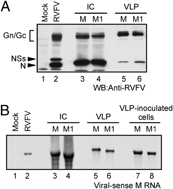
To determine the packaging competence of M1 RNA, BSR-T7/5 cells were cotransfected with protein-expression plasmids, each expressing L protein, N protein, and the viral envelope Gn and Gc proteins (30), and plasmid expressing antiviral-sense M1 RNA. As a control, plasmid-expressing M RNA was used in place of that expressing M1 RNA. Cell extracts and culture fluids were collected at 3 d posttransfection. We observed similar levels of accumulation of intracellular viral proteins (Fig. 2A, lanes 3 and 4) and the replicating M and M1 RNAs (Fig. 2B, lanes 3 and 4). Judging from the abundance of Gn/Gc proteins in purified VLPs, similar amounts of VLPs were produced from both samples (Fig. 2A). Similar levels of M RNA and M1 RNA were detected in the purified VLPs, demonstrating that both RNAs were packaged into VLPs with similar efficiencies (Fig. 2B, lanes 5 and 6). Inoculation of these VLPs into cells coexpressing L and N proteins resulted in the efficient replication of both M RNA and M1 RNA (Fig. 2B, lanes 7 and 8), demonstrating that the VLP carrying viral RNA was biologically active to initiate RNA replication after inoculation into cells. Thus, M1 RNA was competent for efficient RNA replication and packaging into VLPs.
Fig. 2.
Characterization of VLPs carrying M RNA or M1 RNA. Cells were cotransfected with plasmids encoding N protein, L protein, M gene ORF, and antiviral-sense M RNA or M1 RNA. Intracellular proteins, intracellular RNAs, and VLPs were harvested at 3 d posttransfection and the released VLPs were purified by sucrose gradient centrifugation. (A) Intracellular proteins from mock-infected cells (lane 1), RVFV-infected cells (lane 2), cells expressing M RNA (lane 3), cells expressing M1 RNA (lane 4), purified VLPs from cells expressing M RNA (lane 5), and those from cells expressing M1 RNA (lane 6) were subjected to Western blot analysis using anti-RVFV mouse antibody. IC, intracellular samples. Gn/Gc, Gn/Gc proteins; NSs, NSs protein; and N, N protein. (B) RNA samples corresponding to the samples in A were subjected to Northern blot analysis using an RNA probe that hybridizes with viral-sense M or M1 RNA (lanes 1–6). Released VLPs carrying M RNA (lane 7) or M1 RNA (lane 8) were inoculated to cells coexpressing L and N proteins. Intracellular RNAs were extracted at 24 h postinoculation and subjected to Northern blot analysis.
The 5′ UTR of M RNA Is Important for the Copackaging of M RNA with S RNA into VLPs.
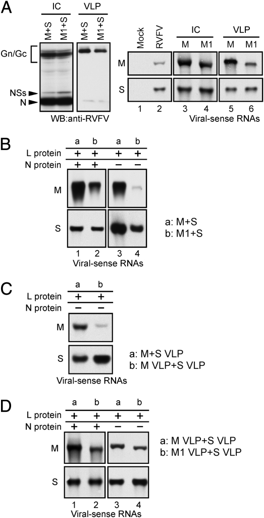
The above data led us to hypothesize that M1 RNA was not efficiently copackaged with S RNA and/or L RNA to generate infectious virus particles. To determine whether M1 and S RNAs could be copackaged into VLPs, we prepared VLPs from BSR-T7/5 cells, supporting the replication of S and M1 RNAs and coexpressing all of the viral structural proteins, i.e., L, N, and Gn/Gc proteins. As a control, M RNA was used in place of M1 RNA (M+S VLP sample). Similar levels of VLPs were released in both groups and both VLPs had similar amounts of S RNA (Fig. 3A). The amount of M RNA in the M+S VLP sample was approximately three times more than the M1 RNA in the experimental VLP sample (Fig. 3A). Inoculation of these VLPs into cells coexpressing L and N proteins resulted in the efficient replication of M RNA and M1 RNA, demonstrating that L and N proteins exerted the replication of incoming M RNA and M1 RNA in VLPs (Fig. 3B, Left). Importantly, very poor M1 RNA accumulation occurred after inoculation of the experimental group VLPs into L protein-expressing cells, whereas inoculation of the control M+S VLP sample into cells expressing L protein resulted in M RNA replication (Fig. 3B, Right). We inferred from these data that M and S RNAs, but not M1 and S RNAs, were copackaged into VLPs. Specifically, we considered that after inoculation of the control group VLP, L and N proteins, which are incorporated into the VLP, initiated N mRNA transcription using the incoming S RNA as the template. The newly synthesized N protein from the N mRNA transcripts acted in concert with the expressed L protein to amplify M RNA. M1 RNA was unable to replicate efficiently, because M1 and S RNAs were independently packaged into VLPs and accumulation of N protein from the incoming S RNA did not occur in cells inoculated with VLP carrying M1 RNA. To exclude a possibility that high titers of VLPs carrying only M RNA and those carrying only S RNA were produced in the control M+S VLP sample and coinfection of these two putative VLPs resulted in M RNA accumulation in L protein-expressing cells, we prepared three VLP samples: M VLP sample and S VLP sample, obtained from viral protein-expressing cells supporting M RNA and S RNA replication, respectively, and the control M+S VLP sample. Like M RNA (Fig. 2), S RNA was also efficiently packaged into VLPs in the absence of any other viral RNA(s) (Fig. S2). Using a portion of the three VLP samples, the abundance of packaged viral RNAs in the purified VLPs was determined by Northern blot analysis. Subsequently, we prepared a mixture of M VLP and S VLP samples such that the same amount of M RNA was present in the mixed VLP sample and in the M+S VLP sample. The S RNA amount was also the same in both these VLP samples. Then, L protein-expressing cells were inoculated with either the M+S VLP sample or the mixture of M VLP and S VLP samples. If VLPs carrying only M RNA and those carrying only S RNA were produced in the M+S sample and if the coinfection of these two putative VLPs resulted in efficient M RNA replication in L protein-expressing cells as shown in Fig. 3B, then it is reasonable to expect that the inoculation of L protein-expressing cells with the mixture of M VLP and S VLP samples would also result in efficient M RNA replication. However, efficient M RNA replication occurred only in cells inoculated with the M+S VLP sample, but not in those inoculated with the mixture of M VLP and S VLP samples (Fig. 3C), suggesting that most of the VLPs in the M+S sample carried both M and S RNAs, which resulted in efficient M RNA replication in L protein-expressing cells. To confirm that M1 RNA is indeed competent for RNA replication using N protein, synthesized from the copackaged S RNA, we concentrated the S VLP sample ≈10 times using Amicon Ultra, Ultracel-100K (Millipore) to substantially increase its infectivity. Consistent with our assumption, efficient M1 RNA replication occurred after coinoculation of cells expressing L protein with the VLP carrying M1 RNA and the concentrated S VLP sample (Fig. 3D). Taken together, we concluded that replicating M and S RNAs were copackaged into VLPs and that the 146-nt-long region in the 5′ UTR of M RNA, which corresponded to the deletion site in the M1 RNA, was required for S RNA copackaging.
Fig. 3.
Copackaging of M and S RNAs into VLPs. (A) Intracellular proteins, intracellular RNAs, and released VLPs were collected from cells expressing all of the viral structural proteins, M RNA, and S RNA (M+S) at 3 d posttransfection. M1+S represent samples in which M1 RNA was used in place of M RNA. Intracellular proteins (IC) and proteins in purified VLPs were subjected to Western blot analysis using anti-RVFV antibody (Left two panels). Intracellular RNAs (IC) and RNAs in purified VLPs were detected by Northern blot analysis using an RNA probe that hybridizes to viral-sense M RNA or M1 RNA (Right Top) or viral-sense S RNA (S) (Right Bottom). Lane 1 and lane 2 represent intracellular samples from mock-infected cells and RVFV-infected cells, respectively. (B) VLPs produced from cells supporting the replication of M and S RNAs (M+S) or from those supporting the replication of M1 and S RNAs (M1+S) were inoculated into cells coexpressing L and N proteins (lanes 1 and 2) or L protein-expressing cells (lanes 3 and 4). Intracellular RNAs were extracted at 24 h postinoculation and subjected to Northern blot analysis to detect viral-sense M RNA (Top, lanes 1 and 3), M1 RNA (Top, lanes 2 and 4), or viral-sense S RNA (Bottom). (C) Cells expressing L protein were inoculated with either M+S VLP sample, obtained from cells supporting the replication of M and S RNAs, or a mixture of M VLP sample and S VLP sample (M VLP+S VLP), obtained from cells supporting M RNA replication and S RNA replication, respectively. Both VLP samples carried the same amount of M RNA. Similarly, the amount of S RNA was also the same in both VLP samples. Intracellular RNAs were extracted at 24 h postinoculation and subjected to Northern blot analysis to detect viral-sense M RNA (Top) or viral-sense S RNA (Bottom). (D) VLPs produced from cells supporting S RNA replication were concentrated ∼10 times (S VLP). Cells coexpressing L and N proteins (lanes 1 and 2) and those expressing L protein (lanes 3 and 4) were coinoculated with VLPs carrying M RNA (M VLP) and S VLP (lanes 1 and 3) or those carrying M1 RNA (M1 VLP) and S VLP (lanes 2 and 4). Intracellular RNAs were collected at 24 h postinoculation and subjected to Northern blot analysis to detect viral-sense M or M1 RNA (Top) or viral-sense S RNA (Bottom).
M RNA Is Required for Efficient L RNA Packaging.
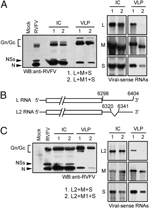
Next, we examined the effect of the 146-nt-long deletion in M1 RNA on L RNA packaging. In the experimental group, VLPs, intracellular RNAs, and intracellular proteins were collected from cells, supporting the replication of L, M1, and S RNAs and coexpressing all of the viral structural proteins, at 3 d posttransfection, whereas in the control group, we used M RNA in place of M1 RNA. We observed similar accumulations of intracellular viral proteins, intracellular viral-sense RNAs, and released particles in both groups (Fig. 4A). The levels of M RNA and M1 RNA were similar in the purified particles, whereas S RNA amount in the control group particles was approximately three times more than that in the experimental group VLPs. L RNA was efficiently packaged into the control group particles, but not into the experimental group particles (Fig. 4A), suggesting the importance of the deletion site in M1 RNA for efficient L RNA packaging.
Fig. 4.
Effect of M1 RNA on L RNA packaging. (A) VLPs, intracellular RNAs, and intracellular proteins were collected from cells supporting the replication of L, M, and S RNAs (lane 1) or from those supporting the replication of L, M1, and S RNAs (lane 2) at 3 d posttransfection. (Left) Western blot analysis of virus-specific intracellular (IC) and VLP proteins. Mock, mock-infected cells; RVFV, RVFV-infected cells. (Right) Northern blot analysis of virus-specific intracellular RNAs (IC) and viral RNAs in VLPs. Top, Middle, and Bottom represent viral-sense L RNA, viral-sense M RNA, and viral-sense S RNA, respectively. (B) Schematic diagram of antiviral-sense L and L2 RNAs. The numbers in L RNA represent the location of the 5′ UTR (in viral-sense) from the 5′ end of antiviral-sense L RNA and those in L2 RNA represents the deletion site. The boxed region represents the L gene ORF. (C) Experiments were performed as described in A except that L2 RNA was used in place of L RNA.
In the experiments described above, the production of infectious virus in the control group was most probably higher than in the experimental group, according to the titers of the recovered infectious viruses using M RNA 5′ UTR mutants. To determine whether the results observed in the above experiments for L RNA packaging were due to the production of higher titer of infectious virus in the control group, we used an L RNA mutant, L2 RNA, which lacked 22 nt at nucleotide positions 6320–6342 in the viral-sense L RNA (Fig. 4B). The deletion in L2 RNA was detrimental for efficient virus replication; 2.3 × 10⁷ pfu/mL infectious virus particles are recovered from cells supporting the replication of L, M, and S RNAs and expressing all of the viral structural proteins at 5 d posttransfection, whereas replacing the plasmid-expressing L RNA with that encoding L2 RNA resulted in the production of only 5.8 × 10² pfu/mL infectious virus particles. In the presence of replicating M and S RNAs, the accumulation of L2 RNA was lower than that of L RNA (Fig. S3), suggesting that the recovery of low titer of infectious virus could be due to the reduced level of L2 RNA replication. Because the 3′ end of bunyaviral mRNAs affects viral protein translation (31), it is also possible that the L2 mRNA carrying a deletion within the 3′ UTR failed to serve as a proper template for efficient L protein translation, resulting in the poor recovery of infectious viruses. We performed a similar experiment described above using L2 RNA and examined L2 RNA packaging (Fig. 4C). L2 RNA was efficiently packaged into VLPs in the presence of M and S RNAs, whereas efficient packaging of L2 RNA did not occur in the presence of M1 RNA and S RNA. Thus, we observed similar results in experiments using L2 RNA, where very few infectious viruses were released, and in those using L RNA.
To determine the effect of the deletion site in M1 RNA on L RNA packaging into the infectious virus particles, we characterized the efficiency of L RNA packaging into the M1 virus (Fig. S4). Due to the production of a low titer of M1 virus, the level of intracellular viral RNA accumulation was lower in M1 virus-infected cells compared with the WT virus-infected cells. Both viruses had similar buoyant densities in sucrose gradients and the viral protein contents were also similar. In contrast, compared with S and M RNAs, the relative amount of packaged L RNA was lower in purified M1 virus than in WT virus, establishing the importance of the deletion site in M1 RNA for efficient L RNA packaging into infectious virus.
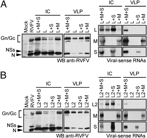
To identify the viral RNA segment(s) that is required for efficient L RNA packaging, cells were cotransfected with plasmids expressing L, Gn/Gc, and N proteins along with the plasmids expressing the following combination of viral RNAs: L RNA only, L and M RNAs, L and S RNAs, or L, M, and S RNAs. At 3 d posttransfection, intracellular proteins, intracellular RNAs, and VLPs were collected. The production of VLPs did not substantially differ among the samples (Fig. 5A). Efficient L RNA amplification occurred in cells expressing L RNA and in those supporting the replication of L, M, and S RNAs (Fig. 5A). Unexpectedly, an increased L RNA abundance was observed in the cells supporting the replication of S and L RNAs and a reduced L RNA abundance was observed in those supporting the replication of M and L RNAs; M RNA might have suppressed L RNA amplification, like defective interfering RNAs suppress helper virus RNA synthesis (32). L RNA was packaged efficiently into only those VLPs released from cells supporting the replication of L, M, and S RNAs (Fig. 5A). Due to the low abundance of L RNA in the cells supporting the replication of L and M RNAs, we were unable to determine whether M RNA alone exerts the efficient packaging of L RNA into VLPs. Nevertheless, these data showed that S RNA alone could not promote L RNA packaging into VLPs and suggested that either M RNA promoted efficient L RNA packaging or both M and S RNAs worked in concert to facilitate efficient L RNA packaging.
Fig. 5.
Analysis of L RNA packaging into VLPs from cells supporting the replication of various combinations of viral RNAs. (A) VLPs, intracellular RNAs, and intracellular proteins were collected from cells supporting the replication of various combinations of viral RNAs as shown at the Top of the panels at 3 d posttransfection. (Left two panels) Western blot analysis of virus-specific intracellular proteins (IC) and VLP proteins. Mock, mock-infected cells; RVFV, RVFV-infected cells. (Right two panels) Northern blot analysis of virus-specific intracellular RNAs (IC) and viral RNAs packaged into VLPs using RNA probes, each of which hybridizes with viral-sense L RNA (L), viral-sense M RNA (M), or viral-sense S RNA (S). (B) Experiments were performed as described in A except that L2 RNA was used in place of L RNA.
We performed similar experiments using L2 RNA in place of L RNA (Fig. 5B). Although we observed a low level of intracellular L2 RNA accumulation in cells supporting the replication of the three viral RNA segments, L2 RNA was packaged efficiently into only those VLPs released from cells supporting the replication of L2, M, and S RNAs; hence, the experiments using L2 RNA and those using L RNA showed similar results.
Discussion
We explored the mechanisms that govern the copackaging of the three genomic RNA segments into RVFV particles. RVFV S and M RNAs were efficiently copackaged into VLPs (Fig. 3), whereas M1 RNA was not copackaged with S RNA, strongly suggesting that the deletion site in M1 RNA carries an RNA signal that promotes the copackaging of M and S RNAs. The efficient copackaging of M and S RNAs into VLPs also implies that the likelihood of generating noninfectious virus carrying only S RNA or M RNA from infected cells is low. Although the current study did not provide direct physical evidence for the copackaging of L RNA with M and S RNAs, we demonstrated that the efficient packaging of L RNA or L2 RNA into virus particles occurred only in the presence of replicating M and S RNAs (Fig. 5). These data strongly suggested the copackaging of L RNA with M and S RNAs into virus particles and pointed toward the low likelihood of producing noninfectious virus particles carrying only L RNA from infected cells. S RNA alone could not facilitate the efficient packaging of L or L2 RNA (Fig. 5), implying the involvement of M RNA in efficient L RNA packaging. Although the poor accumulation of L RNA in cells supporting the replication of L and M RNAs prevented the direct assessment of the role of M RNA alone in L RNA packaging (Fig. 5), the data that efficient L RNA packaging did not occur in cells supporting the replication of L, S, and M1 RNA further highlighted the importance of M RNA for L RNA packaging (Fig. 4 and Fig. S4). Collectively, our data are consistent with the possibility that direct or indirect interactions among the three viral RNA segments drive the viral RNA copackaging to produce infectious RVFV. The data shown in the present study are also consistent with a previous report demonstrating the low particle-to-pfu ratio of Bunyamwera virus (24); these studies suggest that bunyaviruses exert the efficient copackaging of the three viral RNA segments to generate infectious virus particles.
We propose two models for the copackaging of the three viral RNA segments into RVFV. In the first model, M RNA works as a central regulator for the packaging of S and L RNAs into the virion. M RNA has two RNA elements, one of which interacts with L RNA and the other with S RNA, and these M RNA–L RNA and M RNA–S RNA interactions facilitate the copackaging of three RNAs into virus particles. M1 RNA may lack both of these putative RNA elements; hence, S and M1 RNA were not copackaged into VLPs (Fig. 3) and L RNA was poorly packaged into VLPs from cells supporting the replication of L, S, and M1 RNAs (Fig. 4). In the second model, the coordinated functions of M and S RNAs are important for efficient L RNA packaging. We hypothesize that the interaction between M RNA and S RNA induces a conformational change in S RNA that results in the exposure or presentation of a putative RNA element that interacts with L RNA and facilitates L RNA packaging. According to this model, L RNA was not efficiently packaged into VLPs released from cells supporting the replication of L and S RNAs because a putative M RNA-mediated conformational change was not induced in S RNA. S and M1 RNAs were not copackaged into VLPs, implying that M1 RNA did not interact with S RNA (Fig. 3). Hence, the lack of S RNA–M1 RNA interaction in cells supporting the replication of L, S, and M1 RNAs may have prevented efficient L RNA packaging.
Our data also suggest that the proposed intermolecular interactions among the three viral RNA segments affect the efficiency of RNA packaging. We observed that M RNA and M1 RNA were packaged into VLPs with similar efficiencies in the absence of other replicating RNAs (Fig. 2), but in the presence of S RNA, the packaging efficiency of M RNA was higher than that of M1 RNA (Fig. 3A). We suspect that the intermolecular interaction between M RNA and S RNA facilitated the efficient packaging of both RNAs; in the absence of the intermolecular interaction between S RNA and M1 RNA, the S RNA, which probably accumulated more abundantly than M1 RNA due to its smaller size, outcompeted the M1 RNA for the resources that are needed for RNA packaging, resulting in inhibition of M1 RNA packaging into VLPs. Also, we observed a trend that the amount of S RNA in the VLPs produced from cells supporting the replication of L, M, and S RNAs was higher than in those produced from cells supporting the replication of L, M1, and S RNAs (Fig. 4A). A possible interpretation of the data could be that the putative intracellular interactions among the three viral RNAs in cells supporting the replication of L, M, and S RNAs control the optimal packaging efficiency of S RNA.
Materials and Methods
Cells and Viruses.
Vero E6 cells and BSR-T7/5 cells (33), which stably express T7 RNA polymerase, were maintained as described previously (30, 33). arMP-12, a recombinant MP-12 strain of RVFV (34), was used as a control virus.
Plasmid Constructions.
Synonymous mutations carrying a unique BgIII site were introduced at the nucleotide positions 3275 and 3276 in the plasmid-encoding antiviral-sense M RNA (30) using Quickchange II site-directed mutagenesis kit (Agilent Technologies) to create pProT7-M(BgIII). Using pProT7-M(BgIII) as a template, DNA fragments of M segment carrying a series of deletions in the 5′ UTR were amplified by PCR using primers containing BglII and BspEI sites. After digestion of the PCR products with BglII and BspEI, the PCR fragments were cloned into the BglII and BspEI sites in pProT7-M(BgIII). The DNA fragment carrying a deletion between nucleotide positions 6320 and 6341 in the L segment was amplified by PCR using primers containing SpeI and BamHI sites and the plasmid pProT7-L encoding antiviral-sense L RNA (30) as a template. After digestion of the PCR products with SpeI and BamHI, the PCR fragments were cloned into the SpeI and BamHI sites in pProT7-L. The sequences of all of the constructs were confirmed.
Virus Rescue.
RVFV mutants were rescued from cDNAs as described previously (30), except that BSR-T7/5 cells were used in place of BHK/T7-9 cells. The infectivity of the rescued viruses was determined by plaque assay (30).
Expression of Viral Proteins and Viral RNAs.
BSR-T7/5 cells grown in 6-well plates were transfected with 1 μg each of plasmids expressing N protein and L protein and 2 μg of plasmid expressing Gn/Gc proteins (30) per well for viral protein expression. For viral RNA expression, 1 μg of each RNA expression plasmid was added to the mixture of protein expression plasmids. The transfected DNA amounts among the samples were kept the same by adding an empty plasmid pProT7 (30).
Western Blot Analysis.
Cells were harvested by cell scraper and washed with PBS. After incubation of the harvested cells on ice for 20 min in cell lysis buffer (20 mM Tris-HCl, 150 mM NaCl, 1% Triton X-100), the cell lysate was centrifuged at 2,000 rpm for 3 min using a microcentrifuge. The resultant supernatant was mixed with the same amount of 2× sample buffer and boiled for 10 min. Equal amounts of samples were subjected to SDS-polyacrylamide gel electrophoresis. Proteins were electroblotted onto polyvinylidene difluoride membranes (immobilon P; Millipore). After blocking overnight at 4 °C, the membranes were incubated with the primary antibody for 1 h at room temperature and with the secondary antibody for 1 h at room temperature. The proteins on the membrane were detected using an ECL kit (Amersham Biosciences). Anti-RVFV mouse polyclonal antibody was used to detect the virus-specific proteins as described previously (11).
Northern Blot Analysis.
Total RNAs were extracted using TRIzol reagent (Invitrogen) and subjected to Northern blot analysis as described previously (11). Strand-specific digoxigenin-labeled RNA probes and a digoxigenin system (Roche) were used for the detection of viral RNAs. The L RNA probe hybridizes with viral-sense L RNA at nucleotide positions 19–756, the M RNA probe at nucleotide positions 1297–2102, and the S RNA probe at nucleotide positions 39–776 from the 3′ ends of the viral-sense RNA segments.
VLP Purification.
Culture medium was harvested at 3 d posttransfection and clarified by centrifugation at 3,000 rpm for 15 min using a tabletop centrifuge. Intracellular RNAs and proteins were also collected as described above. The clarified supernatant was layered on top of a step sucrose gradient consisting of 20, 30, 50, and 60% (wt/vol) and centrifuged for 3 h at 26,000 rpm at 4 °C using a Beckman SW28 rotor (11). The VLPs at the interface of 30 and 50% sucrose were collected, diluted, and then further applied on top of a step sucrose gradient consisting of 20, 30, 50, and 60% (wt/vol) and centrifuged for 18 h at 26,000 rpm at 4 °C using the same rotor. The VLPs at the interface of 30 and 50% sucrose were collected, diluted, and then pelleted down through a 20% sucrose cushion at 26,000 rpm for 3 h at 4 °C. VLPs in the pellet were resuspended in TRIzol reagent and RNAs were extracted for Northern blot analyses. For Western blot analysis, VLPs in the pellet were dissolved in sample buffer.
Supplementary Material
Acknowledgments
We thank Karl-Klaus Conzelmann (Ludwig-Maximilians-University, Munich) and Andrew Pekosz (Johns Hopkins University, Baltimore) for BSR-T7/5 cells, Robert Tesh (University of Texas Medical Branch, Galveston, TX) for anti-RVFV antibody, Cheng Huang for virus purification analysis, and Krishna Narayanan for critical reading of the manuscript. This work was supported by a grant from the National Institute of Allergy and Infectious Diseases through the Western Regional Center of Excellence for Biodefense and Emerging Infectious Diseases Research, National Institutes of Health Grant U54 AI057156. K.T. and S. Murakami were supported by the James W. McLaughlin fellowship fund. S. Murakami was also supported by research fellowships from the Japan Society for the Promotion of Science.
Footnotes
The authors declare no conflict of interest.
This article is a PNAS Direct Submission.
This article contains supporting information online at www.pnas.org/lookup/suppl/doi:10.1073/pnas.1013155108/-/DCSupplemental.
References
- 1.Balkhy HH, Memish ZA. Rift Valley fever: An uninvited zoonosis in the Arabian peninsula. Int J Antimicrob Agents. 2003;21:153–157. doi: 10.1016/s0924-8579(02)00295-9. [DOI] [PubMed] [Google Scholar]
- 2.Peters CJ, Meegan JM. Rift Valley fever. In: Beran G, editor. CRS Handbook Series in Zoonoses, Section B: Viral Zoonoses. Boca Raton, FL: CRC; 1989. [Google Scholar]
- 3.Bird BH, Ksiazek TG, Nichol ST, Maclachlan NJ. Rift Valley fever virus. J Am Vet Med Assoc. 2009;234:883–893. doi: 10.2460/javma.234.7.883. [DOI] [PubMed] [Google Scholar]
- 4.Gargan TP, 2nd, Clark GG, Dohm DJ, Turell MJ, Bailey CL. Vector potential of selected North American mosquito species for Rift Valley fever virus. Am J Trop Med Hyg. 1988;38:440–446. doi: 10.4269/ajtmh.1988.38.440. [DOI] [PubMed] [Google Scholar]
- 5.Schmaljohn C, Nichol ST. Bunyaviruses. In: Knipe DM, et al., editors. Fields Virology. 5th Ed. Philadelphia, PA: Lippincott Williams & Wilkins; 2007. pp. 1741–1789. [Google Scholar]
- 6.Accardi L, et al. Activity of Toscana and Rift Valley fever virus transcription complexes on heterologous templates. J Gen Virol. 2001;82:781–785. doi: 10.1099/0022-1317-82-4-781. [DOI] [PubMed] [Google Scholar]
- 7.Blakqori G, Kochs G, Haller O, Weber F. Functional L polymerase of La Crosse virus allows in vivo reconstitution of recombinant nucleocapsids. J Gen Virol. 2003;84:1207–1214. doi: 10.1099/vir.0.18876-0. [DOI] [PubMed] [Google Scholar]
- 8.Dunn EF, Pritlove DC, Jin H, Elliott RM. Transcription of a recombinant bunyavirus RNA template by transiently expressed bunyavirus proteins. Virology. 1995;211:133–143. doi: 10.1006/viro.1995.1386. [DOI] [PubMed] [Google Scholar]
- 9.Flick K, et al. Rescue of Hantaan virus minigenomes. Virology. 2003;306:219–224. doi: 10.1016/s0042-6822(02)00070-3. [DOI] [PubMed] [Google Scholar]
- 10.Lopez N, Muller R, Prehaud C, Bouloy M. The L protein of Rift Valley fever virus can rescue viral ribonucleoproteins and transcribe synthetic genome-like RNA molecules. J Virol. 1995;69:3972–3979. doi: 10.1128/jvi.69.7.3972-3979.1995. [DOI] [PMC free article] [PubMed] [Google Scholar]
- 11.Ikegami T, Peters CJ, Makino S. Rift valley fever virus nonstructural protein NSs promotes viral RNA replication and transcription in a minigenome system. J Virol. 2005;79:5606–5615. doi: 10.1128/JVI.79.9.5606-5615.2005. [DOI] [PMC free article] [PubMed] [Google Scholar]
- 12.Freiberg AN, Sherman MB, Morais MC, Holbrook MR, Watowich SJ. Three-dimensional organization of Rift Valley fever virus revealed by cryoelectron tomography. J Virol. 2008;82:10341–10348. doi: 10.1128/JVI.01191-08. [DOI] [PMC free article] [PubMed] [Google Scholar]
- 13.Sherman MB, Freiberg AN, Holbrook MR, Watowich SJ. Single-particle cryo-electron microscopy of Rift Valley fever virus. Virology. 2009;387:11–15. doi: 10.1016/j.virol.2009.02.038. [DOI] [PMC free article] [PubMed] [Google Scholar]
- 14.Overby AK, Pettersson RF, Grünewald K, Huiskonen JT. Insights into bunyavirus architecture from electron cryotomography of Uukuniemi virus. Proc Natl Acad Sci USA. 2008;105:2375–2379. doi: 10.1073/pnas.0708738105. [DOI] [PMC free article] [PubMed] [Google Scholar]
- 15.Fujii K, et al. Importance of both the coding and the segment-specific noncoding regions of the influenza A virus NS segment for its efficient incorporation into virions. J Virol. 2005;79:3766–3774. doi: 10.1128/JVI.79.6.3766-3774.2005. [DOI] [PMC free article] [PubMed] [Google Scholar]
- 16.Fujii Y, Goto H, Watanabe T, Yoshida T, Kawaoka Y. Selective incorporation of influenza virus RNA segments into virions. Proc Natl Acad Sci USA. 2003;100:2002–2007. doi: 10.1073/pnas.0437772100. [DOI] [PMC free article] [PubMed] [Google Scholar]
- 17.Liang Y, Hong Y, Parslow TG. cis-Acting packaging signals in the influenza virus PB1, PB2, and PA genomic RNA segments. J Virol. 2005;79:10348–10355. doi: 10.1128/JVI.79.16.10348-10355.2005. [DOI] [PMC free article] [PubMed] [Google Scholar]
- 18.Muramoto Y, et al. Hierarchy among viral RNA (vRNA) segments in their role in vRNA incorporation into influenza A virions. J Virol. 2006;80:2318–2325. doi: 10.1128/JVI.80.5.2318-2325.2006. [DOI] [PMC free article] [PubMed] [Google Scholar]
- 19.Ozawa M, et al. Contributions of two nuclear localization signals of influenza A virus nucleoprotein to viral replication. J Virol. 2007;81:30–41. doi: 10.1128/JVI.01434-06. [DOI] [PMC free article] [PubMed] [Google Scholar]
- 20.Watanabe T, Watanabe S, Noda T, Fujii Y, Kawaoka Y. Exploitation of nucleic acid packaging signals to generate a novel influenza virus-based vector stably expressing two foreign genes. J Virol. 2003;77:10575–10583. doi: 10.1128/JVI.77.19.10575-10583.2003. [DOI] [PMC free article] [PubMed] [Google Scholar]
- 21.Ozawa M, et al. Nucleotide sequence requirements at the 5′ end of the influenza A virus M RNA segment for efficient virus replication. J Virol. 2009;83:3384–3388. doi: 10.1128/JVI.02513-08. [DOI] [PMC free article] [PubMed] [Google Scholar]
- 22.Liang Y, Huang T, Ly H, Parslow TG, Liang Y. Mutational analyses of packaging signals in influenza virus PA, PB1, and PB2 genomic RNA segments. J Virol. 2008;82:229–236. doi: 10.1128/JVI.01541-07. [DOI] [PMC free article] [PubMed] [Google Scholar]
- 23.Noda T, et al. Architecture of ribonucleoprotein complexes in influenza A virus particles. Nature. 2006;439:490–492. doi: 10.1038/nature04378. [DOI] [PubMed] [Google Scholar]
- 24.Lowen AC, Boyd A, Fazakerley JK, Elliott RM. Attenuation of bunyavirus replication by rearrangement of viral coding and noncoding sequences. J Virol. 2005;79:6940–6946. doi: 10.1128/JVI.79.11.6940-6946.2005. [DOI] [PMC free article] [PubMed] [Google Scholar]
- 25.Kohl A, Lowen AC, Léonard VH, Elliott RM. Genetic elements regulating packaging of the Bunyamwera orthobunyavirus genome. J Gen Virol. 2006;87:177–187. doi: 10.1099/vir.0.81227-0. [DOI] [PubMed] [Google Scholar]
- 26.Flick K, et al. Functional analysis of the noncoding regions of the Uukuniemi virus (Bunyaviridae) RNA segments. J Virol. 2004;78:11726–11738. doi: 10.1128/JVI.78.21.11726-11738.2004. [DOI] [PMC free article] [PubMed] [Google Scholar]
- 27.Gauliard N, Billecocq A, Flick R, Bouloy M. Rift Valley fever virus noncoding regions of L, M and S segments regulate RNA synthesis. Virology. 2006;351:170–179. doi: 10.1016/j.virol.2006.03.018. [DOI] [PubMed] [Google Scholar]
- 28.Habjan M, et al. Efficient production of Rift Valley fever virus-like particles: The antiviral protein MxA can inhibit primary transcription of bunyaviruses. Virology. 2009;385:400–408. doi: 10.1016/j.virol.2008.12.011. [DOI] [PubMed] [Google Scholar]
- 29.Ikegami T, Won S, Peters CJ, Makino S. Characterization of Rift Valley fever virus transcriptional terminations. J Virol. 2007;81:8421–8438. doi: 10.1128/JVI.02641-06. [DOI] [PMC free article] [PubMed] [Google Scholar]
- 30.Ikegami T, Won S, Peters CJ, Makino S. Rescue of infectious rift valley fever virus entirely from cDNA, analysis of virus lacking the NSs gene, and expression of a foreign gene. J Virol. 2006;80:2933–2940. doi: 10.1128/JVI.80.6.2933-2940.2006. [DOI] [PMC free article] [PubMed] [Google Scholar]
- 31.Blakqori G, van Knippenberg I, Elliott RM. Bunyamwera orthobunyavirus S-segment untranslated regions mediate poly(A) tail-independent translation. J Virol. 2009;83:3637–3646. doi: 10.1128/JVI.02201-08. [DOI] [PMC free article] [PubMed] [Google Scholar]
- 32.Holland JJ, et al. Defective interfering RNA viruses and the host-cell response. In: Fraenkel-Conrat H, Wagner RR, editors. Comprehensive Virology. New York: Plenum; 1980. pp. 137–192. [Google Scholar]
- 33.Buchholz UJ, Finke S, Conzelmann KK. Generation of bovine respiratory syncytial virus (BRSV) from cDNA: BRSV NS2 is not essential for virus replication in tissue culture, and the human RSV leader region acts as a functional BRSV genome promoter. J Virol. 1999;73:251–259. doi: 10.1128/jvi.73.1.251-259.1999. [DOI] [PMC free article] [PubMed] [Google Scholar]
- 34.Won S, Ikegami T, Peters CJ, Makino S. NSm and 78-kilodalton proteins of Rift Valley fever virus are nonessential for viral replication in cell culture. J Virol. 2006;80:8274–8278. doi: 10.1128/JVI.00476-06. [DOI] [PMC free article] [PubMed] [Google Scholar]
Associated Data
This section collects any data citations, data availability statements, or supplementary materials included in this article.


