

Abstract
The new Solid State X-Ray Image Intensifier (SSXII) has the unique ability to operate in single photon counting (SPC) mode, with improved resolution, as well as in traditional energy integrating (EI) mode. The SSXII utilizes an electron-multiplying CCD (EMCCD), with an effective pixel size of 32μm, which enables variable signal amplification (up to a factor of 2000) prior to digital readout, providing very high-sensitivity capabilities. The presampled MTF was measured in both imaging modes using the standard angulated-slit method. A measured detector entrance exposure of 24μR per frame was used to provide approximately 0.8 interaction events per pixel in the 10μm-wide slit area. For demonstration purposes, a simple thresholding technique was used to localize events in SPC mode and a number of such frames were summed to provide an image with the same total exposure used for acquiring the EI image. The MTF for SPC mode, using a threshold level of 15% of the maximum 12-bit signal and 95% of the expected events, and for EI mode (in parentheses) was 0.67 (0.20), 0.37 (0.07), 0.20 (0.03), and 0.11 (0.01) at 2.5, 5, 7.5, and 10 cycles/mm, respectively. Increasing the threshold level resulted in a corresponding increase in the measured SPC MTF and a lower number of detected events, indicating a tradeoff between resolution and count efficiency is required. The SSXII in SPC mode was shown to provide substantial improvements in resolution relative to traditional EI mode, which should benefit applications that have demanding spatial resolution requirements, such as mammography.
Keywords: single photon counting (SPC), energy integrating (EI), MTF, SSXII, resolution, image quality
1. INTRODUCTION
Single photon counting (SPC) imaging has gained popularity over the past several years as a result of having the ability to provide improved spatial and contrast resolution relative to traditional energy integrating (EI) detectors.1 SPC is potentially beneficial to many different imaging applications that require high-contrast and high-spatial resolution (e.g., mammography and orthopedics) and is widely used in nuclear imaging (e.g., SPECT).2 In this work, we investigate the unique ability of a new Solid State X-Ray Image Intensifier (SSXII) to operate in both EI mode and SPC mode, by investigating the resolution performance in each respective mode.
2. MATERIALS AND METHODS
2.1 The Solid State X-ray Image Intensifier (SSXII)
The SSXII is a new high-resolution, high-sensitivity x-ray imager that has been developed by our group to improve upon the inherent limitations of current state-of-the-art radiographic and fluoroscopic detectors, such as flat panel detectors and x-ray image intensifiers.3–6 Superior imaging performance has been demonstrated for traditional EI applications, in particular for endovascular image guided interventions.7 The prototype SSXII module, shown in Fig. 1, is composed of an electron-multiplying CCD (EMCCD) camera system (Photonic Science LTD, East Sussex, UK) in which a custom fiber optic input window has been installed. The camera utilizes an EMCCD sensor (Texas Instruments, Dallas, TX), featuring 1000 x 1000 8 μm pixels, the capability to operate real-time (at 30 frames-per-second), and a standard 12-bit camera-link data output. It views a 350 μm thick CsI(Tl) scintillating phosphor grown on an additional fiber optic plate via a removable 4:1 fiber-optic taper (FOT) (Incom, Inc., Charlton, MA) providing an effective pixel size of 32 μm. Larger pixel sizes can be achieved with variable binning, which in turn, increases the maximum frame rates achievable. The detector has been constructed with two fiber optic plates to facilitate the use of different imaging components. A study of different FOT ratios and phosphor thicknesses is currently under way for further optimization under a variety of specific imaging tasks.
Figure 1.

Figure 1(a). Schematic of the solid-state x-ray image intensifier (SSXII) module which includes a CsI(Tl) scintillating plate, a removable fiber-optic taper (FOT) and an EMCCD camera with fiber-optic input window. The FOT holder [seen in Fig. 1(b)] has been omitted so that the inner components can be seen.
Figure 1(b). A photograph of the assembled SSXII detector module with the power supply unit attached.
The EMCCD sensor operates like a standard frame-transfer CCD, with the addition of a unique multiplication register prior to the readout of the signal. This multiplication register provides a built-in adjustable gain which amplifies the signal (up to a factor of 2000), in solid-state, prior to the addition of read-noise — allowing for the effective elimination of read-noise degradations at very low incident x-ray exposure rates and hence enabling the detection of single interacting x-ray photons. A Peltier cooler is used to cool the sensor (to less than 0 °C), reducing thermal electron noise to negligible levels. Easily interchangeable front-end components allow for task-based optimization of the detector system (e.g., using a thinner phosphor for mammography and a thicker phosphor for SPECT). An array of such modules, currently under development, will be used to provide a larger field-of-view.
2.2 Comparison Images
To qualitatively and quantitatively assess resolution improvements when in SPC mode, relative to the standard EI mode, images (gain and offset corrected) were acquired of a mammographic line-pair phantom (with the 4:1 FOT removed from the imaging chain) and an angulated slit with a width of 10 μm (with the 4:1 FOT in the imaging chain). The test objects were placed very close to the detector input to minimize geometric unsharpness. Two different x-ray spectra were used for this investigation. Both were quasi-monoenergetic and are shown in Fig. 2 and Fig. 3, as determined using spectrum generating software.9 The mammographic line-pair phantom was imaged using a 50 kVp x-ray beam on a clinical c-arm system (Toshiba Medical Systems Corp., Tustin CA), with a source-to-detector distance of 80 cm. The x-ray tube was specified to have 1.1 mm Al inherent filtration and a total of 1.8 mm Al and 1 cm of iodine contrast media (density 350 mg/ml) filtration were added at the tube output, providing an average energy of 32.8 keV. Slit images were acquired on the same c-arm system, using a 120 kVp x-ray beam and a source-to-image distance of 95 cm. An additional 1.8 mm Al and 1.7 mm Pb filtration were placed at the tube output, providing an average energy of 81.5 keV. The x-ray fluence per unit exposure was calculated using standard methods to be 147 and 284 incident x-ray photons (inc xph) / mm2 / μR, respectively.8 The exposure per frame was measured according to IEC guidelines9 using a calibrated ionization chamber (Unidos Webline Universal dosimeter with model 34060-2 ionization chamber, PTW, Frieburg, Germany) and the mAs was adjusted such that there was less than one absorbed x-ray photon per pixel. To enable detection of single photons, the EMCCD gain was set to a factor of approximately 1,000 to ensure sufficient signal amplification above the read-noise floor.
Figure 2.

Figure 2(a). X-ray spectrum used for imaging of the mammographic line-pair phantom.
Figure 2(b). X-ray spectrum used for imaging of the slit.
Figure 3.
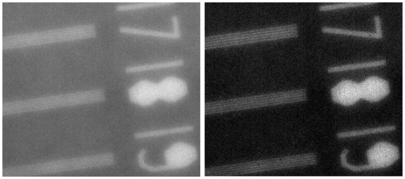
Comparison images of a mammographic line-pair phantom in EI (left) and SPC (right) modes. Background pixel intensity values were normalized and the same windowing width and level were used for display.
For initial demonstration purposes, a simple thresholding technique was used for localization of events in SPC mode; however, more sophisticated methods are available — the implications of which will be discussed further in the results and discussion section.2 In general, threshold levels were chosen such that the measured number of events in the resulting SPC image closely matched the calculated number of events. The effect of using different threshold levels was also investigated. Individual images were summed both before and after thresholding. These summed images were used to provide the final EI and SPC images, respectively, which were used for further analysis. Images of a mammographic line-pair phantom were used to provide a qualitative demonstration of differences in image quality between the two modes. Integrated line profiles, taken perpendicularly across the line pairs, were used to quantify differences. Images of an angulated slit were analyzed using standard methods to obtain finely sampled line spread functions (LSFs) and the detector modulation transfer function (MTF) for both operational modes.10 A new simple and accurate method for measuring the two-dimensional MTF using only the detector noise-response (i.e., no test object) will be implemented in future measurements.11–12
2.3 Simulated Detector System
Experimental images were used to quantify resolution differences in SPC and EI modes. However, a complete picture of detector performance must also take into consideration detector noise characteristics. To provide insight into potential differences in signal-to-noise ratio performance between SPC and EI modes, images were simulated for a simple high-resolution indirect detector model based on the SSXII detector, using MATLAB (version 7.9.0, Mathworks, Natick, MA). The image matrix contained 1000 x 1000, 8 μm square pixels and images were generated as follows. First, a number of incident (monoenergetic) x-ray photons were generated per pixel using a random number generator with a mean value of 0.2 and a variance of 0.2. The number of absorbed x-ray photons was then determined using binomial selection with a success rate of 0.3. The absorbed x-ray photons were then converted to light using a conversion gain of 500 (assumed for simplicity to be Poisson distributed). The blur associated with this conversion process was taken to be a single Gaussian with a full width half maximum (FWHM) of 8.2 pixels (corresponding to a standard deviation of 28 μm). Light photons were then converted to electrons using binomial selection with a success rate of 0.5. Additional spreading of light due to the fiber optics was considered using a second Gaussian blurring function with a FWHM of 4.7 pixels (standard deviation of 16 μm). Electrons were subsequently “digitized” using an electron-to-digital number conversion factor of 0.8 electrons per DN. Finally electronic noise was added (Gaussian with zero-mean) with a standard deviation of 10 DN. Both flat-field and angulated edge images were generated for this simulated detector system and the detective quantum efficiency (DQE) was determined using the output images to quantify the performance of EI mode. The absorbed x-ray image (or event image) was also recorded and used to provide the DQE of the detector in SPC mode and is representative of the implementation of a “perfect” localization technique that accurately determines all event locations. In practice, misregistration of events will likely be unavoidable and will occur with differing degrees depending on the imaging conditions and the localization algorithm used. Potential influences of event misregistration will be discussed.
3. RESULTS AND DISCUSSION
3.1 Mammographic Line-Pair Phantom Images
To localize events in SPC mode, the threshold level was selected based on an approximate estimate of the number of expected events (i.e., the calculated number of absorbed x-ray photons), which was determined using standard methods, described as follows.8 The incident x-ray fluence was calculated to be 147 incident x-ray photons (inc xph) / mm2 / uR and the detector entrance exposure was measured to be 3.8 μR / frame at 0.4 mAs. Hence, there were an estimated 0.036 inc xph / pixel. The quantum detection efficiency of the CsI phosphor was calculated to be 0.87, resulting in approximately 0.031 absorbed x-ray photons (abs xph) / pixel. A background region containing 500 x 200 pixels was selected in the images with an expected number of events in this region of 3,100 (500 x 200 pixels x 0.031 abs xph / pixel = 3,100 abs xph). The threshold value was varied until the measured number of events closely matched this value. A threshold of 830 digital numbers (DN) resulted in 3,060 counts in this region or 99% of the estimated number of total counts.
Figure 3 shows the mammographic line-pair phantom images for both EI and SPC modes, which are the sum of 10,000 frames, giving an effective exposure of 38 mR. The averaged background signals of these images were normalized and the same windowing width and level were used to enable a fair comparison of the inherent image quality differences. Exceptional resolution is evident in either image, with both modes clearly demonstrating 19 line-pairs per mm imaging capabilities. However, the line-pairs are more clearly resolved in the image acquired using SPC mode. SPC mode is also shown to provide a substantial improvement in contrast. Integrated line profiles were taken perpendicularly across the 17 line-pairs per mm pattern to quantify these differences and are shown in Fig. 4(a). The profile of the lines rises and falls more sharply in SPC mode, indicative of an improvement in resolution as compared to EI mode. An increase in contrast is also evident in the image acquired using SPC mode and was a factor of 2.8 larger than that of the EI image.
Figure 4.

Figure 4(a). Comparison of the integrated line profiles taken perpendicularly across the 17 lines-pairs per mm pattern for EI and SPC modes after normalizing the averaged background pixel intensities.
Figure 4(b). Comparison of the integrated line profiles taken perpendicularly to the 17 lines-pairs per mm pattern for EI mode after digital contrast enhancement (through histogram matching) and SPC mode.
Digital images may be contrast enhanced by manipulating the windowing width and level, and therefore increased contrast is not a complete description. To investigate further, the contrast of the EI image was digitally enhanced using a histogram matching technique to that of the SPC image. Figure 4(b) shows the line profiles (zoomed in on the bar pattern) of the EI image after contrast enhancement and the SPC image. Even after enhancement of the EI image to provide comparable image contrast, significant improvements are still evident with SPC mode. The peaks rise and fall more sharply and the trough-to-peak heights are greater by more than a factor of two. The signal-to-noise ratio (SNR) provides a more concrete measure of image quality and was also measured for the different line-pair patterns. SPC mode provided an improvement in SNR that increased with an increasing number of line-pairs per mm and was a factor of 1.4 and 1.75 times larger at 14 and 19 line-pairs per mm, respectively.
3.2 Slit Images, LSFs, and MTFs
As with the mammographic line-pair images, event localization for the slit images was accomplished using a simple thresholding technique and the threshold value was selected based on the expected number of events. The incident x-ray fluence was calculated to be 284 inc xph / mm2 / μR and the detector entrance exposure was measured to be 24 μR / frame at 2 mAs. Hence, there were an estimated 7.0 inc xph / pixel / frame. The quantum detection efficiency of the CsI phosphor was calculated to be 0.37, resulting in approximately 2.6 abs xph / pixel. Incorporating the slit width, which was 10 μm, there were approximately 0.8 abs xph / pixel (i.e., the slit allows photons to pass through only 1/3 of the pixel area). The length of the slit was measured to be ~ 7.8 mm or 244 pixels, resulting in ~195 abs xph (events) per image. To provide sufficient statistics for the slit MTF measurement, a sequence of 1,340 images were acquired, providing 270,400 events for the entire image sequence.
Table 1 shows the corresponding number of counts based on the threshold value used for SPC mode. The 630 digital number (DN) threshold resulted in 257,100 total counts, which was within 95% of the expected theoretical value of 270,400. Using a higher threshold value resulted in a lower counting efficiency (i.e., a decrease in the percent of measured counts relative to the estimated number of counts). Figure 5 shows a 3x magnified region of the slit images for both EI mode (standard operation) and for SPC mode using a threshold of 630 DN. The corresponding line spread functions (LSFs) are shown in Fig. 6. The full-width-at-half-maximum (FWHM) was 4.3 pixels (140 μm) for EI mode and 2.2 pixels (70 μm) for SPC mode. Figure 7 shows the resulting MTF for EI mode and for SPC mode using various thresholds (630, 900, 1100, and 1250 DN). The MTF for SPC mode, using a threshold level of 630 DN (15% of the maximum 12-bit signal) and 95% of the estimated number of total counts, and for EI mode (in parentheses) was 0.67 (0.20), 0.37 (0.07), 0.20 (0.03), and 0.11 (0.01) at 2.5, 5, 7.5, and 10 cycles/mm, respectively. Increasing the threshold level resulted in a corresponding increase in the measured SPC MTF and a lower number of detected events, indicating a tradeoff between resolution and count efficiency is required. Increasing the threshold level resulted in an increase in the measured SPC MTF, in a general trend towards the ideal MTF (sinc function of the pixel size), as a result of rejecting an increasing number of blurred photons. However, this also resulted in a corresponding decrease in the counting efficiency, indicating a tradeoff between resolution and count efficiency is required with a simple thresholding technique. Above a threshold of 1250 DN, there were not enough counts to provide a reliable measurement.
Table 1.
Measured total counts for various threshold values. The last column shows the percentage of the estimated total number of expected counts which was 270,400.
| Threshold Value (DN) | Threshold Value (% of 12-bit signal) | Measured Total Counts in 1,340 Images After Thresholding | % of Estimated Total Counts |
|---|---|---|---|
| 630 | 15 | 257,100 | 95 |
| 900 | 22 | 37,700 | 14 |
| 1100 | 27 | 8,150 | 3.0 |
| 1250 | 31 | 3,000 | 1.1 |
Figure 5.
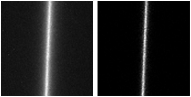
Comparison between slit images acquired in EI mode (left) and SPC mode using a threshold of 630 DN (right). The images have been digitally magnified by a factor of 3 and cropped for display purposes.
Figure 6.

Plot of the line spread function (LSF) using EI mode and SPC mode with a threshold of 630 DN.
Figure 7.

MTF determined from the slit images for EI mode and for SPC mode using various thresholds of 630, 900, 1100, and 1250 DN. Also shown is the sinc function of the pixel size (32 μm), which is the theoretical maximum.
Significant resolution and contrast improvements were observed using a simple thresholding technique, indicating that the SSXII can successfully operate in SPC mode by detecting individual x-ray interaction events. The advantage of a simple thresholding technique is that it is fast and can be readily implemented in real-time. For simplicity, the x-ray spectra used in this study were quasi-monoenergetic, which facilitates the use of such a localization technique. More advanced event localization techniques will be required for more realistic polychromatic x-ray spectra. Detected signal intensities vary proportionally with the absorbed x-ray energy and a polychromatic spectrum will thereby increase the complexity of the analysis. Implementation of a multiple thresholding technique may enable energy resolving capabilities. Other more advanced localization techniques, such as centroiding, should provide a further increase in MTF while maintaining a high counting efficiency.13
3.3 Investigation of Signal-to-Noise Performance Using Simulated Images
The normalized noise power spectrum (NNPS) of the simulated detector system is shown in Fig. 8 for both EI and SPC modes. The NNPS of both modes was equivalent at zero spatial frequency. However, the NNPS in EI mode was significantly lower at higher spatial frequencies due to the blurring processes in the detector imaging chain suppressing high-frequency content. The NNPS in SPC mode was constant, which is expected for an aperture-limited detector and is representative of a detector with no additional blurring (i.e., with the effects of phosphor and fiber optic blur removed).14 DQEs of both modes are shown in Fig. 9. The DQE in SPC mode was approximately constant as a function of spatial frequency and was equal to the quantum detection efficiency of the phosphor. This illustrates, in theory, that with an accurate localization technique, the degradations in SNR performance typically observed with standard EI detectors (most significantly due to the x-ray converting phosphor) can be reduced or even eliminated. SPC images were taken to be the actual event (absorbed x-ray photon) images and are representative of a perfect localization technique. Hence, these SPC results represent a “best-case” scenario.
Figure 8.

The NNPS of a simulated detector based on the SSXII, for EI and SPC modes. The SPC mode shown assumes perfect localization of events.
Figure 9.

The DQE of a simulated detector based on the SSXII, for EI and SPC modes. The SPC mode shown assumes perfect localization of events.
This analysis suggests that the perceived noise in SPC mode should be greater than EI mode as a result of having less blurring or smoothening of the high-frequency content of the image. When also incorporating the signal, SPC mode is shown to have the capability of providing large improvements relative to EI mode, as illustrated with an increased DQE. Event misregistration (i.e., falsely counting or missing events) would tend to lower the DQE relative to this theoretical maximum. Use of a simple thresholding technique has its limitations, including the inability to handle moderate variations in signal intensity, and more intelligent localization techniques are to be preferred. Significant event misregistration could lead to little or no improvement in the MTF and/or a disproportionate increase in noise, ultimately resulting in degradation of the DQE. Measuring the DQE in EI and SPC modes will be required to fully assess performance differences and the practical benefits to be realized. Further investigations will also be required to determine the effect of inaccuracies in event localization and to develop more robust localization techniques that limit such inaccuracies.
4. SUMMARY AND CONCLUSION
The unique ability of a new Solid State X-Ray Image Intensifier (SSXII) to operate in both traditional energy integrating (EI) and single photon counting (SPC) modes was demonstrated by quantifying the resolution performance of each operational mode. A simple thresholding technique was used to localize events in SPC mode. Images of a mammographic line-pair phantom qualitatively demonstrated resolution and contrast improvements of SPC mode. To quantify resolution differences, the MTF was measured using the standard slit method. A substantial improvement was observed in SPC mode as compared to traditional EI mode. More sophisticated localization techniques should provide even greater improvements in performance, maintaining high count efficiency and offering significant improvements in resolution.
Acknowledgments
This work was supported by NIH R01 Grants EB002873 and EB008425 and an equipment grant from Toshiba Medical Systems Corporation.
References
- 1.Amendolia SR, Bisogni MG, Delogu P, Fantacci ME, Paternoster G, Rosso V, Stefanini A. Characterization of a mammographic system based on single photon counting pixel arrays coupled to GaAs x-ray detectors. Med Phys. 2009;36:1330–1339. doi: 10.1118/1.3097284. [DOI] [PubMed] [Google Scholar]
- 2.DeVree GA, Westra AH, Moody I, VanDerHave F, Ligtvoet KM, Beekman FJ. Photon-counting gamma camera based on an electron-multiplying CCD. IEEE Trans Nucl Sci. 2005;52:580–588. [Google Scholar]
- 3.Kuhls-Gilcrist A, Bednarek DR, Rudin S. Component analysis of a new solid state x-ray image intensifier (SSXII) using photon transfer and instrumentation noise equivalent exposure (INEE) measurements. Proc SPIE. 2009;7258:725842. doi: 10.1117/12.813957. [DOI] [PMC free article] [PubMed] [Google Scholar]
- 4.Kuhls-Gilcrist A, Yadava G, Patel V, Jain A, Bednarek DR, Rudin S. The solid state x-ray image intensifier (SSXII): An EMCCD-based x-ray detector. Proc of SPIE. 2008;6913:19130K. doi: 10.1117/12.772724. [DOI] [PMC free article] [PubMed] [Google Scholar]
- 5.Kuhls-Gilcrist A, Yadava G, Patel V, Bednarek DR, Rudin S. Progress in electron-multiplying CCD (EMCCD) based, high-resolution, high-sensitivity x-ray detector for fluoroscopy and radiography. Proc SPIE. 2007;6510:651047. doi: 10.1117/12.713140. [DOI] [PMC free article] [PubMed] [Google Scholar]
- 6.Rudin S, Kuhls AT, Yadava GK, Josan GC, Wu Y, Chityala RN, Rangwala HS, Ionita CN, Hoffmann KR, Bednarek DR. New ligh-amplifier-based detector designs for high spatial resolution and high sensitivity CBCT mammography. Proc SPIE. 2006;6142:614263. doi: 10.1117/12.649501. [DOI] [PMC free article] [PubMed] [Google Scholar]
- 7.Rudin S, Bednarek DR, Hoffmann KR. Endovascular image-guided interventions EIGIs. Med Phys. 2008;35:301–309. doi: 10.1118/1.2821702. [DOI] [PMC free article] [PubMed] [Google Scholar]
- 8.Boone JM. X-ray Production, Interaction, and Detection in Diagnostic Imaging. In: Beutel J, Kundel HL, Metter RLV, editors. Handbook of Medical Imaging: Physics and Psychophysics. Vol. 1. SPIE; Bellingham: 2000. pp. 1–81. [Google Scholar]
- 9.62220-1, IEC. Medical electrical equipment: Characteristics of digital x-ray imaging devices - Part 1: Determination of the detective quantum efficiency. International Electrotechnical Commission; Geneva, Switzerland: 2003. [Google Scholar]
- 10.Fujita H, Tsai DY, Itoh T, Doi K, Morishita J, Ueda K, Ohtsuka A. A simple method for determining the modulation transfer function in digital radiography. IEEE Trans Med Imaging. 1992;11:34–39. doi: 10.1109/42.126908. [DOI] [PubMed] [Google Scholar]
- 11.Kuhls-Gilcrist A, Jain A, Bednarek D, Hoffmann K, Rudin S. Accurate MTF measurement in digital radiography using noise response. Med Phys. 2010;37:724–735. doi: 10.1118/1.3284376. [DOI] [PMC free article] [PubMed] [Google Scholar]
- 12.Kuhls-Gilcrist A, Bednarek DR, Rudin S. A method for the determination of the two-dimensional MTF of digital radiography systems using only the noise response. Proc of SPIE. 2010 doi: 10.1117/12.843918. [DOI] [PMC free article] [PubMed] [Google Scholar]
- 13.Michel R, Fordham J, Kawakami H. Fixed pattern noise in high–resolution, CCD readout photon-counting detectors. Mon Not R Astron Soc. 1997;292:611–620. [Google Scholar]
- 14.Zhao W, Rowlands JA. Digital radiology using active matrix readout of amorphous selenium: theoretical analysis of detective quantum efficiency. Med Phys. 1997;24:1819–1833. doi: 10.1118/1.598097. [DOI] [PubMed] [Google Scholar]