
Figure 1. Representative intracranial aneurysms in hypertensive mice that received a single injection of elastase into the cerebrospinal fluid.
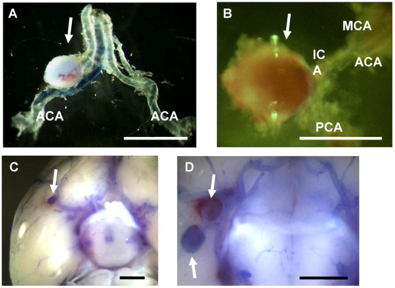
Arrows indicate aneurysms. Large aneurysm formation was found along the Circle of Willis or its major branches (A-D). Dissection of aneurysms revealed saccular shape of the aneurysms (A, B). Some of the mice had multiple aneurysms (D). Bar = 1 mm, ACA: anterior cerebral artery, MCA: middle cerebral artery, PCA: posterior cerebral artery, ICA: internal carotid artery.