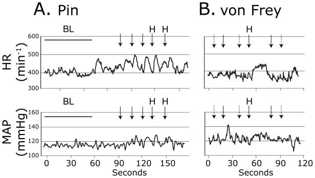
Figure 1.
Sample traces of heart rate (HR, top panels) and mean arterial pressure (MAP, bottom panels) during sensory testing with Pin in an H group animal 3 days following spinal nerve ligation injury (A.), and with von Frey fibers in a non-hyperalgesic animal 3d following spinal nerve ligation (B.). Arrows indicate timing of the application of the stimuli. A dashed arrow represents an application that produced no behavioral response. A solid arrow indicates a withdrawal of the stimulated paw. “H” indicates a hyperalgesia-type withdrawal with sustained elevation with shaking and grooming. “BL” indicates baseline recording interval.

