Abstract
In myocardial ischemia, adrenergic nerves release excessive amounts of norepinephrine (NE), causing dysfunction and arrhythmias. With anoxia and the concomitant ATP depletion, vesicular storage of NE is impaired, resulting in accumulation of free NE in the axoplasm of sympathetic nerves. Intraneuronal acidosis activates the Na+/H+ exchanger (NHE), leading to increased Na+ entry in the nerve terminals. These conditions favor availability of the NE transporter to the axoplasmic side of the membrane, causing massive carrier-mediated efflux of free NE. Neuronal NHE activation is pivotal in this process; NHE inhibitors attenuate carrier-mediated NE release. We previously reported that activation of histamine H3 receptors (H3R) on cardiac sympathetic nerves also reduces carrier-mediated NE release and alleviates arrhythmias. Thus, H3R activation may be negatively coupled to NHE. We tested this hypothesis in individual human SKNMC neuroblastoma cells stably transfected with H3R cDNA, loaded with the intracellular pH (pHi) indicator BCECF. These cells possess amiloride-sensitive NHE. NHE activity was measured as the rate of Na+-dependent pHi recovery in response to an acute acid pulse (NH4Cl). We found that the selective H3R-agonist imetit markedly diminished NHE activity, and so did the amiloride derivative EIPA. The selective H3R antagonist thioperamide abolished the imetit-induced NHE attenuation. Thus, our results provide a link between H3R and NHE, which may limit the excessive release of NE during protracted myocardial ischemia. Our previous and present findings uncover a novel mechanism of cardioprotection: NHE inhibition in cardiac adrenergic neurons as a means to prevent ischemic arrhythmias associated with carrier-mediated NE release.
Myocardial ischemia and infarction are associated with excessive norepinephrine (NE) release from sympathetic nerve endings (1). Cardiac dysfunction and arrhythmias ensue, resulting in high morbidity and mortality (2, 3). The increased secretion of NE is caused by an imbalance in the processes governing NE release and its reuptake into nerve endings. The mechanism normally responsible for the reuptake of NE is a Na+-dependent cotransporter (NET) (4, 5). Any condition that elevates intracellular Na+ in sympathetic nerve endings can trigger the reversal of the NET, i.e., can cause the transport of NE out of the neuron (“carrier-mediated” NE release) (6, 7). In protracted myocardial ischemia, all cells become depleted of ATP. Inasmuch as ATP is necessary for the vesicular storage of NE in sympathetic nerve endings, a lack of ATP will force NE to accumulate in the axoplasm instead of being stored in vesicles. Moreover, intracellular acidosis, a typical feature of myocardial ischemia, activates the Na+/H+ exchanger (NHE), which exchanges intracellular H+ for extracellular Na+. Consequently, intracellular Na+ concentration will rise. Ultimately, the increases in intraneuronal Na+ and unbound NE will contribute to the reversal of NET and thus, to an excessive, “carrier-mediated,” release of NE.
Several lines of evidence demonstrate that an increase in NHE activity enhances NE release via the NET and, conversely, that a decrease in NHE activity attenuates NE release. In LLC-PK1 cells stably transfected with the human NET, lowering intracellular pH (pHi) activates NHE with a consequent increase in intracellular Na+, and a rise in the efflux of NET substrates (8). Moreover, in ischemic guinea pig and human myocardium, inhibition of NHE with amiloride analogs attenuates the NHE-dependent accumulation of Na+ in sympathetic nerve endings. This reduces NE release via the NET and thereby alleviates reperfusion arrhythmias (9, 10). These studies provide a link between changes in NHE activity and carrier-mediated NE release.
Histamine H3 receptors (H3R) were discovered by Schwartz and colleagues (11) as inhibitory autoreceptors in central histaminergic pathways. We identified H3R as inhibitory heteroreceptors in cardiac adrenergic nerve endings (12–14) and demonstrated that H3R activation decreases carrier-mediated NE release in guinea pig and human heart (9, 10). Because H3R agonists and NHE inhibitors attenuate carrier-mediated NE release synergistically (9, 10), we proposed that H3R activation may decrease NHE activity (15) and thereby regulate NE release via the NET.
The purpose of the present study was to examine whether H3R activation reduces NHE activity. With the recent cloning of human, rat, and guinea pig H3R (16–18), it has become possible to test this hypothesis directly. In this study, we used human SKNMC neuroblastoma cells stably transfected with H3R cDNA (SKNMC-H3 cells) (16). These cells possess amiloride-sensitive NHE. NHE activity was assayed in the absence and presence of the H3R agonist imetit (19). Because NHE is not active at neutral pHi (20), its activity was measured as the rate of Na+-dependent pHi recovery in response to an acute acid pulse. We demonstrate that stimulation of H3R indeed inhibits NHE activity. This discovery provides a mechanism for endogenous histamine to attenuate the release of cardiotoxic NE in myocardial ischemia. It also suggests that H3R agonists are likely to be useful cardioprotective agents.
Materials and Methods
Cell Preparation and BCECF Loading.
SKNMC-H3 cells were grown to confluence (2 days after plating) on 22-mm-square standard glass coverslips (No. 1) and maintained in α-MEM supplemented with 10% FBS, 2 mM l-glutamine, 450 μg/ml geneticin, 50 units/ml penicillin, and 50 μg/ml streptomycin at 37°C, 5% CO2. Cells were loaded with the membrane-permeant form of the pHi indicator 2′,7′-bis(carboxyethyl)-5(6)-carboxyfluorescein (BCECF) ester (5 μM) for 20 min at room temperature. After loading with the dye, cells were rinsed with Hepes-buffered Na+ Ringer's solution (140 mM NaCl, 5 mM KCl, 10 mM Hepes, 2 mM CaCl2, 1 mM MgCl2, pH 7.4). The coverslip with the BCECF-loaded cells was attached to the bottom of a flow-through superfusion chamber and mounted on the stage of an inverted epifluorescence microscope (Nikon Diaphot). The cells in the chamber were superfused and maintained at 37°C as described (21, 22). Cells were visualized under transmitted light with a Nikon CF Fluor oil immersion objective (×40/1.3 numerical aperture) before starting the fluorescence measurements. Calibration of the emitted fluorescence signal from each cell in the field was performed at the end of each experiment according to the nigericin/high K+ method (23). Cells in the experimental field of view were analyzed singularly and independently from their neighbors.
Solutions and Reagents.
The experimental solutions were based on the Na+ Ringer's composition described above with the following substitutions: for the NH4Cl solution, NaCl and KCl were replaced with 20 mM NH4Cl and 120 mM N-methyl-d-glucamine (NMDG/Cl); for the Na+-free solution, NaCl and KCl were replaced with 145 mM NMDG/Cl. The Na+-free solutions were titrated to pH 7.4 with NMDG powder. The composition of the high K+-calibration solutions was similar to that of Na+-Ringer, except that NaCl was replaced with KCl, and titrated with KOH to pH 6.5 and pH 7.8, respectively, as described (24). All chemicals were obtained from Sigma unless otherwise stated. Imetit (Research Biochemicals, Natick, MA) was prepared in distilled water and then diluted 1:10,000 to yield a final concentration of 100 nM in the experimental superfusates. Thioperamide (300 nM), an H3R antagonist (25), and 5-(N-ethyl-N-isopropyl)amiloride (EIPA; 10 μM), an inhibitor of NHE (26), were both diluted in dimethyl sulfoxide. Nigericin, a K+/H+ exchanger, was added to the K+ calibration solutions from a 20-mM stock made up in ethanol to yield a final concentration of 10 μM. Individual vials (50 μg) of the acetoxymethyl derivative of BCECF (Molecular Probes) were stored dry at 0°C and reconstituted in dimethyl sulfoxide, at a concentration of 10 mM, for each experiment. At the concentrations used, dimethyl sulfoxide and ethanol had no effect on any preparation in these studies.
Equipment.
The basic components of the experimental apparatus have been described (21, 22). The imaging work station was controlled with the METAFLUOR software package (Universal Imaging, Westchester, PA). Image pairs were obtained every 15 s for the duration of the experiment at 490 nm and 440 nm excitation with emission at 520 nm. The fluorescence excitation was shuttered off except during the brief periods required to record an image. To check for interference from intrinsic autofluorescence and background, images were obtained on cells by using the same exposure time and filter combination used for the experiments and found to be a minor component of the fluorescence signal.
Statistics.
Results are expressed as means (±SEM), where n refers to the number of individually analyzed cells. Significant differences were determined by one-way ANOVA. Significance was asserted if P < 0.05.
Results
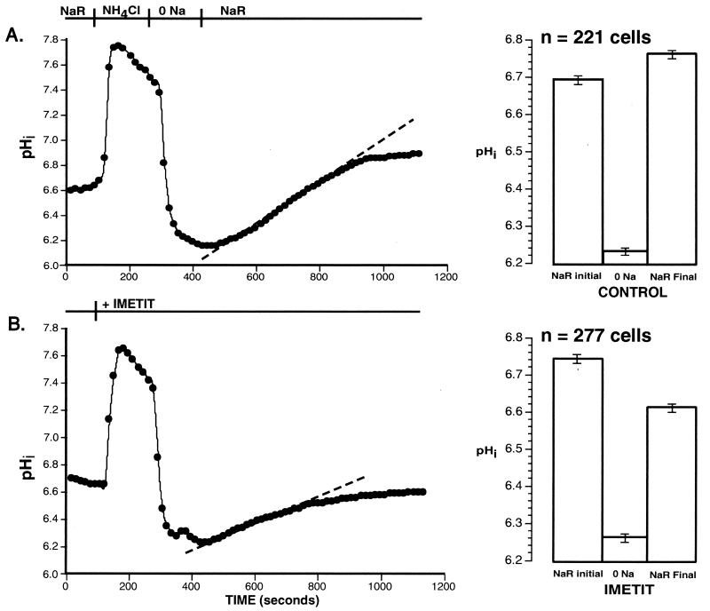
To test the hypothesis that H3R activation diminishes NHE activity, we assayed NHE in individual SKNMC-H3 cells loaded with the pHi indicator, BCECF. The response of these cells to an acute acid load was assessed in the absence or presence of the selective H3R agonist imetit (19), either alone or in combination with the selective H3R antagonist thioperamide (25). Fig. 1 shows the pHi response to a pulse of NH4Cl in a control cell (A) and in a cell exposed to 100 nM imetit (B), and illustrates the protocol used for these experiments. The EC50 for imetit to activate the H3R is 2 nM (19). As indicated in the figure, cells were initially superfused with Ringer's solution (NaR). Acute acidosis was then induced in the cells by superfusion with 20 mM NH4Cl. In B, imetit was present in the superfusate from the NH4Cl pulse through the end of the experimental protocol. As shown in A and B, when NH4Cl was replaced with the Na+-free Hepes-buffered solution (0 Na), the pHi fell ≈0.45 pH units to pHi 6.2. In the absence of extracellular Na+, the pHi remained low. When the Na+-free superfusate solution was replaced with the Na+-Ringer solution, the pHi began to recover because of H+ extrusion by means of NHE. The rate (slope) of this Na+-dependent intracellular alkalinization, calculated from the point at which recovery started, as indicated by the dotted line in each trace, represents the NHE activity. As shown, the rate of Na+-dependent pHi recovery was markedly lower in the presence of imetit (B) than in control conditions (A) (0.05 vs. 0.09 pHi units/min). Overall, imetit significantly reduced the rate of Na+-dependent pHi recovery (0.048 ± 0.002, n = 277 cells vs. 0.074 ± 0.003 pHi units/min, n = 221 cells; P < 0.0001; see Figs. 2 and 3).
Figure 1.
Representative experimental traces from individual SKNMC-H3 cells showing the Na+-dependent pHi recovery after acute exposure to an NH4Cl acid pulse. (A) A control cell: The y axis represents the pHi as determined from the intracellular calibration of the dye in this cell. The cell was initially superfused with Na+-Ringer's solution (NaR), then with 20 mM NH4Cl. Acute exposure to NH4Cl resulted in acidification of the cytosol to ≈pHi 6.2 after its removal. In the absence of extracellular Na (0 Na), there was no measurable pHi recovery. With the reintroduction of extracellular Na+ (NaR), pHi increased with intracellular alkalinization occurring at a rate of 0.094 pHi units/min (dotted line). The final pHi, achieved in the cell shown in this trace, was higher than the starting pHi (6.8 vs. 6.6). This overshoot of the final pHi relative to the initial resting pHi was seen in the majority of control cells studied and is shown in the bar graph accompanying A. For the 221 control cells studied, the initial pHi was 6.69 ± 0.01 and the final pHi was significantly higher at 6.76 ± 0.01 (P < 0.001). (B) Effect of imetit: The y axis represents pHi as determined from the intracellular calibration of the dye in this cell. Imetit (100 nM) was present in the superfusate from the addition of NH4Cl until the end of the protocol as shown. Acute exposure to NH4Cl resulted in acidification of the cytosol to ≈6.2 after its removal. In the absence of extracellular Na+ (0 Na), there was no measurable pHi recovery. With the reintroduction of extracellular Na+ (NaR), pHi started to increase with intracellular alkalinization occurring at a rate of 0.050 pHi units/min. The mean rate of Na+-dependent pHi recovery was significantly less than that observed in the control cells (P < 0.0001) (Figs. 2 and 3). The final pHi achieved in the cell shown in this trace was lower than the starting pHi (6.7 vs. 6.5). This pHi undershoot relative to the starting pHi was seen in the majority of cells exposed to imetit, as shown in the bar graph of B. Unlike control cells, the final pHi of the 277 imetit-treated cells was significantly lower than the initial pHi (6.61 ± 0.01 vs. 6.74 ± 0.01; P < 0.0001).
Figure 2.
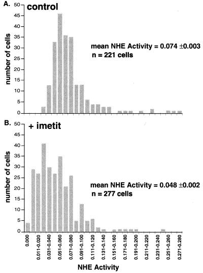
Histogram showing variability of Na+-dependent pHi recovery rates (NHE activity) in control (A) and imetit-treated SKNMC-H3 cells (B). The ordinates represent the number of cells and the abscissae the Na+-dependent pHi recovery rates binned in 0.09 pHi unit/min increments. A illustrates the variability of the response of 221 control cells studied from five coverslips, and B, the response of 277 cells from six coverslips exposed to imetit (100 nM).
Figure 3.
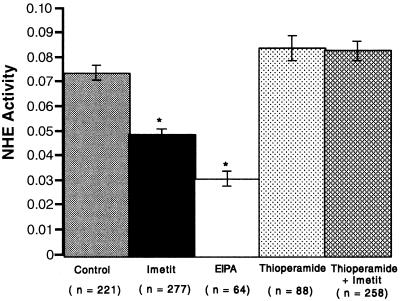
Comparison of NHE activity in the absence and presence of imetit, EIPA, thioperamide, and imetit plus thioperamide in SKNMC-H3 cells. The mean control Na+-dependent pHi recovery rate or NHE activity (pHi units/min) is compared with rates measured in cells exposed to imetit (100 nM), EIPA (10 μM), thioperamide (300 nM), and thioperamide in combination with imetit. *, Significantly different (P < 0.0001) from control NHE activity. Values are means ± SEM, and n refers to the number of cells studied. Imetit, thioperamide, and EIPA were present in the superfusate from the NH4Cl pulse to the end of the experiment. In the experiments with imetit and thioperamide in combination, thioperamide was first introduced in the NH4Cl solution, and, 5 min later, imetit was added to the Na+-free solution, for an additional 5 min, before the reintroduction of Na+ into the superfusate.
In addition, imetit not only slowed the rate of NHE activity, but prevented the full recovery of pHi. This is shown in the bar graphs of Fig. 1, where, in the presence of imetit, the final pHi reached after reintroduction of Na+ was significantly lower (6.61 ± 0.01, n = 277 cells) than the initial pHi (6.74 ± 0.01, n = 277 cells) (P < 0.0001). In contrast, in the control cells, the final pHi was significantly higher (6.76 ± 0.01, n = 221 cells) than the initial pHi (6.69 ± 0.01, n = 221 cells) (P < 0.001). Notably, the pHi measured right before Na+ was reintroduced (0 Na pHi) was the same in control and imetit-treated cells (control 6.23 ± 0.01, n = 221 cells and imetit-treated 6.26 ± 0.01, n = 277 cells). Thus, the imetit-induced attenuation of NHE could not have resulted from a difference in pHi immediately preceding exchanger activation. Collectively, the results in Fig. 1 demonstrate that activation of H3R diminishes NHE activity and thus, the ability of the exchanger to extrude H+.
We next examined the variability in NHE activity of control and imetit-treated cultured cell populations. Fig. 2 is a histogram for the pHi recovery rates of individual cells (A, control; B, imetit-treated). The Na+-dependent recovery rates are binned in increments of 0.09 pHi units/min. All of the cells exhibited a Na+-dependent recovery response from the acid load. For the control cells (n = 221), the rate of recovery ranged from 0.025 to 0.275 pHi units/min, with a mean of 0.074 ± 0.002 pHi units/min. The recovery rate of imetit-treated cells (n = 277) ranged from 0 to 0.198 pHi units/min, with a mean of 0.048 ± 0.002 pHi units/min (P < 0.0001). This difference in the distribution of NHE activity is clearly reflected in the imetit-induced leftward shift of the histogram (compare A and B). Indeed, 77% of the 277 imetit-treated cells exhibited rates lower than the mean control rate of 0.074 pHi units/min.
Fig. 3 shows that the selective H3R antagonist thioperamide (300 nM) (KB 4 nM; ref. 27) did not significantly modify NHE activity but abolished the imetit-induced NHE attenuation. This suggests that the effect of imetit results specifically from H3R activation. Fig. 3 also shows that the amiloride analog EIPA (10 μM) reduced the rate of Na+-dependent intracellular alkalinization from 0.074 to 0.031 pHi units/min, confirming that the Na+-dependent pHi recovery from the acid load is due to NHE. EIPA was significantly more effective than imetit in inhibiting NHE activity (50% vs. 35% inhibition, P < 0.001).
Discussion
The results of this study demonstrate that H3R activation in human neuroblastoma cells, stably transfected with H3 cDNA, inhibits NHE activity. Cultured neuroblastoma cells are a model of cardiac adrenergic nerve endings (28). Thus, our results provide a link between H3R and NHE, which may limit the excessive release of NE during protracted myocardial ischemia. Indeed, we had found that H3R activation attenuates carrier-mediated NE release and alleviates reperfusion arrhythmias (9, 10).
Previous studies show that NHE activation plays a pivotal role in the Na+-dependent carrier-mediated NE release from adrenergic nerve endings that occurs during protracted myocardial ischemia (15). The ischemia-induced acidosis activates NHE (20), and the extrusion of H+ in exchange for extracellular Na+ leads to excessive accumulation of intracellular Na+. The elevated intracellular Na+, together with the increased free NE in the axoplasm, causes the NET to reverse its direction and release massive amounts of NE from cardiac adrenergic nerve endings (carrier-mediated NE release) (15).
We have shown that H3R activation is linked to a decrease in NHE activity (Fig. 1), and that the amiloride derivative EIPA markedly inhibits the rate of Na+-dependent intracellular alkalinization in SKNMC-H3 cells (Fig. 3). The latter result proves that the recovery from NH4Cl-induced acidosis is due to NHE activation (26). The selective H3R agonist imetit (19) inhibited the rate of recovery from an acid load, and the action of imetit was blocked by the selective H3R antagonist thioperamide (25). These results indicate that H3R activation triggers a decrease in NHE activity.
pHi plays an important role in modulating NHE activity (20) and differences in pHi could account for variability in the rate of Na+/H+ exchange. But the pHi measured in control and imetit-treated cells, when Na+ was reintroduced to the superfusate after the NH4Cl pulse, was similar for both (≈pHi 6.2, Fig. 1). This finding suggests that H3R activation modifies NHE activity by means of a mechanism other than a change in pHi. H3R activation also appeared to limit the pHi range over which NHE functions, because the Na+-dependent pHi recovery from the acid load was less complete in imetit-treated cells than in controls. Thus, once Na+ had been reintroduced to the superfusate and NHE activated, the final pHi reached in the cell was significantly lower in the imetit-treated cells than in controls (Fig. 1). The significance of this finding remains to be explored; but it may represent a compensatory effort by the cell to curb excess accumulation of intracellular Na+, which could translate into a potentially beneficial mechanism, because carrier-mediated NE release and intracellular acidosis both would be attenuated.
Fig. 2 provides additional evidence linking H3R activation to diminished NHE activity. Indeed, by plotting the Na+-dependent recovery rates in response to an acid load (NHE activity) against the number of cells displaying such a rate, it becomes evident that imetit-treated cells exhibit a markedly different distribution than control cells, with a marked shift toward lower NHE rates.
Our findings highlight the importance of presynaptic H3R as negative modulators of excessive NE release in protracted myocardial ischemia and their coupling to neuronal NHE. Because of their very high affinity for histamine (KD = 5 nM), in comparison with the much lower affinity of H1R and H2R (KD ≈ 10 μM) (27), H3R are easily activated in myocardial ischemia by histamine released from mast cells juxtaposed to adrenergic nerve endings (15). Thus, our findings advocate a protective role for endogenous cardiac histamine in myocardial ischemia.
Ischemia-induced acidosis activates NHE in cardiac myocytes and NHE inhibitors are known to provide beneficial anti-ischemic effects, such as reduction in infarct size and reperfusion arrhythmias (29–31). This cardioprotection has been attributed to the ability of NHE inhibitors to prevent Ca2+ overload in cardiomyocytes. Indeed, NHE blockade will limit the influx of Na+, so that less intracellular Na+ will be available to the Na+-Ca2+ exchanger and intracellular Ca2+ will fail to accumulate (29). In contrast, our previous (9, 10) and present findings uncover a novel mechanism of cardioprotection: NHE inhibition in cardiac adrenergic neurons as a means to prevent ischemic arrhythmias associated with carrier-mediated NE release. Inasmuch as sympathetic overactivity in myocardial ischemia is associated with severe arrhythmias and sudden cardiac death (1, 32–34), our findings provide a rationale for the use of selective H3R agonists to alleviate dysfunctions associated with myocardial ischemia.
Acknowledgments
We thank Kumar Poonwasi for his technical help in some of the experiments. This work was supported by Grants HL 34215, HL46403, and DK-45828 from the National Institutes of Health and by the Underhill and Wild Wings Foundations.
Abbreviations
- NE
norepinephrine
- NET
norepinephrine transporter
- NHE
Na+/H+ exchanger
- BCECF
2′,7′-bis(carboxyethyl)-5(6)-carboxyfluorescein ester
- EIPA
5-(N-ethyl-N-isopropyl)amiloride
- H3R
histamine H3 receptors
- pHi
intracellular pH
- SKNMC-H3 cells
human SKNMC neuroblastoma cells stably transfected with H3R cDNA
Footnotes
This paper was submitted directly (Track II) to the PNAS office.
References
- 1.Schömig A, Richardt G, Kurz T. Herz. 1995;20:169–186. [PubMed] [Google Scholar]
- 2.Braunwald E, Sobel B E. In: Heart Disease: A Textbook of Cardiovascular Medicine. Braunwald E, editor. Philadelphia: Saunders; 1988. pp. 1191–1221. [Google Scholar]
- 3.Airaksinen K E. Ann Med. 1999;31:240–245. doi: 10.3109/07853899908995886. [DOI] [PubMed] [Google Scholar]
- 4.Amara S G, Kuhar M J. Annu Rev Neurosci. 1993;16:73–93. doi: 10.1146/annurev.ne.16.030193.000445. [DOI] [PubMed] [Google Scholar]
- 5.Rudnick G, Clark J. Biochim Biophys Acta. 1993;1144:249–263. doi: 10.1016/0005-2728(93)90109-s. [DOI] [PubMed] [Google Scholar]
- 6.Schömig A, Haass M, Richardt G. Eur Heart J. 1991;12, Suppl. F:38–47. doi: 10.1093/eurheartj/12.suppl_f.38. [DOI] [PubMed] [Google Scholar]
- 7.Dart A M, Du X-J. Cardiovasc Res. 1993;27:906–914. doi: 10.1093/cvr/27.6.906. [DOI] [PubMed] [Google Scholar]
- 8.Smith N C E, Levi R. J Pharmacol Exp Ther. 1999;291:456–463. [PubMed] [Google Scholar]
- 9.Imamura M, Lander H M, Levi R. Circ Res. 1996;78:475–481. doi: 10.1161/01.res.78.3.475. [DOI] [PubMed] [Google Scholar]
- 10.Hatta E, Yasuda K, Levi R. J Pharmacol Exp Ther. 1997;283:494–500. [PubMed] [Google Scholar]
- 11.Arrang J M, Garbarg M, Schwartz J C. Nature (London) 1983;302:832–837. doi: 10.1038/302832a0. [DOI] [PubMed] [Google Scholar]
- 12.Endou M, Poli E, Levi R. J Pharmacol Exp Ther. 1994;269:221–229. [PubMed] [Google Scholar]
- 13.Imamura M, Poli E, Omoniyi A T, Levi R. J Pharmacol Exp Ther. 1994;271:1259–1266. [PubMed] [Google Scholar]
- 14.Imamura M, Seyedi N, Lander H M, Levi R. Circ Res. 1995;77:206–210. doi: 10.1161/01.res.77.1.206. [DOI] [PubMed] [Google Scholar]
- 15.Levi R, Smith N C E. J Pharmacol Exp Ther. 2000;292:825–830. [PubMed] [Google Scholar]
- 16.Lovenberg T W, Roland B L, Wilson S J, Jiang X, Pyati J, Huvar A, Jackson M R, Erlander M G. Mol Pharmacol. 1999;55:1101–1107. [PubMed] [Google Scholar]
- 17.Lovenberg T W, Pyati J, Chang H, Wilson S J, Erlander M G. J Pharmacol Exp Ther. 2000;293:771–778. [PubMed] [Google Scholar]
- 18.Tardivel-Lacombe J, Rouleau A, Héron A, Morisset S, Pillot C, Cochois V, Schwartz J C, Arrang J M. NeuroReport. 2000;11:755–759. doi: 10.1097/00001756-200003200-00020. [DOI] [PubMed] [Google Scholar]
- 19.Garbarg M, Arrang J M, Rouleau A, Ligneau X, Tuong M D, Schwartz J C, Ganellin C R. J Pharmacol Exp Ther. 1992;263:304–310. [PubMed] [Google Scholar]
- 20.Aronson P S, Nee J, Suhm M A. Nature (London) 1982;299:161–163. doi: 10.1038/299161a0. [DOI] [PubMed] [Google Scholar]
- 21.Cardone M H, Smith B L, Mennitt P A, Mochly-Rosen D, Silver R B, Mostov K E. J Cell Biol. 1996;133:997–1005. doi: 10.1083/jcb.133.5.997. [DOI] [PMC free article] [PubMed] [Google Scholar]
- 22.Silver R B. Methods Cell Biol. 1998;56:237–251. doi: 10.1016/s0091-679x(08)60429-x. [DOI] [PubMed] [Google Scholar]
- 23.Thomas J A, Buchsbaum R N, Zimniak A, Racker E. Biochemistry. 1979;18:2210–2218. doi: 10.1021/bi00578a012. [DOI] [PubMed] [Google Scholar]
- 24.Silver R B, Choe H, Frindt G. Am J Physiol. 1998;275:F94–F102. doi: 10.1152/ajprenal.1998.275.1.F94. [DOI] [PubMed] [Google Scholar]
- 25.Arrang J M, Garbarg M, Lancelot J C, Lecomte J M, Pollard H, Robba M, Schunack W, Schwartz J C. Nature (London) 1987;327:117–123. doi: 10.1038/327117a0. [DOI] [PubMed] [Google Scholar]
- 26.Vigne P, Frelin C, Cragoe E J, Jr, Lazdunski M. Biochem Biophys Res Commun. 1983;116:86–90. doi: 10.1016/0006-291x(83)90384-4. [DOI] [PubMed] [Google Scholar]
- 27.Hill S J, Ganellin C R, Timmerman H, Schwartz J C, Shankley N P, Young J M, Schunack W, Levi R, Haas H L. Pharmacol Rev. 1997;49:253–278. [PubMed] [Google Scholar]
- 28.Vaughan P F, Peers C, Walker J H. Gen Pharmacol. 1995;26:1191–1201. doi: 10.1016/0306-3623(94)00312-b. [DOI] [PubMed] [Google Scholar]
- 29.Karmazyn M, Gan X H T, Humphreys R A, Yoshida H, Kusumoto K. Circ Res. 1999;85:777–786. doi: 10.1161/01.res.85.9.777. [DOI] [PubMed] [Google Scholar]
- 30.Théroux P. Circulation. 2000;101:2874–2876. doi: 10.1161/01.cir.101.25.2874. [DOI] [PubMed] [Google Scholar]
- 31.Gumina R J, Daemmgen J, Gross G J. Eur J Pharmacol. 2000;396:119–124. doi: 10.1016/s0014-2999(00)00200-4. [DOI] [PubMed] [Google Scholar]
- 32.Podrid P J, Fuchs T, Candinas R. Circulation. 1990;82:I103–I113. [PubMed] [Google Scholar]
- 33.Barron H V, Lesh M D. J Am Coll Cardiol. 1996;27:1053–1060. doi: 10.1016/0735-1097(95)00615-X. [DOI] [PubMed] [Google Scholar]
- 34.Kennedy H L. Am J Cardiol. 1997;80:29J–34J. doi: 10.1016/s0002-9149(97)00836-9. [DOI] [PubMed] [Google Scholar]


