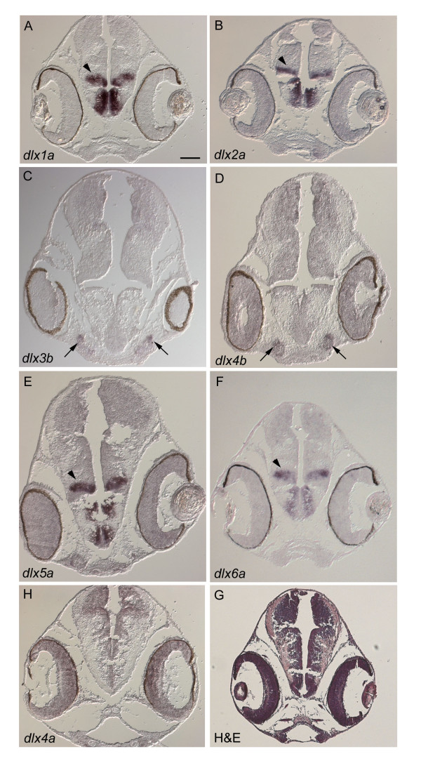
Figure 3.
A. burtoni dlx expression patterns in the brain. In situ hybridization on transverse sections at 7 dpf (A to G). (H) Hematoxylin-eosin staining. dlx1a (A), dlx2a (B), dlx5a (E) and dlx6a (F) show similar expression patterns in the diencephalon of the forebrain (arrowheads). dlx3b (C) and dlx4b (D) also share expression signals in the region where the olfactory placodes are developing (arrows). No expression is seen for dlx4a (G). Scale bar in A: 100 μm. Anteroposterior levels of these photos are indicated in Additional file 9. Note that pigmentation persists in the eye.

