Abstract
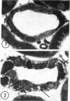
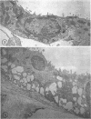
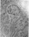
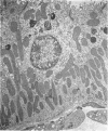
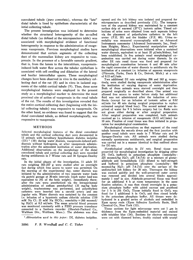
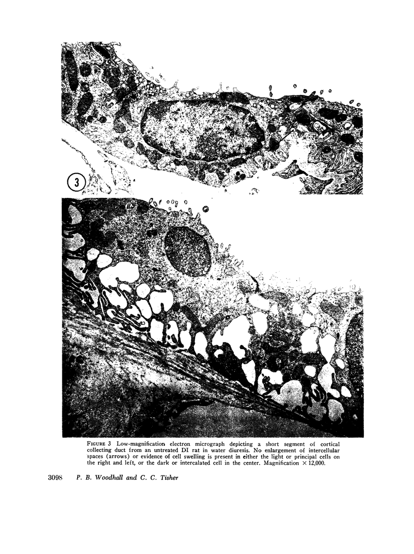
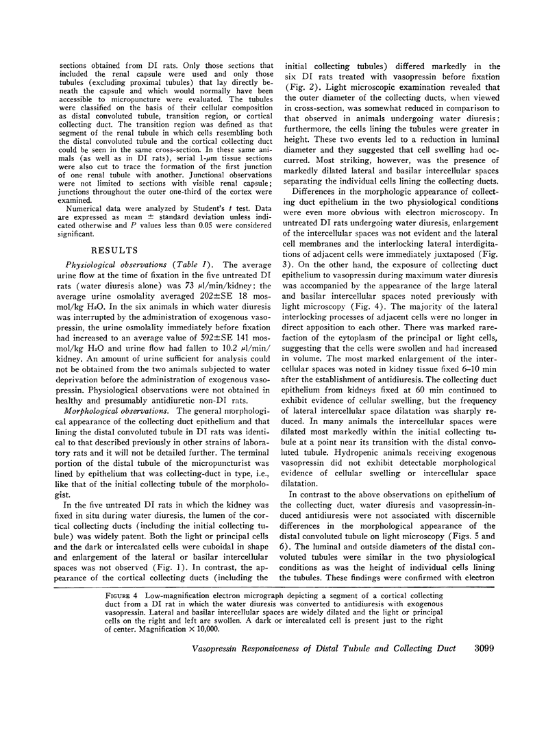
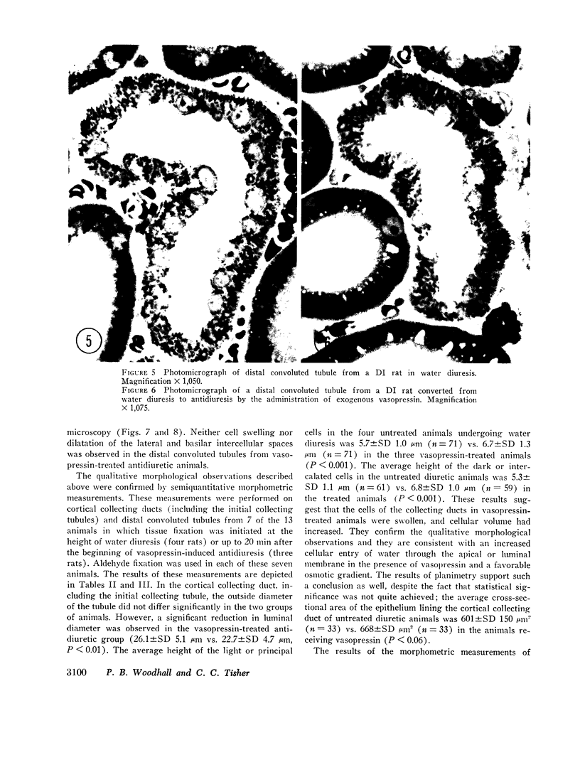
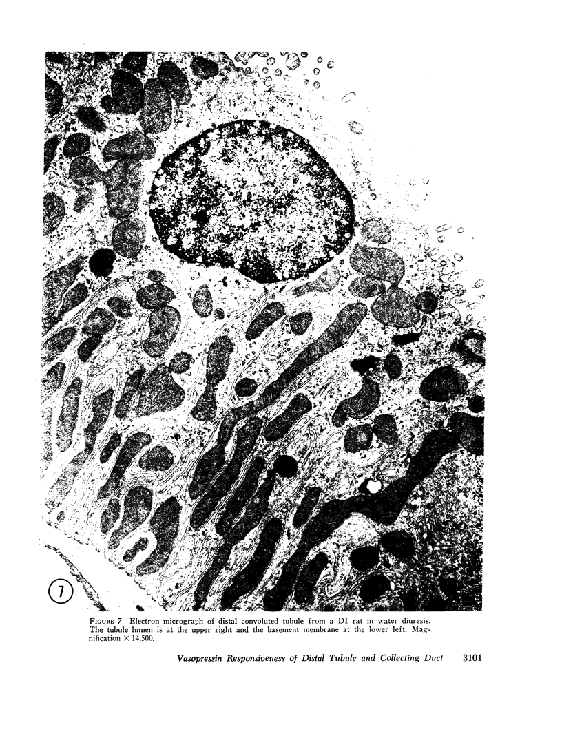
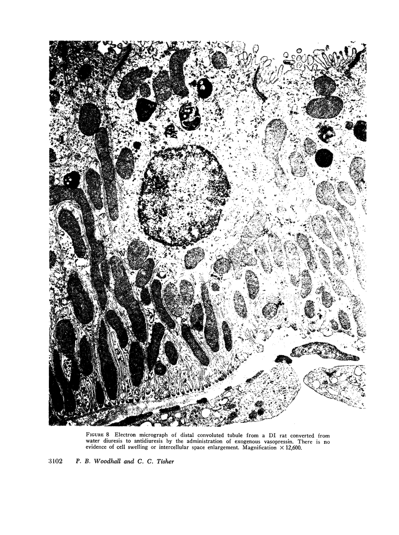
Renal micropuncture observations in the rat suggest that the entire “distal tubule” (defined by the micropuncturist as that portion of the renal tubule extending between the macula densa and its first junction with another (renal tubule) may be responsive to vasopressin. However, this portion of the renal tubule contains two segments that are morphologically dissimilar. The “early” distal tubule is lined by epithelium characteristic of the distal convoluted tubule, while the “late” distal tubule is lined by epithelium characteristic of the cortical collecting duct. Thus, the present study was initiated to identify the most proximal site of action of vasopressin in the distal renal tubule. A water diuresis was established in rats with hereditary hypothalamic diabetes insipidus. In one-half of the animals the diuresis was interupted by an i.v. infusion of exogenous vasopressin. Morphological preservation of the kidneys was initiated after induction of vasopressin-induced antidiuresis or during maximum water diuresis. Cell swelling and dilatation of intercellular spaces, morphological findings indicative of vasopressin responsiveness, were observed in the cortical collecting duct including the late segment of the distal tubule, a segment that has also been described by morphologists as the initial collecting tubule. Morphological evidence of vasopressin-responsiveness was not observed in the early distal tubule (distal convoluted tubule). Additional morphological studies in Wistar, Long-Evans, and Sprague-Dawley rats demonstrated a marked difference in the random availability of distal convoluted tubules versus initial collecting tubules potentially available for micropuncture just beneath the renal capsule. The results suggest that hypotonic tubular fluid entering the early distal tubule (distal convoluted tubule) remains hypotonic to plasma until it enters the late distal tubule (initial collecting tubule) and that vasopressin-induced osmotic equilibration is a function of the latter segment alone. The findings emphasize the importance of morphological characterization of those segments of the renal tubule that are subjected to physiological investigation.
Full text
PDF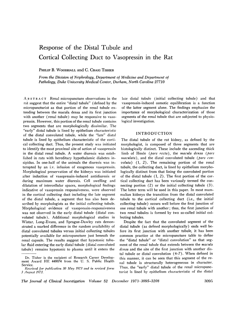
Images in this article
Selected References
These references are in PubMed. This may not be the complete list of references from this article.
- Bennett C. M., Brenner B. M., Berliner R. W. Micropuncture study of nephron function in the rhesus monkey. J Clin Invest. 1968 Jan;47(1):203–216. doi: 10.1172/JCI105710. [DOI] [PMC free article] [PubMed] [Google Scholar]
- Clapp J. R., Robinson R. R. Osmolality of distal tubular fluid in the dog. J Clin Invest. 1966 Dec;45(12):1847–1853. doi: 10.1172/JCI105488. [DOI] [PMC free article] [PubMed] [Google Scholar]
- Colindres R. E., Lechene C. Technical problems associated with collection of distal tubular fluid in the rat. Yale J Biol Med. 1972 Jun-Aug;45(3-4):233–239. [PMC free article] [PubMed] [Google Scholar]
- GOTTSCHALK C. W., MYLLE M. Micropuncture study of the mammalian urinary concentrating mechanism: evidence for the countercurrent hypothesis. Am J Physiol. 1959 Apr;196(4):927–936. doi: 10.1152/ajplegacy.1959.196.4.927. [DOI] [PubMed] [Google Scholar]
- Ganote C. E., Grantham J. J., Moses H. L., Burg M. B., Orloff J. Ultrastructural studies of vasopressin effect on isolated perfused renal collecting tubules of the rabbit. J Cell Biol. 1968 Feb;36(2):355–367. doi: 10.1083/jcb.36.2.355. [DOI] [PMC free article] [PubMed] [Google Scholar]
- LASSITER W. E., GOTTSCHALK C. W., MYLLE M. Micropuncture study of net transtubular movement of water and urea in nondiuretic mammalian kidney. Am J Physiol. 1961 Jun;200:1139–1147. doi: 10.1152/ajplegacy.1961.200.6.1139. [DOI] [PubMed] [Google Scholar]
- LUFT J. H. Improvements in epoxy resin embedding methods. J Biophys Biochem Cytol. 1961 Feb;9:409–414. doi: 10.1083/jcb.9.2.409. [DOI] [PMC free article] [PubMed] [Google Scholar]
- Lechène C., Morel F., Guinnebault M., De Rouffignac C. Etude par microponction de l'élaboration de l'urine. I. Chez le rat dans différents états de diurèse. Nephron. 1969;6(4):457–477. doi: 10.1159/000179745. [DOI] [PubMed] [Google Scholar]
- REYNOLDS E. S. The use of lead citrate at high pH as an electron-opaque stain in electron microscopy. J Cell Biol. 1963 Apr;17:208–212. doi: 10.1083/jcb.17.1.208. [DOI] [PMC free article] [PubMed] [Google Scholar]
- Schnermann J., Valtin H., Thurau K., Nagel W., Horster M., Fischbach H., Wahl M., Liebau G. Micropuncture studies on the influence of antidiuretic hormone on tubular fluid reabsorption in rats with hereditary diabetes insipidus. Pflugers Arch. 1969;306(2):103–118. doi: 10.1007/BF00586878. [DOI] [PubMed] [Google Scholar]
- TRUMP B. F., SMUCKLER E. A., BENDITT E. P. A method for staining epoxy sections for light microscopy. J Ultrastruct Res. 1961 Aug;5:343–348. doi: 10.1016/s0022-5320(61)80011-7. [DOI] [PubMed] [Google Scholar]
- Tisher C. C., Bulger R. E., Valtin H. Morphology of renal medulla in water diuresis and vasopressin-induced antidiuresis. Am J Physiol. 1971 Jan;220(1):87–94. doi: 10.1152/ajplegacy.1971.220.1.87. [DOI] [PubMed] [Google Scholar]
- Tisher C. C., Cirksena W. J., Arstila A. U., Trump B. F. Subcellular localization of sodium in normal and injured proximal tubules of the rat kidney. Am J Pathol. 1969 Nov;57(2):231–251. [PMC free article] [PubMed] [Google Scholar]
- Tisher C. C., Clapp J. R. Intraluminal latex injection: an aid to the histological identification of renal tubules. Kidney Int. 1972 Jul;2(1):54–56. doi: 10.1038/ki.1972.69. [DOI] [PubMed] [Google Scholar]
- ULLRICH K. J., RUMRICH G., FUCHS G. WASSERPERMEABILITAET UND TRANDTUBULAERER WASSERFLUSS CORTICALER NEPHRONABSCHNITTE BEI VERSCHIEDENEN DIURESEZUSTAENDEN. Pflugers Arch Gesamte Physiol Menschen Tiere. 1964 Jul 1;280:99–119. [PubMed] [Google Scholar]
- ULLRICH K. J., SCHMIDT-NIELSON B., O'DELL R., PEHLING G., GOTTSCHALK C. W., LASSITER W. E., MYLLE M. Micropuncture study of composition of proximal and distal tubular fluid in rat kidney. Am J Physiol. 1963 Apr;204:527–531. doi: 10.1152/ajplegacy.1963.204.4.527. [DOI] [PubMed] [Google Scholar]
- Valtin H. Hereditary hypothalamic diabetes insipidus in rats (Brattleboro strain). A useful experimental model. Am J Med. 1967 May;42(5):814–827. doi: 10.1016/0002-9343(67)90098-8. [DOI] [PubMed] [Google Scholar]
- WATSON M. L. Staining of tissue sections for electron microscopy with heavy metals. J Biophys Biochem Cytol. 1958 Jul 25;4(4):475–478. doi: 10.1083/jcb.4.4.475. [DOI] [PMC free article] [PubMed] [Google Scholar]


