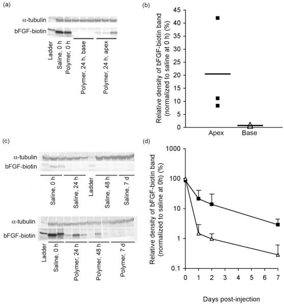
Figure 3.
Quantification of bFGF-biotin retention by western blot analysis following injection into infarcted rat myocardium. Localization of bFGF to apex but not base half of heart at 24 h demonstrated by (a) western blot and (b) densitometry results. Quantification of bFGF release kinetics demonstrated by (c) western blot and (d) densitometry results. Heart samples were harvested at 0 h, 24 h, 48 h, or 7 days post-injection then homogenized to facilitate detection of bFGF-biotin by western blot. Samples injected with either phosphate buffered saline (PBS) (△in panels b and d) or polymer (■in panels b and d) (28 kDa, 5 w/v % in PBS) as the delivery vehicle. Each 40 μL injection contained 2.5 μg of bFGF-biotin. Values reported as mean±SD are normalized to α-tubulin and to bFGF band density of saline control at 0 h, n=3 for each time point.

