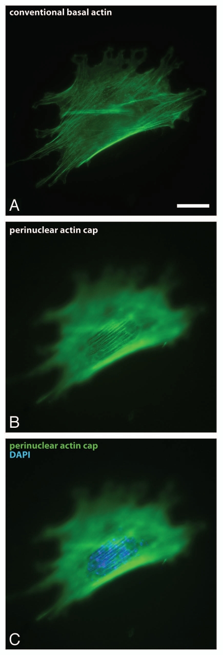
Figure 1.
The perinuclear actin cap. (A–C) Fluorescence micrographs showing conventional basal stress fibers confined to the basal surface of the cell (A) and the perinuclear actin cap located at the apical surface of the nucleus (B and C). F-actin was stained using phalloidin (red); nuclear DNA was stained using DAPI (blue). Scale bar, 20 µm.

