Abstract
Spinal epidural haematoma (SEH) is a rare complication of haemophilia. A 3-month-old boy presenting with non-traumatic acute onset quadriparesis was found to have SEH on MRI scan. On further investigations, diagnosis of severe haemophilia A was confirmed. He responded well to conservative treatment with replacement of factor VIII without any need for surgical decompression. Neurological recovery was complete. We believe this is the youngest reported case of haemophilia presenting with spontaneous SEH. Another peculiarity of this case is absence of excessive bleeding due to forceps and vacuum application, circumcision and intramuscular injection, even in the presence of severe haemophilia. This case calls attention to the clinical features, radiological appearances and management options of this rare complication of SEH in people with haemophilia.
Background
Spinal epidural haematoma (SEH) is a rare complication of haemophilia. We believe this is the youngest reported case of haemophilia presenting with spontaneous SEH. Another peculiarity of this case is absence of excessive bleeding due to forceps and vacuum application, circumcision and intramuscular injection, even in the presence of severe haemophilia.
Case presentation
A 3-month-old boy, born out of a non-consanguineous marriage, presented with a history of decreased upper and lower limb movements for the past 3 days. He had not passed faeces for 3 days and had dribbling micturition. There was no history of trauma, fever, vomiting, altered sensorium, convulsions and bleeding tendencies. Perinatal history revealed that it was a difficult delivery done at a private nursing home in a remote village. There was a history of prolonged labour and the boy was delivered by forceps due to failure of vacuum extraction. His birth weight was 3.5 kg. The boy had caput at birth, which resolved spontaneously in 3 days. He was exclusively breast fed from day 1 and had received intramuscular vitamin K at birth. There was no history of neonatal jaundice or neonatal encephalopathy. There was no history of bleeding from the umbilical cord. He underwent circumcision at home for religious reasons when he was 7 days old, but there was no history of excessive bleeding. The boy was vaccinated for BCG and oral polio on the 7th day and for DPT, hepatitis B and oral polio at 6 weeks. There was no history suggestive of ecchymosis or haematoma at vaccination site. There was no family history of bleeding tendencies.
Investigations
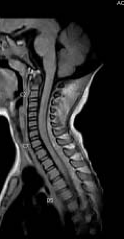
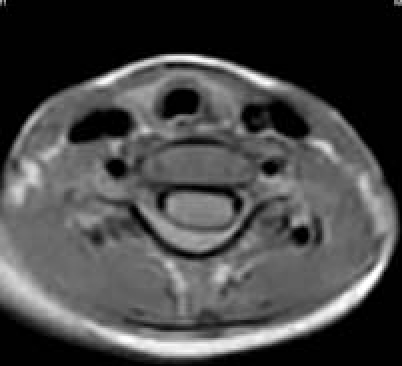
On examination, the boy was afebrile with normal anterior fontanelle and absence of neck stiffness. His vital signs were normal. He was accepting breast feeds. There were no bruises or telangiectasia. Spine examination was normal. On central nervous system (CNS) examination, higher functions, cranial nerves and social smile was normal. He had flaccid quadriparesis with exaggerated deep tendon reflexes. Sensations in all four limbs were diminished, plantars were not elicitable and superficial reflexes (cremasteric and anal) were absent. Abdominal examination revealed distended bladder up to the umbilicus. The rest of the examination was normal. A clinical diagnosis of cervical myelopathy was made. His complete blood counts, including platelet count, C reactive protein, serum calcium, sodium, potassium, hepatic transaminases, blood urea, serum creatinine and routine urine, analysis were normal. His cranial ultrasound and cervical spine radiographs were done and were normal. Examination of cerebrospinal fluid (obtained by lumber puncture) was also normal. His cervical spine MRI revealed posterior acute epidural haematoma extending from C2 to visualised D5 vertebra with maximum thickness of haematoma being 6 mm significantly displacing the spinal cord anteriorly. (figure 1). Coagulation profile revealed bleeding time of 4 min (normal 3–9 min), clotting time of 14 min (normal 3–7 min), prothrombin time of 11 s (control 11 s) and partial thromboplastin time of 75 s (control 26 s), which normalised after adding normal plasma (1:1) and incubating the mixture for 1 h.
Figure 1.


Cervical spine MRI images: (A) saggital and (B) transverse.
Differential diagnosis
Differential diagnoses of traumatic, vascular and infectious aetiologies were made.
Treatment
Samples for factor VIII and IX assay were sent and the patient was given cryoprecipitate. Factor VIII level was <1% of normal. Recombinant factor VIII concentrate was started with the initial dose of 50 U/kg followed by 30 U/kg twice daily for 7 days and 15 U/kg twice daily for the next 7 days. Thereafter, he was advised prophylactic factor VIII treatment for 6 months in the dose of 30 U/kg three times weekly.
He started showing neurological recovery from the 4th day of treatment with factor VIII. At the end of 3 weeks, his neurological examination was normal with good bladder and bowel control.
Outcome and follow-up
Complete recovery and normal examination on follow-up.
Discussion
Haemophilia A (factor VIII deficiency) and haemophilia B (factor IX deficiency) are hereditary bleeding disorders transmitted in an X-linked manner. Severity of haemophilia is classified on the basis of patient's baseline factor level. Factor level <1% of normal is severe, between 1% and 5% is moderate and >5% is mild haemophilia.1 The majority of patients with haemophilia are diagnosed at birth due to family history, although one-third of patients with haemophilia represent a new mutation,2 as in our case. About 1–2% of neonates with haemophilia may develop spontaneous intracranial haemorrhage (ICH), while massive ICH occurs in cases of difficult deliveries;3 hence, vacuum extraction and forceps delivery are to be avoided. Surprisingly, our patient with severe haemophilia developed no clinical evidence of ICH despite multiple adverse factors like prolonged labour, presence of caput, failed vacuum extraction and forceps delivery. Even circumcision on day 7 and intramuscular injection at birth and at 6 weeks did not lead to excessive bleeding or intramuscular haematoma. The stress of delivery and other neonatal problems may transiently elevate factor VIII levels into the normal or near normal range1 and this could possibly explain the absence of intracranial bleeding, intramuscular haematoma and bleeding from circumcision in this case.
CNS bleeding is the most dreaded life-threatening haemorrhage seen in people with haemophilia. Incidence of CNS bleeding in patients with haemophilia is variously quoted as 2.2–7.8%.4 In a series of 2500 patients with haemophilia over a period of 11 years, the prevalence rate of CNS bleeding for factor VIII and factor IX deficiency was 2.7% and 3.6%, respectively.5 Out of 71 patients in this series who had CNS bleeding, 65 had intracranial while only 6 (8.4%) had intraspinal bleeding. History of trauma to the spine was present in five patients and, hence, occurrence of spontaneous intraspinal bleeding is an extremely rare event in haemophiliacs. Intraspinal bleed could be intramedullary, subdural or epidural (ie, extradural). Most of the cases reported have been in children under 5 years of age;6 the youngest being 3-month-old7 in whom SEH occurred post-lumbar puncture. Hence, we feel our case is the youngest reported spontaneous, non-traumatic, SEH in haemophiliacs. Although older children present with variety of signs and symptoms, like acute onset neck and back pain, walking impairment with urinary retention and acute transverse myelitis, initial symptoms in infants can be non-specific leading to a diagnostic dilemma. Infants can present with irritability without a focus, torticollis or respiratory distress.6 8 Our case had none of these signs and symptoms but presented with quadriparesis along with bladder and bowel involvement.
MRI scan is the modality of choice for the early diagnosis of this potentially reversible cause of spinal cord compression. Epidural haematomas are more commonly posterior to spinal cord than anterior,9 as in our case. Early conservative management with factor replacement is safe and effective leading to complete neurological recovery.10 11 12 Our case also demonstrated this in 3 weeks with factor VIII replacement. As CNS haemorrhages may occur without known trauma and early signs and symptoms can be minimal and non-specific, replacement treatment should be initiated even before radiological evaluation. Treatment of CNS haemorrhage involves replacement treatment to achieve a level of 100 U/dl for factor VIII and 80 U/dl for factor IX, maintenance of adequate haemostatic level (>50 U/dl) for a minimum of 14 days and a more prolonged period of prophylactic treatment for an additional 2–3 weeks or longer to ensure resolution of the underlying event.1 Factor VIII dose in units can be calculated by using the formula: factor VIII in units=U/dl desired rise×body weight in kg×0.5.
Patients with CNS bleed have a risk of recurrence for approximately 6 months after an episode;, hence, these patients often continue to receive prophylactic factor VIII treatment for this period in the dose of 20–40 U/kg alternate day or three times a week. Surgical decompression is considered only in selected patients with progressive symptoms, failure of symptoms to resolve with conservative treatment and delayed diagnosis and treatment.13 14
Learning points.
-
▶
SEH is a rare complication of haemophilia.
-
▶
Excessive bleeding due to forceps and vacuum application, circumcision and intramuscular injection can be absent even in the presence of severe haemophilia.
-
▶
Initial symptoms of SEH in infants can be non-specific leading to diagnostic dilemma and MRI scan is the modality of choice for the early diagnosis of this potentially reversible cause of spinal cord compression.
-
▶
Early conservative management with factor replacement is safe and effective leading to complete neurological recovery, and surgical decompression is considered only in selected patients with progressive symptoms, failure of symptoms to resolve with conservative treatment and delayed diagnosis and treatment.
Footnotes
Competing interests None.
Patient consent Obtained.
References
- 1.Montgomery RR, Gill JC, Paola JD. Hemophilia and von Willebrand disease. In: Orkin SH, Nathan DG, Ginsburg D, eds. Nathan and Oski's Hematology of Infancy and Childhood. Seventh edition Philadelphia, PA: Saunders Elsevier; 2009:1487–524 [Google Scholar]
- 2.Ljung R, Petrini P, Nilsson IM. Diagnostic symptoms of severe and moderate haemophilia A and B. A survey of 140 cases. Acta Paediatr Scand 1990;79:196–200 [DOI] [PubMed] [Google Scholar]
- 3.Lynn MM, Patricia M, James D. Intracranial hemorrhage in neonates with unrecognised hemophilia A: A Persistent Problem. Pediatr Neurosurg 2001;34:94–7 [DOI] [PubMed] [Google Scholar]
- 4.Silverstein A. Intracranial bleeding in hemophilia. Arch Neurol 1960;3:141–57 [DOI] [PubMed] [Google Scholar]
- 5.Eyster ME, Gill FM, Blatt PM, et al. Central nervous system bleeding in hemophiliacs. Blood 1978;51:1179–88 [PubMed] [Google Scholar]
- 6.Hutt PJ, Herold ED, Koenig BM, et al. Spinal extradural hematoma in an infant with hemophilia A: an unusual presentation of a rare complication. J Pediatr 1996;128:704–6 [DOI] [PubMed] [Google Scholar]
- 7.Faillace WJ, Warrier I, Canady AI. Paraplegia after lumbar puncture. In an infant with previously undiagnosed hemophilia A. Treatment and peri-operative considerations. Clin Pediatr (Phila) 1989;28:136–8 [DOI] [PubMed] [Google Scholar]
- 8.Cuvelier GD, Davis JH, Purves EC, et al. Torticollis as a sign of cervico-thoracic epidural haematoma in an infant with severe haemophilia A. Haemophilia 2006;12:683–6 [DOI] [PubMed] [Google Scholar]
- 9.Patel H, Boaz JC, Phillips JP, et al. Spontaneous spinal epidural hematoma in children. Pediatr Neurol 1998;19:302–7 [DOI] [PubMed] [Google Scholar]
- 10.Iwamuro H, Morita A, Kawaguchi H, et al. Resolution of spinal epidural haematoma without surgery in a haemophilic infant. Acta Neurochir (Wien) 2004;146:1263–5 [DOI] [PubMed] [Google Scholar]
- 11.Balkan C, Kavakli K, Karapinar D. Spinal epidural haematoma in a patient with haemophilia B. Haemophilia 2006;12:437–40 [DOI] [PubMed] [Google Scholar]
- 12.Tetsuo K, Yuji M. Spinal extradural haematoma due to haemophilia A. Arch Dis Child 2007;92:498. [DOI] [PMC free article] [PubMed] [Google Scholar]
- 13.Rois PV, López MR, de Vergara BC, et al. Spinal epidural hematoma in hemophilic children: controversies in management. Childs Nerv Syst 2009;25:987–91; discussion 993, 995 [DOI] [PubMed] [Google Scholar]
- 14.Lee JS, Yu CY, Huang KC, et al. Spontaneous spinal epidural hematoma in a 4-month-old infant. Spinal Cord 2007;45:586–90 [DOI] [PubMed] [Google Scholar]


