Abstract
Adenomyoepitheliomas are uncommon breast tumours. By definition they have a prominent component of myoepithelial cells, in addition to glandular elements lined by epithelial cells. Malignant adenomyoepithelioma of the breast is even more rare, characterised by malignant proliferation of epithelial and myoepithelial cells that show characteristic histological and immunohistochemical features. Only 11 cases have been reported to date. A case of malignant adenomyoepithelioma of the breast is reported.
BACKGROUND
Adenomyoepithelioma is an uncommon breast tumour, the defining feature being a prominent component of myoepithelial cells in addition to glandular elements lined by epithelial cells.1–4 Adenomyoepithelioma of the breast was first described by Hamperl in 1970 and further classified by Tavassoli in 1991.4–7 According to the most recent World Health Organization (WHO) classification these tumours have an organoid, biphasic pattern which is combination of easily recognisable tubular formation and multi-layered spindle cell (myoepithelial elements).8 These tumours usually behave indolently, however more aggressive forms do exist.4,5,9
Malignant adenomyoepithelioma of the breast is a rare lesion characterised by malignant proliferation of epithelial and myoepithelial cells that show characteristic histological and immunohistochemical features.6 Only 11 cases have been reported to date.6,10
Easily recognised malignant features include irregular invasive margins (with surrounding stromal reaction), cellular atypia and pleomorphism, necrosis, and a high mitotic count throughout the tumour. Overgrowth of myoepithelial cells, high cellularity and satellite foci are also considered features of malignancy.4,10,11
Local recurrence is very common if the excision margin is narrow or incomplete and in such cases, re-excision is recommended.5
The metastatic potential of this entity is not well studied. Metastases appear to be via the haematogenous rather than lymphatic route and usually occur in primary tumours that are more than 2 cm in size. Common sites of metastasis are lung, brain and thyroid.6,12
CASE PRESENTATION
A 65-year-old woman had a history of a gradually increasing palpable left breast mass for the past 4 years. She had a normal mammogram and there was no family history of breast cancer. A 3×2×1.5 cm nodule was removed by excisional (wedge) biopsy; the histological diagnosis was lobulated adenomyoepithelioma.
At 2 months later she again presented with a left breast mass, 13×11×10 cm, with skin ulceration. Again, an excisional biopsy was done. At 6 months later radical mastectomy with axillary clearance was performed (description and diagnosis of the next two cases will follow).
INVESTIGATIONS
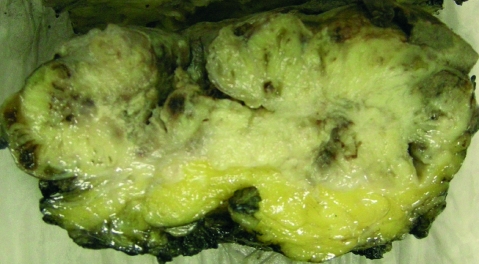
The initial specimen coded as breast cyst comprised multiple fragments measuring 3×2×1.5 cm at its greatest dimension and was grossly described as tan and firm to rubbery. The recurrence (ie, the second specimen) was an irregular shaped skin-covered piece of tissue which measured 13×11×10 cm and was tan. The cut surface showed a firm nodular lesion, which was focally ulcerating the overlying skin (gross photograph, fig 1). The third specimen received 6 months later, coded as mastectomy with axillary clearance, showed biopsy cavity as well as firm foci within the wall of the biopsy cavity.
Figure 1.
Gross photograph showing a tan grey firm lobulated mass with ulcerated skin.
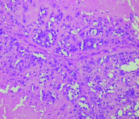
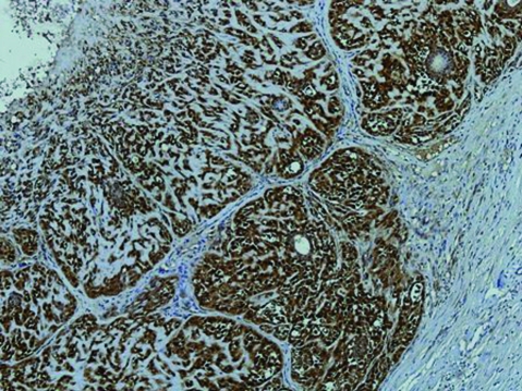
The initial specimen exhibited a tumour composed of a solitary, well circumscribed, nodular proliferation of epithelial and myoepithelial cells. Epithelial cells were flattened or cuboidal and appeared toward the central or luminal portion. Myoepithelial cells are spindled to polygonal in shape with clear cytoplasm. Mitoses and necrosis were present (fig 2). The myoepithelial cells showed diffuse positivity for α-smooth muscle actin (ASMA) immunohistochemical stain (fig 3).
Figure 2.
H&E stained slide 20× magnification showing epithelial forming glandular structures and myoepithelial spindle cell areas.
Figure 3.
α-Smooth muscle actin (ASMA) immunohistochemical stain showing diffuse positivity in tumour cells.
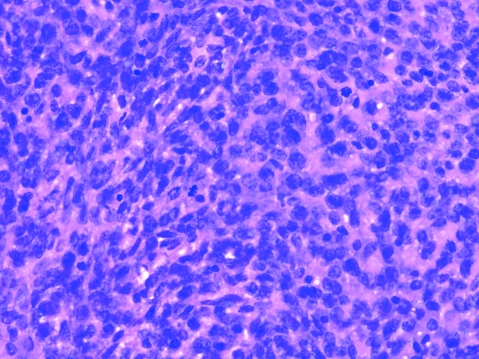
Wide local excision performed for tumour recurrence displayed the same biphasic, lobulated pattern. Epithelial and myoepithelial cells showed pleomorphism and atypia with increased mitotic activity ,with a maximum mitotic count of approximately 22 mitosis/10 high power field (HPF). Microscopic areas of necrosis were also seen. Skin ulceration was present (fig 4).
Figure 4.
H&E stained slide 40× magnification showing spindle cell areas with increased mitotic activity.
Sections taken from the mastectomy specimen revealed foci of spindle cells as well as areas reminiscent of the original lesion.
In the original tumour and recurrence specimens almost all myoepithelial cells were diffusely positive for S100 and ASMA. The epithelial cells reacted uniformly with AE1/AE3 and anti-cytokeratin (CAM 5.2) reagents.
TREATMENT
Lumpectomy followed by mastectomy with clear margins.
DISCUSSION
Myoepithelial cells are a normal component of breast tissue, and their presence in neoplastic lesions has been considered a hallmark of benignity. Recently, however, breast neoplasms have been described that are entirely or partially composed of myoepithelial cells. Neoplasm of purely myoepithelial origin have been called myoepitheliomas and may be benign or malignant in approximately equal propotions.4,6 Tumours with bicellular proliferation of epithelial and myoepithelial cells are called adenomyoepithelioma.3,6,11,13 Aenomyoepitheliomas of the breast are rare neoplasms first described by Hamperl in 1970 and further classified by Tavassoli in 1991.4,7 The majority of adenomyoepitheliomas are considered benign with the potential at most for local recurrence and their behaviour is believed to be similar to that of benign mixed tumours of salivary glands.4
Adenomyoepitheliomas of the breast are characterised histologically by aggregated lobules of small epithelial lined ducts, each surrounded by a mantle of myoepithelial cells. These may have polygonal shape with clear cytoplasm, or spindle shaped1,4 epithelial cells exhibiting positivity for AE1/E3 and CAM 5.2 and myoepithelial cells positive for S100 and ASMA.6,12
Rare examples of malignant adenomyoepithelioma with metastasis have been reported in literature.4,6 Most of these demonstrated malignant transformation of only one cellular component, mostly epithelial rather than myoepithelial. Only 11 (biphasic) adenomyoepitheliomas have been reported in which epithelial and myoepithelial cells were malignant.5,6
Adenomyoepithelial tumours have been reported in patients aged between 26 to 76 (mean age 60 years). The tumours can be peripheral or centrally located near the areola.5,6,13 Grossly adenomyoepitheliomas are well circumscribed, firm to hard and nodular, but may be soft or have ill-defined margins and occasionally contain small cystic areas.13 Tumour sizes range from 1 to 15 cm.6
Malignant change in an adenomyoepithelioma has been previously reported. Malignant change may involve only one cellular element, more often the epithelial component than the myoepithelial component. Malignant transformation of both elements may also occur, but it is extremely rare and fewer than 11 cases have been published in the literature to date.5,6,12,13 The presence of necrosis, marked cytological atypia and mitotic activity is usually associated with malignant transformation.6,8,13 In the case presented, the mitotic rate was greater than 22 per 10 high-power fields with large areas of necrosis and cytological atypia. The tumour exhibited irregular margins.
Metastases may occur in malignant adenomyopiythlioma.6,12 In a review of 12 cases of biphasic malignant adenomyoepithelioma, 1 study found that lung metastases were presented in 3 patients and brain metastases in 2 patients, and 1 patient presented with thyroid metastasis.6,12 Metastasis occur through haematogenous rather then lymphatic spread.3,6,12,13
The best predictor for local recurrence is the state of the surgical margin. If the excision is narrow or incomplete, re-excision to gain adequate margins is recommended.5
LEARNING POINTS
Biphasic malignant adenomyoepitheliomas of breast are very rare tumours.
Metastasis may occur; common sites of metastases include lung, brain and thyroid.
Complete excision with long-term follow-up appears to be an appropriate measure in all malignant adenomyoepitheliomas.
Footnotes
Competing interests: None.
Patient consent: Patient/guardian consent was obtained for publication.
REFERENCES
- 1.Simpson RHW, Cope N, Skalove A, et al. Malignant adenomyoepithelioma of the breast with mixed osteogenic, spindle cell and carcinomatous differentiation. Am J Surg Pathol 1998; 22: 631–6 [DOI] [PubMed] [Google Scholar]
- 2.McLarn BK, Smith J, Schuyler PA, et al. Adenomyoepithelioma: clinical, histologic, and immunohistologic evaluation of a series of related lesions. Am J SurgPathol 2005; 29: 1294–9 [DOI] [PubMed] [Google Scholar]
- 3.Rasbridge SA, Millis RR. Adenomyoepithelioma of the breast with malignant features. Virchow Arch 1998; 432: 123–30 [DOI] [PubMed] [Google Scholar]
- 4.Celina M, Nadelman, Kevin O, et al. Benign, matastasizing adnomyoepithelioma of the breast: report of 2 cases. Arch Pathol Lab Med 2006; 130: 1349–53 [DOI] [PubMed] [Google Scholar]
- 5.Suresh Attili VS, Kamal Saini, Laskshmaiah KC, et al. Malignant adenomyoepithelioma of the breast. Indian J Surg 2007; 69: 14–16 [Google Scholar]
- 6.Ahmed AA, Heller DS. Malignant adenomyoeoithelioma of the breast with malignant proliferation of epithelial elements: a case review of the literature. Arch Pathol Lab Med 2000; 124: 632–5 [DOI] [PubMed] [Google Scholar]
- 7.Tavassoli FA. Myoepithelial leasion of the breast. Myoepitheliosis, adenomyoepithelioma, and myoepithlial carcinoma. Am J Surg Pathol 1991; 15: 554–68 [DOI] [PubMed] [Google Scholar]
- 8.Hungermann D, Buerger H, Oehlschlegel C, et al. Adenomyoepithelioma tumours and myoepithelial carcinomas of breast –a spectrum of monophasic tumours dominated by immature myoepithelial cells. BMC Cancer 2005; 5: 92. [DOI] [PMC free article] [PubMed] [Google Scholar]
- 9.Hock Y-L, Chan S-Y. Aenomyopithelioma of the breast: a case report correlating cytologic and histologic features. Acta Cytol 1994; 38: 953–6 [PubMed] [Google Scholar]
- 10.Ng WK. Adenomyoepithelioma of the breast. A review of three cases with reappraisal of the fine needle aspiration bioposy findings. Acta Cytol 2002; 46: 317–24 [DOI] [PubMed] [Google Scholar]
- 11.Loose JH, Patchefsky AS, Hollander IJ, et al. Adenomyoepithelioma of the breast: a spectrum of biologic behavior. Am J Surg Pathol 1992; 16: 868–76 [DOI] [PubMed] [Google Scholar]
- 12.Howlett DC, Mason CH, Biswas S, et al. Adenomyoepithelioma of the breast: spectrum of disease with associated imaging and pathology. AJR Am J Roentgenol 2003; 180: 799–803 [DOI] [PubMed] [Google Scholar]
- 13.Harigopal M, Park K, Chen X, et al. Pathological quiz case: a rapidly increasing breast mass in a postmenopausal woman. Arch Pathol Lab Med 2004; 128: 235–6 [DOI] [PubMed] [Google Scholar]


