Abstract
The most common clinical presentation of chronic Q fever is endocarditis with infections of aneurysms or vascular prostheses being the second most common presentation. Here, the authors report a case of vascular chronic Q fever. In this patient, a renal artery aneurysm was discovered by abdominal and pelvic CT during a systematic investigation to identify predisposing factors to chronic Q fever because of high antibody titres in a patient with valve disease.
Background
Chronic Q fever is detected by an increase in phase I antigen-specific antibodies against Coxiella burnetii. It has been recommended to test serum samples 3 and 6 months after acute Q fever both to detect the progression to chronic Q fever and to investigate cardiac valve lesions using echocardiography.
The infection of aneurysms and vascular prostheses is the second most common form of chronic Q fever. Chronic Q fever is potentially fatal and therefore needs be diagnosed early to enable adequate treatment and avoid more severe complications. If chronic Q fever is suspected, a systematic search may allow for the discovery of small aneurysms.
Case presentation
A 72-year-old man was admitted to the Jacques Coeur Hospital in Bourges, France, on September 1, 2008, for polyarthralgia associated with biologic inflammatory syndrome. The symptoms had started 3 weeks earlier. On examination, he complained of shoulder pain, myalgia and neck stiffness. He was afebrile, asthenic and reported a weight loss of a few kilograms. Cardiopulmonary and neurological examinations were normal.
Investigations
The patient's leukocyte count was 6.5 g/l. The erythrocyte sedimentation rate and C-reactive protein were elevated at 95 mm, first hour and 54 mg/ml, respectively. Moreover, polyclonal hypergammaglobulinaemia (17.3 g/l) and hyper-α2globulinaemia (11.6 g/l) were detected. The serum level of liver enzymes was normal. Because a diagnosis of polymyalgia rheumatica was suspected, treatment with 40 mg per day of prednisone was started on September 4, 2008. Concomitantly, Q fever serology was found to be positive, both for immunoglobulins to phase II (1:800, 1:200 and 1:100 for IgG, IgM and IgA, respectively) and to phase I antigens (1:400, 1:100 and 1:50, respectively). Such titres were consistent with acute Q fever. Upon subsequent questioning, the patient acknowledged that he was in frequent contact with farm animals, the usual source of C burnetii, the aetiologic agent of Q fever. Treatment with doxycycline, 200 mg per day orally, was prescribed for 14 days, and the prednisone was decreased to 5 mg every 14 days. One month later, new serology showed increasing antibody titres consistent with chronic Q fever, with the titres of phase I antigen-specific antibodies being 1:6400, 1:100 and 1:50 for IgG, IgM and IgA, respectively (table 1). No C burnetii could be detected in the serum using a previously described PCR-based protocol.1 In our patient, both transthoracic and transoesophageal echocardiography ruled out the diagnosis of endocarditis, or pre-existing valvulopathy. Radiologic exploration was completed with abdominal and pelvic CT, and, as part of our systematic investigation of patients with chronic Q fever, a small, right-renal-artery aneurysm measuring 13 mm in diameter was detected. On the basis of these findings, the diagnosis of chronic vascular Q fever was made.
Table 1.
Evolution of Q fever serology (indirect immunofluoresence assay) and PCR in our patient
| Phase I | Phase II | PCR | |||||
|---|---|---|---|---|---|---|---|
| IgG | IgM | IgA | IgG | IgM | IgA | ||
| 17/09/2008 | 400 | 100 | 50 | 800 | 200 | 100 | |
| 18/10/2008 | 6400 | 0 | 50 | 12,800 | 0 | 100 | Negative |
| 18/12/2008 | 6400 | 0 | 50 | 12,800 | 0 | 100 | Negative |
Ig, immunoglobulin.
Treatment
Treatment with a combination of doxycycline (200 mg per day) and hydroxychloroquine (600 mg per day) was started for a minimum of 18 months. Surveillance of this treatment consists of both drugs dosages on serum samples and an ophthalmologic examination every 6 months to detect possible ocular toxicity due to hydroxychloroquine. Surgery was planned for the patient shortly after diagnosis.
Discussion
Q fever is an ubiquitous zoonosis caused by C burnetii. Infected aerosols generated by farm animals are the usual source of human infection.2 Our patient reported frequent contact with farm animals, specifically goats. C burnetii is an obligate intracellular bacterium that may cause acute and chronic infections in humans. Although most acute infections (60%) are asymptomatic, frequently observed symptoms include isolated fever, atypical pneumonia and hepatitis.3 Recovery is spontaneous in most cases. However, acute Q fever may evolve to chronic infection, that is, an infection persisting for more than 6 months, in 1 to 5% of patients.4 Such a progression occurs most frequently in patients with a valve disease, a vascular prosthesis or aneurysm, immunocompromised patients or in pregnant women.5 Serologically, chronic Q fever is characterised by an IgG titre to phase I antigen greater than 1:800. Clinically, chronic Q fever presents as endocarditis, vascular infections, osteoarticular infections and chronic hepatitis.6 Infective aneurysms and infection of vascular prostheses account for 9% of chronic Q fever cases.7 The risk of progression from acute to chronic infection is estimated to be 40% in patients with valvular defects,5 but it is as yet undetermined in patients with arterial diseases.
The delay between acute and chronic infection is variable. In 2007, Landais et al8 demonstrated that 50% of patients developed chronic Q fever within 3 months of acute infection, and 75% within 6 months. Here, the development of chronic Q fever may have been facilitated by the use of corticosteroids for polymyalgia rheumatica. Indeed, exacerbation of chronic Q fever with corticosteroid therapy has been previously reported.9 Despite the severity of chronic Q fever, its diagnosis is often delayed due to the absence of specific clinical symptoms and because the initial infection is often asymptomatic.10
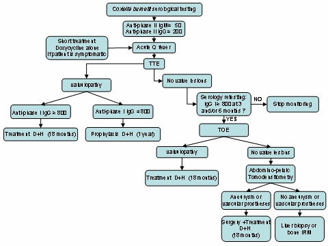
Q fever is mainly diagnosed through serology with the reference method being the indirect immunofluorescence assay.11 In our laboratory in Marseille (Southern France), the French National Reference Centre for Rickettsial Diseases, we use a microimmunofluorescence technique. Chronic Q fever was defined by a cut-off titre of phase I antigen-specific IgG greater than 1:800. The serological diagnosis of chronic Q fever should encourage the search for valve disease by echocardiography. Transthoracic echocardiography is recommended in the first instance; however, in the case of a normal transthoracic echocardiograph, transoephageal echocardiography should be performed in order to detect a bicuspid aortic valve or mitral regurgitation12 if the antibody titre increases. Indeed minor valvulopathies, such as minor valvular insufficiency, mitral valve prolapse or a bicuspid aortic valve, are a predisposing factor for Q fever endocarditis.13 If a valvular lesion is ruled out, abdominal and pelvic CT should be performed to search for an arterial aneurysm.14 (figure 1). The diagnosis of an aneurysm infection can be established by serology with the same profile as endocarditis.6 The documentation of cardiac or arterial abnormalities is crucial to improve the management and prognosis of chronic Q fever. The management of chronic Q fever is complex. Prolonged antibiotic therapy with both doxycycline (200 mg per day) and hydroxychloroquine (600 mg per day) should be administered for a minimum of 18 months.10
Figure 1.

Strategy and management of chronic Q fever diagnosis.
TTE: Transthoracic echocardiography
TOE: Transoesophageal echocardiography
D: doxycycline
H: hydroxychloroquine
C burnetii vascular infection has a poor prognosis. An early diagnosis is necessary to enable early treatment and avoid severe complications. In a recent study of 30 cases of C burnetii infected aortic aneurysms or vascular grafts, vascular surgery was significantly associated with recovery but the associated mortality rate was high (25%). Because rupture of infected aneurysms is the main complication of C burnetii aortic infections, surgery is required in most C burnetii vascular infections.15
It is recommended that serological testing be performed 3 and 6 months following the diagnosis of acute Q fever to allow for the early detection of chronic infection. Delays in diagnosis have been shown to have a significant negative impact on prognosis.8 When phase I antigen-specific antibody titres are increasing rapidly and when echocardiography is negative, we suggest performing a CT scan to identify any arterial aneurysms.
Learning points.
Radiologic examinations, notably echocardiography and CT, have as important a place in chronic Q fever diagnosis as serology because management of Q fever infection depends on the presence or absence of valve disease or vascular infection.
These vascular infections have a poor prognosis, which is why early management of this infection is fundamental, especially because diagnosic delays have a significant impact on prognosis.
Footnotes
Competing interests None.
Patient consent Obtained.
References
- 1.Fournier PE, Marrie TJ, Raoult D. Diagnosis of Q fever. J Clin Microbiol 1998;36:1823–34 [DOI] [PMC free article] [PubMed] [Google Scholar]
- 2.Maurin M, Raoult D. Q fever. Clin Microbiol Rev 1999;12:518–53 [DOI] [PMC free article] [PubMed] [Google Scholar]
- 3.Raoult D. Clinical Features, Diagnosis, Treatment, and Prevention of Q Fever. 2009. http://www.uptodate.com (Accessed 1 January 2010) [Google Scholar]
- 4.Raoult D, Marrie T. Q fever. Clin Infect Dis 1995;20:489–95; quiz 496 [DOI] [PubMed] [Google Scholar]
- 5.Fenollar F, Fournier PE, Carrieri MP, et al. Risks factors and prevention of Q fever endocarditis. Clin Infect Dis 2001;33:312–6 [DOI] [PubMed] [Google Scholar]
- 6.Raoult D, Tissot-Dupont H, Foucault C, et al. Q fever 1985–1998. Clinical and epidemiologic features of 1,383 infections. Medicine (Baltimore) 2000;79:109–23 [DOI] [PubMed] [Google Scholar]
- 7.Raoult D, Marrie T, Mege J. Natural history and pathophysiology of Q fever. Lancet Infect Dis 2005;5:219–26 [DOI] [PubMed] [Google Scholar]
- 8.Landais C, Fenollar F, Thuny F, et al. From acute Q fever to endocarditis: serological follow-up strategy. Clin Infect Dis 2007;44:1337–40 [DOI] [PubMed] [Google Scholar]
- 9.Lev BI, Shachar A, Segev S, et al. Quiescent Q fever endocarditis exacerbated by cardiac surgery and corticosteroid therapy. Arch Intern Med 1988;148:1531–2 [PubMed] [Google Scholar]
- 10.Fournier PE, Casalta JP, Piquet P, et al. Coxiella burnetii infection of aneurysms or vascular grafts: report of seven cases and review. Clin Infect Dis 1998;26:116–21 [DOI] [PubMed] [Google Scholar]
- 11.Dupont HT, Thirion X, Raoult D. Q fever serology: cutoff determination for microimmunofluorescence. Clin Diagn Lab Immunol 1994;1:189–96 [DOI] [PMC free article] [PubMed] [Google Scholar]
- 12.Brouqui P, Dupont HT, Drancourt M, et al. Chronic Q fever. Ninety-two cases from France, including 27 cases without endocarditis. Arch Intern Med 1993;153:642–8 [DOI] [PubMed] [Google Scholar]
- 13.Fenollar F, Thuny F, Xeridat B, et al. Endocarditis after acute Q fever in patients with previously undiagnosed valvulopathies. Clin Infect Dis 2006;42:818–21 [DOI] [PubMed] [Google Scholar]
- 14.Million M, Lepidi H, Raoult D. [Q fever: current diagnosis and treatment options]. Med Mal Infect 2009;39:82–94 [DOI] [PubMed] [Google Scholar]
- 15.Botelho-Nevers E, Fournier PE, Richet H, et al. Coxiella burnetii infection of aortic aneurysms or vascular grafts: report of 30 new cases and evaluation of outcome. Eur J Clin Microbiol Infect Dis 2007;26:635–40 [DOI] [PubMed] [Google Scholar]


