Abstract
A 41-year-old woman had a general health examination and was diagnosed with a non-functioning adrenocortical carcinoma (ACC). Despite surgery and chemotherapy with mitotane, the ACC progressed with metastases to the lymph nodes, liver and lung. Initially, she developed adrenal insufficiency and was treated with hydrocortisone. As the ACC progressed, it produced superabundant cortisol, resulting in clinically overt Cushing’s syndrome. As the liver metastases grew, the patient developed hypoglycaemia with suppression of endogenous insulin secretion. She had to be given large quantities of glucose intravenously to remain normoglycaemic. The serum insulin-like growth factor (IGF)-II/IGF-I ratio had increased to 84. We identified big IGF-II, a primary hormonal mediator of non-islet cell tumour hypoglycaemia (NICTH), in the serum and tumour using western blotting. This is the first case of ACC that showed both Cushing’s syndrome and NICTH associated with big IGF-II.
Background
Adrenocortical carcinoma (ACC) is a rare tumour accounting for <0.2% of all malignancies.1,2 The natural clinical course of ACC is not well known because of its poor prognosis. Excluding insulinoma, tumour associated hypoglycaemia has been reported in various neoplasms and is recognised as non-islet cell tumour hypoglycaemia (NICTH), which is caused by an insulin independent pathway.3,4 Big insulin-like growth factor (IGF)-II secreted from the tumour is thought to be the primary hormonal mediator of NICTH.5–7 Here, we report a patient who presented with adrenal failure, Cushing’s syndrome and hypoglycaemia during the course of ACC progression. This is the first case in which big IGF-II protein was demonstrated in both serum and the tumour.
Case presentation
A 41-year-old woman had a general health examination that revealed an abdominal mass on ultrasonography in November 2005. The patient was diagnosed with stage III ACC (T3N0M0) following a detailed examination. The tumour was resected with her left adrenocortical gland in January 2006. She received chemotherapy with mitotane postoperatively. However, a follow-up fluorodeoxyglucose-positron emission tomography examination integrated with computed tomography (FDG-PET CT) revealed a recurrence in a left adrenal site. She underwent a second tumour resection in July 2006.
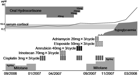
In September 2006, she was admitted to Kanazawa University Hospital to receive additional chemotherapy. The laboratory data on admission suggested anaemia and an increase in ductal enzymes. The endocrinological examination revealed adrenal cortex dysfunction, and plasma adrenocorticotropic hormone (ACTH) was elevated at 45.9 pg/ml. Urinary excretion of dehydroepiandrosterone (DHEAS), 17-hydroxycorticosteroid (17-OHCS) and 17-ketosteroids (17-KS) had decreased to 355 ng/ml, 1.1 mg/day and 0.7 mg/day, respectively. She was given replacement therapy with 15 mg of oral hydrocortisone (fig 1).
Figure 1.
Clinical course.
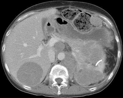
As the ACC progressed, it produced superabundant cortisol, resulting in clinically overt Cushing’s syndrome; her ACTH values were suppressed, and she was Cushingoid with central obesity and red striae. Her serum cortisol concentration was 39.8 μg/dl without oral hydrocortisone (table 1). At this time, a massive left adrenocortical tumour infiltrated the pancreas and left kidney and metastasised to the liver, spleen and central nodes, as revealed on computed tomography (fig 2).
Table 1.
Endocrinological data when the patient developed hypoglycaemia
| Normal range | Units | ||
| Blood glucose | 19 | 69–109 | mg/dl |
| IRI | <1.0 | <17 | μU/ml |
| C-peptide | 0.2 | 0.62–3.50 | ng/ml |
| Glucagon | 100 | 70–160 | pg/ml |
| IGF-I | 14 | 100–300 | ng/ml |
| IGF-II | 1200 | 400–800 | ng/ml |
| GH | 0.38 | <2.1 | ng/ml |
| ACTH | <5 | <46 | pg/ml |
| Cortisol | 39.8 | 6.2–19.4 | μg/dl |
| DHEAS | 355 | ng/ml | |
| Adrenalin | <0.01 | <0.17 | ng/ml |
| Noradrenaline | 0.27 | 0.15–0.57 | ng/ml |
| Dopamine | <0.02 | <0.03 | ng/ml |
ACTH, adrenocorticotropic hormone; DHEAS, dehydroepiandrosterone sulfate; GH, growth hormone; IGF, insulin-like growth factor; IRI, immunoreactive insulin.
Figure 2.
Abdominal computed tomography scan when the patient first exhibited symptoms of hypoglycaemia. A massive adrenocortical tumour infiltrated the pancreas and left kidney, and metastasised to the liver, spleen and central nodes.
One morning in December 2007, she first exhibited symptoms of hypoglycaemia, which progressively became more severe. When the patient developed hypoglycaemia, her serum insulin concentration was <1.0 μU/ml, C-peptide was <0.2 ng/ml, and blood glucose was 19 mg/dl.
Investigations
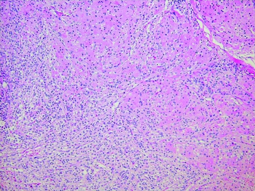
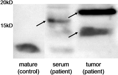
Histopathology showed malignant cells in the adrenocortical site, predominantly eosinophilic, with a diffuse architectural pattern (fig 3). The adrenal tumour was immunohistochemically stained (data not shown) and examined using Western blotting with anti-human IGF-II rabbit polyclonal antibody (Abcam) and mouse monoclonal antibody (Upstate) (fig 4). Western blotting revealed big IGF-II protein in both the tumour and serum. The molecular weight of the big IGF-II was 15–20 kDa, which was greater than that of mature IGF.
Figure 3.
Histopathology showing malignant cells in the adrenocortical site, predominantly eosinophilic, with a diffuse architectural pattern.
Figure 4.
Western blotting revealing big IGF-II in both the tumour and serum. The molecular weight of the big IGF-II was 15–20 kDa, which is heavier than that of mature IGF.
Differential diagnosis
At the point of hypoglycaemia, the serum insulin concentration was <1.0 μU/ml, C-peptide was <0.2 ng/ml, and the plasma glucose was 19 mg/dl (table 1).
Treatment
Although the patient underwent chemotherapy with mitotane 4 g per day, the follow-up abdominal CT revealed growth of the tumour. Additional combined chemotherapy with cisplatin, irinotecan, amrubicin hydrochloride, ketoconazole, etoposide and adriamycin was ineffective (fig 1).
Her hypoglycaemia was so severe that she had to be given large quantities of glucose (>20 g/h) to keep her plasma glucose within normal limits. The somatostatin analogue octreotide did not improve her hypoglycaemia.
Outcome and follow-up
The patient died from pneumonia and an autopsy was performed.
Discussion
The aetiology of NICTH could be as follows: increased glucose consumption by the tumour, decreased glycogenolysis and gluconeogenesis because of the liver metastases, and suppression of the counter-regulatory hormones for insulin or insulin-like factor secreted by the tumour.
NICTH is mediated via IGF-II (molecular weight 7.5 kDa), which exhibits a high degree of structural homology to proinsulin.5,6 Only one case of ACC with NICTH has been reported.8 In that case, Cushing’s syndrome was not evident: the serum IGF-II concentration was elevated and the hypoglycaemia improved after removing the tumour; ACC tissue was positive for IGF-II immunohistochemically, but no evidence of big IGF-II associated with ACC was seen.
We report the first case of ACC presenting with both Cushing’s syndrome and NICTH in whom big IGF-II protein was demonstrated in both the serum and tumour using western blotting; the molecular weight of the IGF-II was 15–20 kDa, which is heavier than normal.
IGF-II exists in three different forms in serum: 20–30% of total IGF-II is bound to a 50 kDa IGF-binding protein (IGFBP), 70–80% of total IGF-II occurs in a 150 kDa complex, and free IGF-II is <1%. In NICTH, 70% of the patients have high molecular weight IGF-II, so-called “big” IGF-II (11–18 kDa), although the total IGF-II values are within the normal range.9 Big IGF-II forms binary complexes with IGFBP, instead of the normal ternary 150 kDa complex, and these small complexes reach insulin target organs through the capillaries. Delivered big IGF-II could increase glucose uptake and inhibit hepatic gluconeogenesis.10,11
Comprehensive gene expression profiling using DNA microarrays showed that IGF-II is expressed abundantly in ACC.12 As the ACC develops, big IGF-II is generated by abnormal processing of an IGF-II precursor.7,13
In addition, posttranslational modifications such as O-glycosylation of the residues in the first 21 positions of the proIGF-II E-domain have been suggested to be involved in the generation of big IFG-II.14 Indeed, we found variable sized protein blotted with antibody to IGF-II in both the tumour and serum of our patient.
In the clinical setting, diagnosing NICTH is difficult because the total IGF-II is within the normal range. Hizuka et al suggested that the IGF-II/IGF-I ratio in serum is useful for detecting NICTH with big IGF-II.9 They reported that the IGF-II/IGF-I ratio in serum exceeded 20 in patients with NICTH.15 In these patients, IGF-I production could be suppressed by an insulin-like factor, big IGF-II. In our case, the IGF-II/IGF-I ratio increased to 84 in the terminal phase.
Therapeutic treatment with glucocorticoids is reported to suppress big IGF-II production.16 Notably, our case developed Cushing’s syndrome because the ACC produced excessive cortisol autonomously. However, our patient required large amounts of glucose to keep her plasma glucose within normal limits.
This is the first case of ACC in which big IGF-II was identified in both the serum and tumour using western blotting. To our knowledge, the natural clinical course of ACC is not well known because of the poor prognosis. Our case might present the natural clinical course of ACC. In the initial stage, our patient needed oral hydrocortisone; subsequently, the tumour produced superabundant cortisol resulting in Cushing’s syndrome; and finally, it caused hypoglycaemia by NICTH. With the current progress in cancer therapy, patients with ACC will live longer, resulting in more big IGF-II being produced to cause more severe hypoglycaemia. Further study will contribute to the therapy for hypoglycaemia caused by NICTH in ACC.
Learning points
The typical natural course of patients with adrenocortical carcinoma (ACC) might involve adrenal failure, Cushing’s syndrome and hypoglycaemia caused by ACC associated big IGF-II.
Stage specific care is needed for such patients.
Acknowledgments
We thank all the doctors who provided us with opportunity to study the clinical course of this case. This work was supported by Department of Disease Control and Homeostasis, Kanazawa University Graduate School of Medical Science.
Footnotes
Competing interests: None.
Patient consent: Patient/guardian consent was obtained for publication.
REFERENCES
- 1.Schulick RD, Brennan MF. Adrenocortical carcinoma. World J Urol 1999; 17: 26–34 [DOI] [PubMed] [Google Scholar]
- 2.Dackiw AP, Lee JE, Gagel RF, et al. Adrenal cortical carcinoma. World J Surg 2001; 25: 914–26 [DOI] [PubMed] [Google Scholar]
- 3.Nadler WH, Wolfer JA. Hepatogenic hypoglycemia associated with primary liver cell carcinoma. Arch Intern Med 1929; 44: 700–10 [Google Scholar]
- 4.Kahn CR. The riddle of tumour hypoglycaemia revisited. Clin Endocrinol Metab 1980; 9: 335–60 [DOI] [PubMed] [Google Scholar]
- 5.Daughaday WH, Emanuele MA, Brooks MH, et al. Synthesis and secretion of insulin-like growth factor II by a leiomyosarcoma with associated hypoglycemia. N Engl J Med 1988; 319: 1434–40 [DOI] [PubMed] [Google Scholar]
- 6.Daughaday WH, Kapadia M. Significance of abnormal serum binding of insulin-like growth factor II in the development of hypoglycemia in patients with non-islet-cell tumors. Proc Natl Acad Sci U S A 1989; 86: 6778–82 [DOI] [PMC free article] [PubMed] [Google Scholar]
- 7.de Groot JW, Rikhof B, van Doorn J, et al. Non-islet cell tumour-induced hypoglycaemia: a review of the literature including two new cases. Endocr Relat Cancer 2007; 14: 979–93 [DOI] [PubMed] [Google Scholar]
- 8.Eguchi T, Tokuyama A, Tanaka Y, et al. Hypoglycemia associated with the production of insulin-like growth factor II in adrenocortical carcinoma. Intern Med 2001; 40: 759–63 [DOI] [PubMed] [Google Scholar]
- 9.Hizuka N, Fukuda I, Takano K, et al. Serum insulin-like growth factor II in 44 patients with non-islet cell tumor hypoglycemia. Endocr J 1998; 45(Suppl): S61–5 [DOI] [PubMed] [Google Scholar]
- 10.Burvin R, LeRoith D, Harel H, et al. The effect of acute insulin-like growth factor-II administration on glucose metabolism in the rat. Growth Horm IGF Res 1998; 8: 205–10 [DOI] [PubMed] [Google Scholar]
- 11.Rossetti L, Barzilai N, Chen W, et al. Hepatic overexpression of insulin-like growth factor-II in adulthood increases basal and insulin-stimulated glucose disposal in conscious mice. J Biol Chem 1996; 271: 203–8 [DOI] [PubMed] [Google Scholar]
- 12.Giordano TJ, Thomas DG, Kuick R, et al. Distinct transcriptional profiles of adrenocortical tumors uncovered by DNA microarray analysis. Am J Pathol 2003; 162: 521–31 [DOI] [PMC free article] [PubMed] [Google Scholar]
- 13.Ilvesmaki V, Kahri AI, Miettinen PJ, et al. Insulin-like growth factors (IGFs) and their receptors in adrenal tumors: high IGF-II expression in functional adrenocortical carcinomas. J Clin Endocrinol Metab 1993; 77: 852–8 [DOI] [PubMed] [Google Scholar]
- 14.Hizuka N, Fukuda I, Takano K, et al. Serum high molecular weight form of insulin-like growth factor II from patients with non-islet cell tumor hypoglycemia is O-glycosylated. J Clin Endocrinol Metab 1998; 83: 2875–7 [DOI] [PubMed] [Google Scholar]
- 15.Fukuda I, Hizuka N, Ishikawa Y, et al. Clinical features of insulin-like growth factor-II producing non-islet-cell tumor hypoglycemia. Growth Horm IGF Res 2006; 16: 211–6 [DOI] [PubMed] [Google Scholar]
- 16.Teale JD, Wark G. The effectiveness of different treatment options for non-islet cell tumour hypoglycaemia. Clin Endocrinol (Oxf) 2004; 60: 457–60 [DOI] [PubMed] [Google Scholar]


