Abstract
A 31-year-old Moroccan woman with no significant past medical history was seen during her second pregnancy. At 25 weeks gestation she was admitted with a febrile illness associated with a productive cough which was treated as a community acquired pneumonia with oral antibiotics. At 31 weeks gestation she was admitted with a tender swelling in the right groin and underwent incision and drainage of a presumed femoral abscess. At 36 weeks gestation she re-presented with multiple skin lesions on her arms, legs and buttocks. Initial investigation found no obvious cause for her presentation. The decision for induction of labour was taken as the patient was not improving, and resulted in an uncomplicated Caesarean delivery. After delivery, Mantoux and Quantiferon tests were reported to be positive and the patient was diagnosed with papulonecrotic tuberculides.
Background
Papulonecrotic tuberculids (PNT) is a rare cutaneous manifestation of tuberculosis (TB). Here we describe a case of PNT as the main manifestation of TB presenting during pregnancy. The rarity of this condition makes this a difficult diagnosis.
Case presentation
A 31-year-old Moroccan woman booked for her second pregnancy, having conceived spontaneously. Her previous pregnancy 4 years earlier had been the result of in vitro fertilisation (IVF) and she had a normal vaginal delivery at term. She had no significant past medical history. Her booking bloods and infection screen including HIV were normal, as was an oral glucose tolerance test performed at 28 weeks.
The initial antenatal period was uncomplicated. At 25 weeks gestation she was admitted with a febrile illness associated with productive cough and mildly raised inflammatory markers. A chest x-ray was performed and was grossly normal. This episode was treated as a community acquired pneumonia with oral antibiotics.
At 31 weeks gestation she was admitted with a tender swelling in the right groin. It was felt that this was an incarcerated femoral hernia and she underwent groin exploration by the surgical team. This demonstrated a groin abscess which was drained. Histology from the specimen obtained during this procedure demonstrated fibrofatty tissue and lymph nodes showing non-specific reactive changes. The surrounding tissue contained multiple granulomata with necrotic centres, and exuberant fibrosis was also noted. Staining for fungi and acid fast bacilli was negative. A gradual recovery from this episode was made and she was discharged 7 days later.
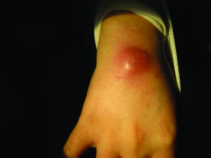
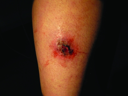
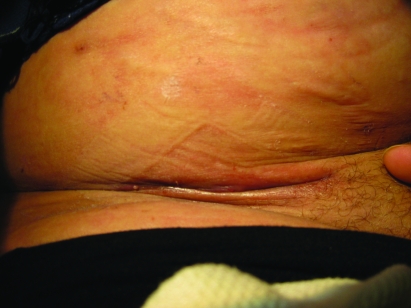
She re-presented at 36 weeks gestation with a history of having developed further “abscesses” initially on the left leg, then on the right buttock, then on the right leg, and finally at the site of a previous cannula insertion on her left forearm. The formation of these skin lesions was associated with symptoms of myalgia, weakness and lethargy. Clinical examination demonstrated multiple skin lesions at different stages of development on the patient’s arms, legs and buttocks. Early lesions comprised firm, tender erythematous papules 1–2 cm in size (fig 1). Older lesions demonstrated central necrosis and ulceration (fig 2). It was also noted that the surgical incision from her previous groin exploration had failed to heal (fig 3).
Figure 1.
An early lesion developing on the dorsal aspect of the patient’s wrist—a tender, firm, erythematous papule.
Figure 2.
A later lesion on patient’s right leg with evidence of central necrosis and ulceration of an earlier lesion.
Figure 3.
Surgical site of groin exploration, 4 weeks previously, which was failing to heal.
The patient was commenced on broad spectrum intravenous antibiotics. She remained persistently afebrile. The only abnormality demonstrated on standard haematological and biochemical investigation was a significantly raised C reactive protein (CRP); white blood cell count (WBC) and differential counts were normal. Blood cultures were performed but demonstrated no growth, and a chest x-ray was performed and reported as normal. An echocardiogram was performed to exclude endocarditis and associated septic emboli, and was also normal. A further oral glucose tolerance test was performed and demonstrated no abnormality. It was considered that this presentation might be explained by underlying immunocompromise; however, no evidence of T cell/B cell or complement component deficiency could be demonstrated following immunological assessment. An HIV test was also repeated and was reported as negative. Culture from skin lesion sites grew only coagulase negative staphylococcus, felt likely to be skin flora. Polymerase chain reaction (PCR) amplification tests for Mycobacterium tuberculosis were performed from skin lesions and were negative.
Because there was uncertainty regarding the diagnosis and the patient’s condition was not improving, after a thorough discussion with the patient the decision was made to induce labour. The induction process was started; however, following administration of the first prostaglandin vaginal pessary, signs of fetal distress developed. Consequently, she proceeded to an uncomplicated emergency Caesarean section and delivered a healthy female baby, weighing 3960 g with normal APGAR scores.
Soon after delivery Mantoux and Quantiferon tests were reported to be positive and therefore advice was sought from pulmonary physicians. It was felt that her presentation was likely that of PNT and she was immediately started on quadruple therapy for TB.
Investigations
Haematological/biochemical indices:
Haemoglobin 10.9 g/dl, WBC 9.1×10⁹/l, neutrophils 6.6×10⁹/l, Na+ 136 mmol/l, K+ 3.8 mmol/l, creatinine 53 mmol/l, urea 4.7 mmol/l, CRP 122 mg/l.
Blood cultures: no growth
HIV test: negative
Oral glucose tolerance test: normal
Imaging:
Chest x-rays: reported as normal
Echocardiogram: reported as normal
Swabs from lesions:
Culture: coagulase negative Staphylococcus.
PCR amplification test for M. tuberculosis: negative
Histology of right femoral abscess sac:
Fibrofatty tissue with lymph nodes showing non-specific reactive changes. The surrounding tissues, however, contain multiple granulomata with necrotic centres. Exuberant fibrosis is also a feature.
Microscopy of right femoral abscess sac:
Staining of specimen for fungi and acid fast bacilli was negative.
Mantoux test:
Positive (16 mm)
Quantiferon test:
Positive (8%).
Differential diagnosis
Bacterial skin infection
Miliary TB with cutaneous involvement
Bacterial endocarditis
Pityriasis lichenoides et varioliformis acuta
Papulopustular syphilid
Drug sensitivity.
Treatment
Quadruple therapy for TB (rifampicin, isoniazid, pyrazinamide, ethambutol)
Delivery of baby.
Outcome and follow-up
Follow-up of her skin lesions showed probable need for skin grafts as healing had occurred with excessive scarring.
Discussion
PNT is a rare cutaneous manifestation of TB, infrequently seen in the Western world but more commonly encountered in areas of higher TB prevalence.1 PNT has been described as a recurrent, symmetric eruption of papules with a necrotic centre, arising in crops, and most commonly involving the arms and legs.1,2 A hallmark of this condition is healing of the lesions with scarring and hyperpigmentation.3
The pathophysiology of this condition is controversial. The most commonly held view is that PNT represents a hypersensitivity reaction to TB antigens released from a distant focus of infection.3 Evidence of TB elsewhere is reported in up to 40% of patients.4 Tubercle bacilli have never been demonstrated from in skin biopsies; however, mycobacterial DNA has been isolated from lesions using PCR techniques in around 50% of cases.5 The eruption normally resolves promptly with antituberculoid therapy.
In this case it was not until the Mantoux and Quantiferon tests were reported to be positive that the diagnosis was made. Other features that that would aid diagnosis include a history of TB or previous exposure to TB, histology suggestive of TB from lesion biopsies, and a response to antituberculoid therapy.1,3 An awareness of this condition is important as failure to make an appropriate diagnosis would result potentially curative treatment being withheld. A review of literature revealed no recent case report of PNT presenting in pregnancy.
Learning points
Papulonecrotic tuberculids (PNT) is a rare cutaneous manifestation of tuberculosis (TB).
TB should be excluded in any patient presenting with skin lesions suggestive of PNT.
PNT is treated with standard antituberculoid therapy.
This is the first recent description of this presentation in pregnancy.
Footnotes
Competing interests: none.
Patient consent: Patient/guardian consent was obtained for publication
REFERENCES
- 1.Tay E, Chinegwundoh J, Sahota A, et al. Papulonecrotic tuberculide: a forgotten manifestation of tuberculosis. Hosp Med 1999; 60: 450–2 [DOI] [PubMed] [Google Scholar]
- 2.Sloan JB, Medenica M. Papulonecrotic tuberculid in a 9-year-old American girl: case report and review of the literature. Pediatr Dermatol 1990; 7: 191–5 [DOI] [PubMed] [Google Scholar]
- 3.Dar NR, Raza N, Zafar O, et al. Papulonecrotic tuberculids associated with uveitis. J Coll Physicians Surg Pak 2008; 18: 236–8 [PubMed] [Google Scholar]
- 4.Jordaan HF, Van Niekerk DJ, Louw M. Papulonecrotic tuberculid. A clinical, histopathological, and immunohistochemical study of 15 patients. Am J Dermatopathol 1994; 16: 474–85 [PubMed] [Google Scholar]
- 5.Freiman A, Ting P, Miller M, et al. Papulonecrotic tuberculid: a rare form of cutaneous tuberculosis. Cutis 2005; 75: 341–6 [PubMed] [Google Scholar]


