Abstract
Bilateral tension pneumothorax occurring as a result of recreational activity is exceedingly rare. A 10-year-old boy with no previous respiratory symptoms was involved in a bicycle-to-bicycle collision during play. He was the only one hurt. A few hours later, he was rushed to the general casualty unit of the emergency department of our institution with respiratory distress, diminished bilateral chest excursions and diminished breath sounds. The correct diagnosis was made after a chest radiograph was obtained in the course of resuscitation at the casualty unit. Pleural space needle decompression was suggestive of tension only on the right. Bilateral tube thoracostomies provided effective relief. He was discharged from hospital after a week in excellent health. This case illustrates the need for children to have safety instruction to reduce the risks of recreational bicycling. Chest radiography may be needed to establish the diagnosis of bilateral tension pneumothorax. Needle thoracostomy decompression is not always effective.
Background
Bilateral tension pneumothorax (BTP) of any cause is rare. Clinical signs alone may not be adequate to establish the diagnosis. Barotrauma occasioned by positive pressure mechanical ventilation is responsible for most cases of BTP. BTP following blunt trauma suffered during recreational sporting activity is exceedingly rare. We report such a case in a young boy following an unsupervised recreational bicycle activity to highlight the diagnostic challenges, the problems with pleural space needle decompression, and the necessity of safety instruction for children engaged in recreational outdoor activities.
Case presentation
A 10-year-old boy was rushed to the general casualty unit of the emergency department of our institution with acute respiratory distress of a few hours’ duration. The boy had been in previous good health with no prior lung disease. He had been involved in an unsupervised bicycle sport with friends at home a few hours before onset of symptoms. The goal of the sport was for two opponents to race their bikes at top speed toward each other and swerve to avoid a head-on collision just before the moment of impact. Unfortunately, during one of such runs, both riders swerved in the same direction and collided. The game was interrupted to attend to this 10-year-old who complained of chest pain and some shortness of breath; the other victim of the collision suffered no ill effects. A few hours later, the boy was noticed to be increasingly breathless and restless. The older siblings were informed, following which the boy was rushed to the casualty unit of our institution. At the casualty unit, the history of the collision during play was withheld; the boy was reported to have been taken ill while at play. The casualty doctor on call found the patient to be restless with laboured breathing, with a tachypnoea of 40/min and bilaterally reduced chest excursions and breath sounds. There was no tracheal deviation. The percussion notes were judged to be resonant bilaterally. On high flow oxygen (12 litres/min) by reservoir bag, the Spo2 was 74%. He had a tachycardia of 134/min and a blood pressure of 86/54 mm Hg. Lacking an accurate history at the time, and on the basis of acute onset of reduced air flow to both lungs, an upper airway obstruction was (mis-) diagnosed. The decision to proceed to a tracheotomy (based on insufficient evidence of upper airway obstruction) was erroneous. The tracheotomy attempt was soon abandoned when subcutaneous emphysema became obvious. The cardiothoracic team was contacted while a chest radiographic examination was arranged at the casualty.
Investigations
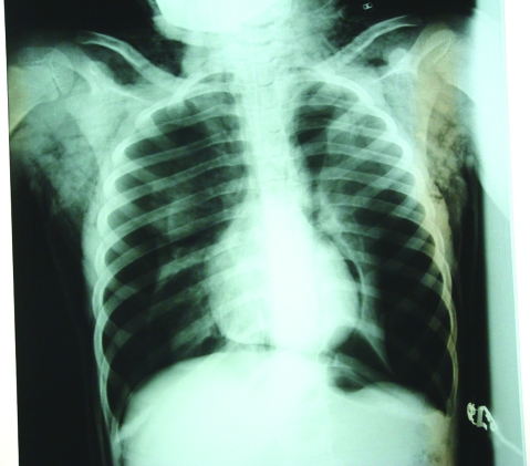
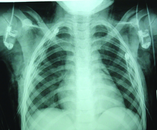
The chest radiographic examination (fig 1) was performed at the casualty unit shortly before pleural space needle decompression was attempted bilaterally. The chest film was ready for viewing while a right sided chest tube insertion was near completion. It showed bilateral tension pneumothorax evidenced by collapse of both lungs, hyperexpansion of both chest cavities, depression of both hemidiaphragms, and compression of the lateral cardiac borders and mediastinum. Rib fractures were not demonstrable. A second chest tube was inserted on the left side. After the bilateral tube thoracostomies, a repeat x-ray showed expansion of both lungs and relief of the features of tension.
Figure 1.
Chest x-ray showing bilateral tension pneumothorax.
Differential diagnosis
Upper airway obstruction was the initial working diagnosis although the basis for this was questionable. When the cardiothoracic team was contacted, spontaneous pneumothorax, possibly bilateral, was suspected though bilateral tension physiology was not considered.
Treatment
Resuscitation included administration of high flow oxygen by reservoir bag. Needle decompression of the pleural space using a 16 gauge over-the-needle cannula (inserted full length, about 4.5 cm into the fifth interspace, mid-axillary line) was performed. This produced a brief hiss of air on the right but not on the left side. Chest tube insertion was then performed on the right and subsequently on the left side (fifth intercostal space mid-axillary line on both sides) with resolution of respiratory distress. Chest tube decompression produced expulsion of air under pressure indicating bilateral tension pneumothorax. Expansion of both lungs was confirmed radiologically (fig 2) afterwards.
Figure 2.
Post-tube thoracostomy chest x-ray showing expansion of both lungs.
Outcome and follow-up
The child made a smooth recovery following tube thoracostomies and was discharged home after seven days of hospitalisation. The full details of the events leading up to the injury were obtained on the third day of admission when the patient himself could be interviewed in detail. The patient remains well 5 years after the event with excellent respiratory function and normal lung fields on chest x-ray.
Discussion
Although BTP of any aetiology is rare, unilateral tension pneumothorax (UTP) is not uncommon following blunt chest trauma. A pneumothorax after blunt chest trauma results when a fractured rib is driven inwards to cause a lung puncture or laceration. It may also result from sudden compression of the chest with a closed glottis without rib fracture. Viano’s group1 estimated that the force for all impact speeds resulting in rib fracture range from 5.5–11.2 kN; the force required to cause a pneumothorax in the absence of rib fracture is unknown. Sports- or recreational activity-related chest trauma is uncommon, representing only 2% of all chest injuries requiring treatment.2 The largest series of sports-related pulmonary air leaks has been reported by Kizer and MacQuarrie.3 In their report, the greatest number of cases of sports related traumatic pneumothorax resulted from martial arts, bicycling, and equestrian sports.3 To the best of our knowledge, this is the first report in the English literature of a bicycle-to-bicycle collision resulting in bilateral tension pneumothorax.
The amount of kinetic energy involved is a significant factor in impact injuries. In the case under discussion, riding the bicycle at top speed was a fundamental determinant of the resulting injury. Barotrauma, the presumed mechanism of injury, is more likely above a transalveolar pressure of 35 mm Hg when alveoli are over-distended and the more fragile ones tend to rupture.4
The most probable mechanical explanation for our patient’s injury is that the moment of impact coincided with a full inspiration against a closed glottis, causing alveolar over-distension and rupture without concomitant rib fracture. Presumably, a one way valve mechanism resulted in both pleurae to give rise to BTP in the interval between impact and presentation at the casualty. Other possible mechanisms include occult rib fractures causing pneumothorax and barotrauma resulting from air forced down an open glottis during high speed cycling.
Wearing of protective gear by children using bicycles as a means of transportation and recreation has been advocated and adopted in several countries. This advocacy has not been paralleled by safety education towards risk reduction by children in the same setting. Although we recognise that risk is an inevitable downside to childhood play and activity, a closer look at childhood injuries displays common patterns from which risk reduction strategies can be derived. In the case under discussion, a simple directive to the sport (for example, each opponent swerving to their right just before impact) could make the game less risky without taking away the activity or the fun from the sport. We certainly encourage childhood play and activity, and cycling is a useful activity both for transportation and recreation. But we believe children need guidance to balance risk and recreation.
While the diagnosis of UTP may be difficult to establish, the occurrence of bilateral tension complicates the diagnosis even further. The diagnosis of BTP using clinical signs alone may be difficult.5 Reduced chest wall excursions and diminished breath sounds occurring bilaterally may be confused with other entities such as severe asthma6 or upper airway obstruction. In the reported case, the diagnosis of upper airway obstruction was based on insufficient evidence; good clinical examination would have indicated the likelihood of pneumothorax. The unsuspecting clinician, however, is unlikely to establish the diagnosis of BTP in this case. Others7 have reported on the inconsistencies in eliciting the commonly taught classical signs of tension pneumothorax in the emergency setting. A prompt chest radiograph may be useful in establishing the diagnosis of BTP and prevent fatality.
Needle decompression followed by tube thoracostomy is widely advocated by many as the optimal approach to the patient with tension pneumothorax. It is also widely conceived as a ‘rule out’ investigation in the patient with suspected tension pneumothorax. However, as our case illustrates, needle decompression may prove ineffective even in established tension pneumothorax. The reasons underlying failure of needle decompression in tension pneumothorax have been described by other workers.5 Among the factors that may result in failure of decompression, chest wall thickness relative to the needle, obstruction of the cannula caused by blood, tissue or kinking of the cannula are important. In this patient, it is likely that the presence of subcutaneous emphysema may have presented a relatively thicker chest wall for penetration, although this does not explain the effectiveness of needle decompression on the right side. Presumably the cannula was obstructed by blood or tissue and could not drain. A larger cannula (14 gauge) may have been effective, although this was not readily available at the time.
The limitations of needle decompression as a ‘rule out’ investigation for tension pneumothorax must be appreciated. Absence of the classic hiss of air with needle thoracostomy does not rule out tension pneumothorax.5,8 Failure to appreciate this fact in a patient with tension pneumothorax is likely to result in unnecessary morbidity and mortality.
Learning points
Bilateral tension pneumothorax may be a difficult diagnosis without chest radiography.
Needle thoracocentesis does not provide consistently effective decompression or confirmation for tension pneumothorax.
Children need to have safety instruction to reduce the risks of recreational bicycling.
Footnotes
Competing interests: none.
Patient consent: Patient/guardian consent was obtained for publication
REFERENCES
- 1.Viano DC, Lau IV, Asbury C, et al. Biomechanics of the human chest, abdomen and pelvis in lateral impact. Accid Anal Prev 1989; 21: 553–74 [DOI] [PubMed] [Google Scholar]
- 2.Patridge RA, Coley A, Bowie R, et al. Sports-related pneumothorax. Ann Emerg Med 1997; 30: 539–41 [DOI] [PubMed] [Google Scholar]
- 3.Kizer KW, MacQuarrie MB. Pulmonary air leaks resulting from outdoor sports: a clinical series and literature review. Am J Sports Med 27: 517–20 [DOI] [PubMed] [Google Scholar]
- 4.Levy AS, Bassett F, Lintner S, et al. Pulmonary barotrauma: diagnosis in American football players. Three cases in three years. Am J Sports Med 1996; 24: 227–9 [DOI] [PubMed] [Google Scholar]
- 5.Leigh-Smith S, Harris T. Tension pneumothorax—time for a re-think? Emerg Med J 2005; 22: 8–16 [DOI] [PMC free article] [PubMed] [Google Scholar]
- 6.Sunam G, Gok M, Ceran S, et al. Bilateral pneumothorax: a retrospective analysis of 40 patients. Surg Today 2004; 34: 817–21 [DOI] [PubMed] [Google Scholar]
- 7.S Leigh-Smith S, Davies G. Tension pneumothorax: eyes may be more diagnostic than ears. Emerg Med J 2003; 20: 495–6 [DOI] [PMC free article] [PubMed] [Google Scholar]
- 8.Castle N, Tagg A, Owen R. Bilateral tension pneumothorax. Resuscitation 2005; 65: 103–5 [DOI] [PubMed] [Google Scholar]


